Ultrasound Obstet Gynecol 2009; 33: 421–426 Published online in Wiley InterScience (www.interscience.wiley.com). DOI: 10.1002/uog.6320
Ultrasonographic measurement of thymus size in IUGR fetuses: a marker of the fetal immunoendocrine response to malnutrition A. CROMI*, F. GHEZZI*, R. RAFFAELLI†, V. BERGAMINI†, G. SIESTO*, and P. BOLIS* *Department of Obstetrics and Gynecology, University of Insubria, Varese †Department of Obstetrics and Gynecology, University of Verona, Verona, Italy
K E Y W O R D S: fetal thymus; immune system; IUGR; organ size; ultrasound
ABSTRACT
INTRODUCTION
Objective To test the hypothesis that intrauterine growth restriction (IUGR) is associated with decreased thymus size in the human fetus.
It has long been known that the thymus, a key organ of the cellular arm of the mammalian immune system, is a sensitive target organ in postnatal malnutrition1 . Deficiency in intake of proteins2,3 , minerals4,5 or vitamins6 consistently results in a dramatic shrinkage of the thymus, as part of the physiological response to starvation. Changes in thymus size and histopathology have been observed both in animal models of intrauterine growth restriction (IUGR)7,8 and on postmortem examination of stillborn infants who were small-for-gestational age9 . However, since the process of dying itself, irrespective of the cause, elicits a stress response, and thymic morphometry is highly susceptible to stress-induced modifications, it is difficult to select appropriate autopsy controls in which the thymus is representative of a healthy, normally nourished fetus. In recent times, with advances in ultrasound technology, prenatal assessment of thymus size has become feasible in the vast majority of fetuses10,11 and nomograms of fetal thymus perimeter have been generated11 . Several studies have shown that thymic involution, as assessed by ultrasound, is an integral part of the fetal inflammatory response to a hostile environment in the context of intrauterine infection in women with preterm labor12 – 14 . The purpose of this study was to test the hypothesis that suboptimal intrauterine conditions leading to impaired fetal growth are associated with decreased thymus size, as demonstrated by ultrasonographically obtained in-utero measurements of the thymus perimeter.
Methods The thymus perimeter was measured in 60 consecutive IUGR fetuses at prenatal ultrasound examination. IUGR was defined as an abdominal circumference (AC) < 5th centile. Sixty controls were identified by selection of the next consecutive appropriately grown fetus of similar gestational age (± 1 week). To exclude fetal size effects, ratios between thymus perimeter and fetal biometry measurements including biparietal diameter (BPD), AC and femur length (FL), as well as estimated fetal weight (EFW) were compared between IUGR fetuses and controls. Results The proportion of fetuses with thymus perimeter < 5th centile for gestation was significantly higher in IUGR fetuses than in controls (58/60 vs. 7/60, P < 0.0001). The mean thymus perimeter/BPD ratio (0.87 ± 0.20 vs. 1.13 ± 0.13, P < 0.0001), thymus perimeter/AC ratio (0.28 ± 0.06 vs. 0.35 ± 0.03, P < 0.0001), thymus perimeter/FL ratio (1.18 ± 0.26 vs. 1.51 ± 0.19, P < 0.001) and thymus perimeter/EFW ratio (0.05 ± 0.01 vs. 0.06 ± 0.01, P = 0.02) were significantly lower in IUGR fetuses than in controls. There was a significant positive correlation between the observed-toexpected mean for gestation thymus perimeter ratio and the enrolment-to-delivery interval (r = 0.44, P < 0.001). Conclusion IUGR is associated with a disproportionately small thymus. This supports the hypothesis that thymic involution may be part of the fetal neuroendocrine response to intrauterine starvation. Copyright 2009 ISUOG. Published by John Wiley & Sons, Ltd.
METHODS Between September 2006 and July 2007, consecutive patients who presented at the ultrasound unit of one
Correspondence to: Dr A. Cromi, Department Obstetrics and Gynecology, University of Insubria, Piazza Biroldi 1, 21100 Varese, Italy (e-mail:
[email protected]) Accepted: 20 August 2008
Copyright 2009 ISUOG. Published by John Wiley & Sons, Ltd.
ORIGINAL PAPER
422
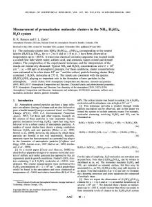
of two academic hospitals (University of Insubria and University of Verona) for routine or clinically indicated examinations and whose pregnancy was complicated by IUGR were considered eligible for the study. Inclusion criteria were: 1) singleton gestation, 2) certain gestational age, 3) gestational age > 20 weeks, 4) absence of known congenital malformation or chromosomal abnormality, and 5) intact membranes. The same criteria applied to the selection of controls that were matched 1 : 1 with each IUGR infant and were identified by selection of the next appropriately grown fetus of similar gestational age (± 1 week) undergoing routine ultrasound evaluation in the same institution as the matched case. All participants gave written informed consent and institutional review board approval was obtained before the beginning of the study. The diagnosis of IUGR was made in the presence of a fetal abdominal circumference (AC) < 5th centile for gestational age. Calculation of gestational age was based on reliable recollection of the last menstrual period and was confirmed or modified by sonographic crown–rump length measurement within the first 14 weeks of gestation. Women enrolled in the study belong to a well-nourished urban population. Ultrasound examinations were performed by one of two operators, both with extensive experience in obstetric ultrasound, using a GE Voluson 730 (GE Medical Systems, Zipf Austria) ultrasound machine. Patients underwent a detailed sonographic evaluation to assess fetal biometry (including measurement of biparietal diameter (BPD), AC and femur length (FL)) and amniotic fluid volume and to exclude major structural fetal anomalies. Estimated fetal weight (EFW) was obtained using the formula proposed by Hadlock et al.15 . Flow velocity waveform patterns of umbilical (UA) and middle cerebral (MCA) arteries and the ductus venosus (DV) were assessed. Oligohydramnios was defined as an amniotic fluid index measuring 5 cm or less. Abnormal UA and DV Doppler indices were defined as a pulsatility index (PI) > 95th centile for gestational age16,17 . Brain sparing was defined as a MCA-PI < 5th centile for gestational age16 . Targeted evaluation of the fetal thymus and a calculation of its perimeter were then performed as described previously (Figure 1)12 . The thymus was visualized on a transverse section of the fetal chest between the sternum anteriorly and the great vessels of the heart posteriorly. The thymus perimeter was measured at maximal magnification three times for each patient with the use of the trace function of the ultrasound machine. The mean of the three measurements was used for statistical analysis. Accuracy of the thymus perimeter measurement has been reported previously12 .
Cromi et al.
Figure 1 Cross-sectional view of the fetal chest at the level of the great vessels of the heart showing the thymus in the anterior mediastinum; measurement of the thymus perimeter is demonstrated in a normally grown fetus (a) and in a growth-restricted fetus (b) of the same gestational age. AO, aorta; PA, pulmonary artery; SVC, superior vena cava.
Statistical analysis Statistical analysis was performed with GraphPad Prism version 4.00 for Windows, (GraphPad Software, San Diego, CA, USA). The t-test and the Mann–Whitney U-test were utilized to compare continuous parametric and non-parametric variables, respectively. Proportions
Copyright 2009 ISUOG. Published by John Wiley & Sons, Ltd.
were analyzed with Fisher’s exact test. Spearman’s rank correlation was used to assess the relationship between continuous variables. In order to exclude a possible influence of fetal size on the dimension of the thymus, the ratio between the thymus perimeter and fetal anthropometric
Ultrasound Obstet Gynecol 2009; 33: 421–426.
Fetal thymus size
423
RESULTS During the study period 63 consecutive patients with growth-restricted fetuses met the inclusion criteria. In three patients, unfavorable fetal position and severe oligohydramnios made visualization of the entire thymus impossible and hindered accurate measurement of the perimeter, leaving 60 IUGR fetuses available for the study. The control group consisted of 60 appropriatefor-gestational age fetuses, whose thymus perimeter was successfully measured in all cases. The clinical characteristics of the study groups are shown in Table 1. Figure 2 displays the fetal thymus perimeter in IUGR fetuses and in controls plotted on the reference ranges published by Zalel et al.11 . The proportion of fetuses with a thymus perimeter < 5th centile for gestational age was significantly higher in IUGR pregnancies compared with controls (58/60 vs. 7/60, P < 0.0001). The ratio of thymus perimeter to BPD, FL, AC and EFW was significantly lower in IUGR fetuses than in appropriately grown fetuses (Figure 3). In IUGR fetuses, the mean O/E thymus perimeter ratio was similar in the subgroup with abnormal UA Doppler indices (n = 20) compared with those with UA-PI within the normal range (n = 40) (0.64 ± 0.40 vs. 0.67 ± 0.16, P = 0.46). Conversely, IUGR fetuses with oligohydramnios (n = 18) had a mean O/E thymus perimeter ratio significantly lower than had those with normal amniotic fluid volume (n = 42) (0.59 ± 0.10 vs. 0.69 ± 0.16, P = 0.02). Similarly, there was a significantly lower mean O/E thymus perimeter ratio in fetuses exhibiting brain sparing (n = 9) compared with those with normal MCA Doppler indices (n = 51) (0.56 ± 0.05 vs. 0.68 ± 0.16, P = 0.03). Among IUGR fetuses, there was no difference in the O/E thymus perimeter ratio between pregnancies complicated by hypertensive disorders (n = 8) Table 1 Clinical characteristics of the intrauterine growthrestricted (IUGR) and control groups
Characteristic GA at enrollment (weeks) Maternal age (years) Nulliparous Hypertensive disorder Oligohydramnios Abnormal UA Doppler Brain sparing Increased DV pulsatility
IUGR (n = 60)
Controls (n = 60)
33 (22.5–39) 31 (17–41) 35 (58.3) 8 (13.3) 18 (30.0) 20 (33.3) 9 (15.0) 2 (3.3)
32 (23.5–38) 30 (19–40) 41 (68.3) 0 (0) 0 (0) 0 (0) 0 (0) 0 (0)
Values are reported as median (range) or n (%). DV, ductus venosus; GA, gestational age; UA, umbilical artery.
Copyright 2009 ISUOG. Published by John Wiley & Sons, Ltd.
140 130 120 Fetal thymus perimeter (mm)
parameters was calculated. Moreover, since the fetal thymus perimeter increases as a function of gestational age11 , the observed-to-expected mean for gestation (O/E) thymus perimeter ratio was calculated to obtain a gestational age-independent assessment of thymus size. P < 0.05 was considered statistically significant.
110 100 90 80 70 60 50 40 30 20 21 22 23 24 25 26 27 28 29 30 31 32 33 34 35 36 37 38 39 40 Gestational age (weeks)
Figure 2 Sonographic fetal thymus perimeter plotted on reference ranges of Zalel et al.11 (5th , 50th , and 95th centiles) in patients with intrauterine growth restriction ( ) and in controls ( ).
°
ž
and those without this complication (n = 52) (0.60 ± 0.13 vs. 0.67 ± 0.15, P = 0.14). Table 2 displays the pregnancy outcome of both cases and controls. There were no chromosomal or structural abnormalities detected after birth. In the IUGR group, there was a significant positive correlation between the O/E thymus perimeter ratio and the enrolment-to-delivery interval (r = 0.44, P < 0.001).
DISCUSSION Our findings suggest that IUGR is associated with a significant decrease in fetal thymic size compared with that in controls. To our knowledge, this is the first study documenting a relationship between inappropriate fetal growth and impaired intrauterine thymic growth, as assessed in-vivo by ultrasound, and our results may provide a mechanism for the association between fetal growth and adult immune function that has been reported in studies drawing attention to the thymus as a potential mediator of the immunological consequences of IUGR18 – 21 . A dramatic decline in thymic weight has been reported consistently both in animal models of malnutrition3 – 6 and in postmortem series of severly malnourished individuals22,23 . A smaller thymus has also been observed in vivo, on ultrasound examination, in infants suffering from nutritional deprivation24 . Malnutrition-related thymic involution seems to result from enhanced thymocytic gene-directed cell death along with decreased T-cell proliferation and alterations in the thymic microenvironment23,25 . The mechanisms by which malnutrition induces thymic involution seem to act via neuroendocrine pathways. Thymic involution was prevented in animals that were adrenalectomized prior to being exposed to a zinc-deficient diet, suggesting a critical
Ultrasound Obstet Gynecol 2009; 33: 421–426.
Cromi et al.
424 P < 0.0001
P < 0.0001 (b) 2.00
(a) 1.50
1.75
Thymus perimeter/FL ratio
Thymus perimeter/BPD ratio
1.25
1.00
0.75
1.50
1.25
1.00
0.50 0.75
0.25
IUGR P < 0.0001
(c) 0.50
0.50
Controls
IUGR
Controls P = 0.02
(d) 0.12
0.45 0.10
Thymus perimeter/EFW ratio
Thymus perimeter/AC ratio
0.40
0.35
0.30
0.25
0.08
0.06
0.04
0.20 0.02 0.15
0.10
IUGR
Controls
0.00
IUGR
Controls
°
Figure 3 Ratio of thymus perimeter to conventional anthropometric measures in patients with intrauterine growth restriction (IUGR) ( ) and in controls ( ). The conventional anthropometric measures were: (a) biparietal diameter (BPD); (b) femur length (FL); (c) abdominal circumference (AC); and (d) estimated fetal weight (EFW).
ž
role of the hypothalamic-pituitary-adrenal (HPA) axis in this process26 . Noteworthy, injection of recombinant leptin prevented thymic shrinkage in mice exposed to acute starvation, and this was associated with a reduction
Copyright 2009 ISUOG. Published by John Wiley & Sons, Ltd.
in glucocorticoid levels27 . This supports the speculation that nutritional deprivation induces thymocytic depletion through an imbalance in the production of leptin (which is decreased) and glucocorticoids (which are increased).
Ultrasound Obstet Gynecol 2009; 33: 421–426.
Fetal thymus size
425
Table 2 Pregnancy outcome of the intrauterine growth-restricted (IUGR) and control groups
Outcome GA at delivery (weeks) Delivery before 30 weeks Enrollment-to-delivery interval (days) Birth weight (g) Birth weight < 1000 g NICU admission Intrauterine fetal demise
IUGR (n = 60)
Controls (n = 60)
33 (24.8–39) 11 (18.3) 11 (0–72)
39.6 (34.4–41.5) 0 (0) 49 (2–116)
1809 ± 740 12 (20) 31 (51.7) 3 (5.0)
3220 ± 470 0 (0) 2 (3.3) 0 (0)
Values are reported as median (range), mean ± SD or n (%). GA, gestational age; NICU, neonatal intensive care unit.
Since the prenatal assessment of the thymus is technically demanding, the hypothesis that intrauterine undernutrition could induce changes in thymic morphometry similar to those observed in infancy and adulthood so far has been tested only in animal experiments7,8 and in pathological studies of growth-restricted babies dying in the perinatal period9 . Studies investigating the metabolic status of growth-restricted fetuses have documented hypoglycemia with a disproportionate degree of hypoinsulinemia, an increased ratio of non-essential to essential amino acids, and enhanced lipolysis, parameters all compatible with intrauterine starvation28 . Accumulating data in humans suggest that intrauterine adversity, including reduced substrate availability, is associated with alterations in the HPA axis29,30 . Concurrent with this view is the speculation that the HPA axis, in directing resource partitioning among competing demands during prenatal nutritional stress, induces thymic involution as part of the adaptative response that is intended to enhance fetal survival. What, if any, is the effect of impaired development of the thymus in growth-restricted fetuses on immunological competence throughout life remains to be elucidated. Prospective studies have provided evidence in support of early programming of later immune function21 , suggesting that the thymus is a mediator of the association between fetal undernutrition and the risk of atopic, autoimmune and infectious disease in infancy and adulthood18 – 20 . Limitations of this investigation include the lack of longitudinal sonographic measurements and the relatively small sample size, which did not allow us to show a significant association between the degree of impaired thymic growth and neonatal morbidity. Given the study design, examining point estimates of different thymus glands, it could be argued that the thymus is small in fetuses with IUGR simply because it stops growing rather than because it shrinks in size. However, the disproportionately large reduction of thymus perimeter relative to conventional anthropometric parameters and EFW as well as biological plausibility support the hypothesis that thymic involution may be part of the spectrum of fetal neuroendocrine responses to reduced substrate availability. The definition of IUGR based
Copyright 2009 ISUOG. Published by John Wiley & Sons, Ltd.
exclusively on AC < 5th centile could be regarded as a further weakness of our study, since it did not allow us to exclude the possibility of constitutionally small fetuses being labeled as growth restricted. However, AC is the measurement that best reflects fetal nutrition, and a 5th centile cutoff for AC is considered relevant in indicating, among small-for-gestational age fetuses, the group with a truly increased risk of adverse perinatal outcome. Lastly, since we did not investigate the presence of chromosome 22q11 microdeletion, we are unable to rule out the possibility that thymic hypoplasia was a manifestation of this condition in some cases31 . Nevertheless, no structural abnormalities characteristic of this syndrome, undiagnosed prenatally, were detected in our study population up to the time of hospital discharge. Additional research is required to explore the physiological processes linking prenatal undernutrition to thymic development and function, and to establish the long-term implications for immunocompetence and risk of adult disease.
REFERENCES 1. Beisel WR. History of nutritional immunology: introduction and overview. J Nutr 1992; 122: 591–596. 2. Konno A, Utsuyama M, Kurashima C, Kasai M, Kimura S, Hirokawa K. Effects of a protein-free diet or food restriction on the immune system of Wistar and Buffalo rats at different ages. Mech Ageing Dev 1993; 72: 183–197. 3. Pallaro AN, Roux ME, Slobodianik NH. Nutrition disorders and immunologic parameters: study of the thymus in growing rats. Nutrition 2001; 17: 724–728. 4. Kuvibidila S, Dardenne M, Savino W, Lepault F. Influence of iron-deficiency anemia on selected thymus functions in mice: thymulin biological activity, T-cell subsets, and thymocyte proliferation. Am J Clin Nutr 1990; 51: 228–232. 5. Malpuech-Brug`ere C, Nowacki W, Gueux E, Kuryszko J, Rock E, Rayssiguier Y, Mazur A. Accelerated thymus involution in magnesium-deficient rats is related to enhanced apoptosis and sensitivity to oxidative stress. Br J Nutr 1999; 81: 405–411. 6. Dhur A, Galan P, Christides JP, Polier de Courcy G, Preziosi P, Hercberg S. Effect of folic acid deficiency upon lymphocyte subsets from lymphoid organs in mice. Comp Biochem Physiol 1991; 98: 235–240. 7. Lansdown AB. Histological observations on thymic development in fetal and newborn mammals subject to intrauterine growth retardation. Biol Neonate 1977; 31: 252–259. 8. Lang U, Baker RS, Khoury J, Clark KE. Effects of chronic reduction in uterine blood flow on fetal and placental growth in the sheep. Am J Physiol Regul Integr Comp Physiol 2000; 279: R53–R59. 9. Hartge R, Jenkins DM, Kohler HG. Low thymic weight in small-for-dates babies. Eur J Obstet Gynecol Reprod Biol 1978; 8: 153–155. 10. Cho JY, Min JY, Lee YH, McCrindle B, Hornberger LK, Yoo SJ. Diameter of the normal fetal thymus on ultrasound. Ultrasound Obstet Gynecol 2007; 29: 34–38. 11. Zalel Y, Gamzu R, Mashiach S, Achiron R. The development of the fetal thymus: an in utero sonographic evaluation. Prenat Diagn 2002; 22: 114–117. 12. Di Naro E, Cromi A, Ghezzi F, Raio L, Uccella S, D’Addario V, Loverro G. Fetal thymic involution: a sonographic marker of the fetal inflammatory response syndrome. Am J Obstet Gynecol 2006; 194: 153–159. 13. El-Haieg DO, Zidan AA, El-Nemr MM. The relationship between sonographic fetal thymus size and the components of
Ultrasound Obstet Gynecol 2009; 33: 421–426.
Cromi et al.
426
14.
15.
16.
17.
18.
19.
20.
21.
22.
the systemic fetal inflammatory response syndrome in women with preterm prelabour rupture of membranes. BJOG 2008; 115: 836–841. Yinon Y, Zalel Y, Weisz B, Mazaki-Tovi S, Sivan E, Schiff E, Achiron R. Fetal thymus size as a predictor of chorioamnionitis in women with preterm premature rupture of membranes. Ultrasound Obstet Gynecol 2007; 29: 639–643. Hadlock FP, Harrist RB, Sharman RS, Deter RL, Park SK. Estimation of fetal weight with the use of head, body, and femur measurements – a prospective study. Am J Obstet Gynecol 1985; 151: 333–337. Arduini D, Rizzo G. Normal values of pulsatility index from fetal vessels: a cross-sectional study on 1556 healthy fetuses. J Perinat Med 1990; 18: 165–172. Hecher K, Campbell S, Snijders R, Nicolaides KH. Reference ranges for fetal venous and atrioventricular blood flow parameters. Ultrasound Obstet Gynecol 1994; 4: 381–390. Ferguson AC. Prolonged impairment of cellular immunity in children with intrauterine growth retardation. J Pediatr 1978; 93: 52–56. Phillips DI, Cooper C, Fall C, Prentice L, Osmond C, Barker DJ, Rees Smith B. Fetal growth and autoimmune thyroid disease. Q J Med 1993; 86: 247–253. Godfrey KM, Barker DJ, Osmond C. Disproportionate fetal growth and raised IgE concentration in adult life. Clin Exp Allergy 1994; 24: 641–648. McDade TW, Beck MA, Kuzawa CW, Adair LS. Prenatal undernutrition and postnatal growth are associated with adolescent thymic function. J Nutr 2001; 131: 1225–1231. Jambon B, Ziegler O, Maire B, Hutin MF, Parent G, Fall M, Burnel D, Duheille J. Thymulin (facteur thymique serique) and
Copyright 2009 ISUOG. Published by John Wiley & Sons, Ltd.
23. 24.
25.
26.
27.
28.
29.
30. 31.
zinc contents of the thymus glands of malnourished children. Am J Clin Nutr 1988; 48: 335–342. Lyra JS, Madi K, Maeda CT, Savino W. Thymic extracellular matrix in human malnutrition. J Pathol 1993; 171: 231–236. Parent G, Chevalier P, Zalles L, Sevilla R, Bustos M, Dhenin JM, Jambon B. In vitro lymphocyte-differentiating effects of thymulin (Zn-FTS) on lymphocyte subpopulations of severely malnourished children. Am J Clin Nutr 1994; 60: 274–278. Poetschke HL, Klug DB, Perkins SN, Wang TT, Richie ER, Hursting SD. Effects of calorie restriction on thymocyte growth, death and maturation. Carcinogenesis 2000; 21: 1959–1964. Fraker PJ, Osati-Ashtiani F, Wagner MA, King LE. Possible roles for glucocorticoids and apoptosis in the suppression of lymphopoiesis during zinc deficiency: a review. J Am Coll Nutr 1995; 14: 11–17. Howard JK, Lord GM, Matarese G, Vendetti S, Ghatei MA, Ritter MA, Lechler RI, Bloom SR. Leptin protects mice from starvation-induced lymphoid atrophy and increases thymic cellularity in ob/ob mice. J Clin Invest 1999; 104: 1051–1059. Economides DL, Nicolaides KH, Gahl WA, Bernardini I, Bottoms S, Evans M. Cordocentesis in the diagnosis of intrauterine starvation. Am J Obstet Gynecol 1989; 161: 1004–1008. Economides DL, Nicolaides KH, Linton EA, Perry LA, Chard T. Plasma cortisol and adrenocorticotropin in appropriate and small for gestational age fetuses. Fetal Ther 1988; 3: 158–164. Fowden AL, Forhead AJ. Endocrine mechanisms of intrauterine programming. Reproduction 2004; 127: 515–526. Chaoui R, Kalache KD, Heling KS, Tennstedt C, Bommer C, ¨ Korner H. Absent or hypoplastic thymus on ultrasound: a marker for deletion 22q11.2 in fetal cardiac defects. Ultrasound Obstet Gynecol 2002; 20: 546–552.
Ultrasound Obstet Gynecol 2009; 33: 421–426.