mvocardium appeared normal, the intercalated disks were straight or ran in a stepwise manner, the sarcomeres were in register (with a wide I band), and the ...
Ultrastructure of the Myocardium After Pulmonary Embolism A Study in the Rat Henri F. Cu6noud, MD, Isabelle Joris, PhD, and Guido Majno, MD
The purpose of this study was to find out whether acute massive pulmonarv embolism can produce myocardial changes visible by light and electron microscopy. We therefore produced pulmonary embolism in rats using plastic microspheres (diameter, 15 ± 5 A). Two experimental protocols were used: lethal embolism, with a dose of microspheres known to kill in 3 to 15 hours (these rats were killed after 1 hour), and sublethal embolism, with a dose compatible with 100% survival (these rats were killed after 24 hours). In both groups, the left ventricle was normal. The right ventricle showed two types of changes: a) A distinctive lesion of the myocytes, more diffuse after lethal embolism and different from the "zonal lesion" of shock. It consisted primarily in a localized shredding of the myofibrillar system; hence, the name shredding is proposed. Earlier stages of this lesion were represented by focal dissolution of the Z line (Z lysis). The pathogenesis of these lesions appeared to be primarily mechanical. b) Necrosis was already apparent at 1 hour and was more extensive after 24 hours. The pathogensis of the necrotic lesions is best explained by a temporary ischemia followed by delayed reflow; a possible potentiating role of endogenous catecholamines cannot be excluded. Most capillaries in the necrotic foci remained functional; this explains the rapid rate of the healing process of such lesions. A comparison is drawn between the observed foci of necrosis and the human myocardial lesions known as "miliary infarcts" and "mvocvtolysis." It is proposed that a factor common to all three is the preservation of the microcirculatory vessels and that our experimental model helps illuminate the pathogenesis of the human lesions. It is concluded that the right ventricle of acute cor pulmonale may develop cellular changes with a complex pathogenesis (mechanical, ischemic, and possibly hormonal). The nature of the changes found in our model could represent the morphologic substrate of right-sided failure; it can be correlated with the electrocardiographic abnormalities found in the comparable human condition. (Am J Pathol 92:421-48, 1978)
THE FUNCTIONAL ASPECTS of acute cor pulmonale have been studied in experimental animals and, to a lesser extent, in humans."2 Not much is known, however, about the structural correlates of right ventricular failure as it occurs after massive pulmonary embolism. Light microscopy has given inconsistent results, ie, that myocardial necrosis mav or may not occur.348 An electron microscopic study, to our knowledge, was From the Department of Pathology. University of \Massachusetts \Medical School. *Worcester. \Massachusetts. Supported in part by Grant HL-16952 from the National Institutes of Health and bx- a grant of the Fonds Rapin. Societe Academique of Geneva. Sswitzerland Accepted for publication April 5. 1978. Address reprint requests to Henri F Cuenoud. MD. Department of Pathology. Uiniversit\ of \lassachusetts Medical Center. 55 Lake Avenue North. W\orcester. MIA 01605 0002-9440/78/0810-0421$01 .00 421
422
CUtNOUD ET AL
American Journal of Pathology
not available. We therefore decided to re-examine this problem by light as well as by electron microscopy, using a convenient model: the heart of the rat after lethal or sublethal embolization with plastic microspheres. Materials and Methlods Animals
Fifty male Wistar rats (Charles River Breeding Laboratories, W'ilmington, Mass.)
weighing 250 to 400 g were used in this study. Intravenous Inectios of Micropheres
Microembolism was produced by injecting microspheres into the saphenous vein with the rats under light ether anesthesia. We used 10 g of insoluble carbonized plastic spheres, 15 ± 5 A in diameter (3M1 Co., Nuclear Products Division, St. Paul, Minn.), suspended in 100 ml of 10%c dextran (molecular weight, 70,000). Pilot experiments had shown that a dose of 0.115 ml/100 g of bodv weight corresponded approximately to an LD,; hence, for the studs of lethal microembolism, we injected 0.130 ml,l100 g and when survival was required we used a dose of 0.100 ml 100 g. Control Soutins
We injected 0.130 ml,/100 g of body w-eight of either dextran (the same as used for the suspension of microspheres obtained from the 3NM Co.) or sterile 0.9%c NaCl intravenouslv. After the injection, the rats were carefully obsenred, and signs of distress were recorded: tachvpnea; dyspnea; weakness; cyanosis of ears and paws; edema of snout, ears, and pau-s; and a group of signs that we interpreted as evidence of "shock," ie, pale paws and ears and cold paws and tail. Fxation, Samlng, and Processing Perfusion With Fixative
The hearts were fixed by retrograde aortic perfusion in situ at a pressure of 100 to 130 mmHg, using 3%c glutaraldehyde in cacodylate buffer. At the appropriate time the rats were overanesthetized with ether and the perfusion needle was inserted upward into the lower abdominal aorta. An exit for the perfusing fluid was provided by cutting the jugular veins. The perfusion was continued for 20 minutes. Thereafter, the heart was dissected out and the following specimens were routinely sampled: three areas of the anterior wall of the right ventricle, two right papillary muscles, the two left papillary muscles, and a transmural section of the midlateral wall of the left ventricle. More samples were added when lesions were macroscopicallv suspected. Every specimen u-as further trimmed into 1 x 1 x 2 mm blocks. The tissues were fixed for a total of a hours (including perfusion time) in 3% glutaraldehyde in cacodylate buffer at room temperature, then washed in cacodylate buffer for 12 hours at 4 C, postfixed in 2%5 osmium tetroxide in collidine buffer for 2 hours at 4 C, dehydrated in graded alcohols, and embedded in Epon 812,9 orienting the blocks to obtain longitudinal sections of the myocardial fibers. For light microscopy, sections 1 u thick u-ere stained with toluidine blue (these will be referred to hereafter as epon sections). Ultrathin sections were cut with a diamond knife on an LKB Ultrotome III, mounted on uncoated grids, stained with uranyl acetate and lead citrate,10'11 and examined with a Philips EM 301 electron microscope.
Vol. 92, No. 2 August 1978
ULTRASTRUCTURE OF THE MYOCARDIUM
423
Fixation by immersion
In glutaraldehyde. At the appropriate time, with the rat under deep anesthesia, the chest was opened, the heart was removed, and the right ventricle was quickly excised and immediately fixed in 3% glutaraldehyde in cacodylate buffer. It was further trimmed into 1 x 1 x 2 mm blocks, which were allowed to fix for a total of 5 hours and processed as described above. Informalin. After the rat died, the heart and lungs were removed as a whole and fixed in 10% buffered formalin for 3 days. The heart was dissected out; coronal sections were taken at three levels and processed toward paraffin embedding. Five-P sections were stained with hematoxylin and eosin.
The 50 rats were subdivided into two control and two experimental groups. Group 1 (Saline Controls)
Four control rats were injected intravenously with saline. One hour later they were anesthetized and fixed by perfusion with 3% glutaraldehyde; the hearts were processed toward epon embedding. Four control rats were treated as above except that the hearts were fixed by immersion in 3% glutaraldehyde. Group 2 (Dextran Controls)
Eight control rats were injected with dextran, killed 1 hour later, and perfused with 3% glutaraldehyde. The hearts were processed for electron microscopy. Four control rats received the same injection and the hearts were fixed by immersion in 3% glutaraldehyde. Group 3 (Lethal Microembolism)
Twenty-one rats were given a lethal dose of microspheres; of these rats: a) Nine were killed 1 hour later, and their hearts were fixed by perfusion as described. b) Four other rats were killed 1 hour after the injection, and the right ventricle was fixed by immersion in 3% glutaraldehyde to assess possible ultrastructural differences due to the different types of fixation. c) The 8 remaining rats were allowed to die spontaneously; this occurred between 3 and 15 hours. The hearts of these animals were fixed in 10% formalin, processed as described above, and used in light microscopy only. Group 4 (Sublethal Microemboism)
Nine rats were given a sublethal dose of microspheres. All the animals were killed 19 to 24 hours after the injection and were perfused with 3% glutaraldehyde. Their hearts were sampled and processed as previously described. Shd of the Ahs To study the possible role of catecholamine secretion in the pathogenesis of mvocardial necrosis, we examined the adrenals of 12 rats: 4 were killed 1 hour after lethal pulmonary embolism; 8 were controls injected with either saline or dextran. The adrenals were fixed by perfusion as described above.
424
CUENOUD ET AL
American Joumal of Pathology
Results Contros (Saline and Dextran, Grou
1 ad 2)
The saline injection had no noticeable effect. Dextran caused a slight edema of the paws and snout, more obvious in rats weighing less than 300 g. The edema subsided rapidly and was barely noticeable after 1 hour. At autopsv the heart appeared normal for both groups. By light microscopy, the mvocardium was normal (Figures 1 and 2); the sarcomeres were contracted in the specimens fixed bv immersion, relaxed (ie, distended) in perfused hearts. By electron microscopy, after perfusion-fixation, the mvocardium appeared normal, the intercalated disks were straight or ran in a stepwise manner, the sarcomeres were in register (with a wide I band), and the mitochondria were aligned as usual in the long axis of the cells. The microvasculature was unremarkable. Specimens fixed bv immersion showed contraction of the sarcomeres with frequent absence of the I band. Lelhal Mire ioism (Group 3)
Immediately after injection, the animals developed tachvpnea, dvspnea, cyanosis of the paws and tail, edema of the paws and snout, and a progressive state that will be referred to hereafter as "shock," evidenced by a delay in the recoverv from the anesthesia, cold paws and tail, and very pale ears. An occasional episode of respiratory arrest was reversed by artificial respiration using a mouth-to-snout tube. The rats that were not killed at 1 hour remained awake during the prolonged "shock" period and suddenly died between 3 and 12 hours. At autopsv, dilatation of the right atrium and ventricle was obvious in almost all rats. A pleural effusion, consisting of 2 to 10 ml of clear fluid, was found in 4 of the 8 rats that were allowed to die. On light microscopy, we found that paraffin sections were difficult to read and far less satisfactorv than epon sections; the different qualitv of the two tvpes is readily apparent also in the controls (Figures 1 and 2). Paraffin sections from the rats that had been allowed to die showed widespread mvocardial lesions throughout the right ventricular wall. Almost every fiber showed a pale area devoid of striations and often adjacent to the intercalated disk (Figure 3). A few scattered fibers were necrotic and contained bands. Leukocytes were sticking here and there to the endocardium; marginating leukocytes were also found in some intramural venules, in areas of interstitial edema. Focal pale lesions of mvocardial fibers were found also in the bundles representing the insertion of the right ventricle onto the left ventricular wall. The entire left ventricle as w%ell as the septum were normal.
Vol. 92, No. 2 August 1978
ULTRASTRUCTURE OF THE MYOCARDIUM
425
Epon section confirmed these findings; in addition, they showed that almost every intercalated disk was distorted and that 20 to 100% of the myocytes contained a pale area devoid of striation and punctated by a scattering of mitochondria (Figure 4) (by "myocyte" we mean single cellular units along the myocardial fibers). Most of these lesions occurred on only one side of the intercalated disk; occasionally they occurred on both sides and, more rarely, within the body of the fiber, away from the intercalated disk. Severely injured myocytes were observed (Figure 5) as well as foci of interstitial edema (Figure 6). Occasional platelet thrombi were found in small veins (Figure 7). The left ventricle and left papillary muscles were entirely normal, except for small foci of poor cellular preservation associated with incomplete perfusion (seen also in controls). Electron microscopy of the right ventricle in the rats submitted to lethal embolism (and perfusion-fixation) showed a range of lesions; some were immediately apparent, others required close comparison with the controls (Figure 8). The pale areas of the myocardial fibers, clearly seen with the light microscope (Figures 3 and 4), corresponded to a condition that may best be described as "shredding"' of the myocyte (Figures 9 through 12). The myofibrillar pattern was stretched out, loose, and disorganized, leaving a clear space in which the mitochondria appeared to float freely (Figures 10 and 11). This focal lesion of the fiber represented an extreme; lesser injuries could be found, which- we interpreted as earlier stages. The earliest stage identified was a disintegration of the Z line ("Z lysis"), in which the Z-line material was pulled toward one or the other of the adjacent sarcomeres (Figure 13): eventually the Z line was replaced by a gap, which could become as wide as a normal sarcomere (Figure 13). This gap contained shreds of fibrils but usuallv also a number of ribosomes; at times it was possible to see a polyribosome arranged in linear fashion along a fibril, at the site of the disorganized Z line (Figure 13). A lesion similar to the disruption of the Z band sometimes affected one or both sides of the intercalated disk, giving rise to a clear space (between the sarcomeres and the distorted disk) in which thick and thin fibrils were dispersed (Figure 14). At a later stage there developed a complete mvofibrillar disorganization on one side (Figures 9 and 10) or, less frequently, on both sides of the intercalated disk (Figure 15). Other changes in the mvocardium of the right ventricle and papillanr muscles were focal: such were the myocytes loaded with lipid droplets smaller than a mitochondrion (Figures 9 and 10), clear vacuoles (Figure 12), focal cellular blebbing (Figure 12), and cell-to-cell herniae across a distorted intercalated disk. These herniations were limited by two cell membranes (with or without nexus-type specializations) and sometimes
426
CU-NOUD ET AL
American Joumal of Pathology
contained ribosomes, myofibrils, and/or mitochondria. Other changes had been seen by light microscopy: necrotic myocytes with contraction bands, extracellular edema, occasional emigrated white blood cells, and platelet thrombi (Figure 12). Despite careful search, no changes were found in the left ventricle and papillary muscles, with the exception of a very occasional distorted intercalated disk. Fixation by Inn
A peculiar difference was noticed after immersion fixation in epon sections of control or lethal microembolism: some myocytes were partially hypercontracted, swollen, and very dense (Figure 16). These changes were often close to the border of the specimen and slightly more frequent in the experimental animals. We interpreted them as fixation artifacts due to lack of tension on the myocardial fibers. By electron microscopy, in the hypercontracted sarcomeres no Z-band disruption or sarcomere disorganization was noticed. The shredding lesions, when present, were still easy to recognize by electron microscopy (Figure 17). S dlth Micros ibolm (Gmup 4)
After a transient episode of dyspnea and edema, the rats of this group recovered and appeared to behave normally, except one which began to develop a secondary bout of dyspnea at the 18th hour and then lapsed into the condition referred to earlier as "shock." All these rats, including the one in "'shock," were killed between 19 and 24 hours after pulmonary embolism. At autopsy the heart appeared normal in all except the animal in "shock." The heart of this rat showed severe dilatation of the right atrium and ventricle, many small pale patches on the surface of the right ventricle, and a very obvious white discoloration with slight swelling of the two right papillary -muscles (Figure 18). There was no pleural effusion. In epon sections of the right ventricle and right papillary muscles, there were no visible lesions in 6 of the 9 rats. Three other rats (including the one that lapsed into "shock") had tiny foci of necrosis infiltrated with leukocytes. Occasionally, next to a distorted intercalated disk, there were lesions of the type we described as "shredding" after lethal microembolism. One rat had a large area of necrosis involving half of the base of the right papillary muscle. In the rat that was killed while in "shock," both papillary muscles of the right ventricle were completely necrotic
Vol. 92, No. 2 August 1978
ULTRASTRUCTURE OF THE MYOCARDIUM
A 0%-F
427
(Figures 18B, 19, and 20); there were patches of necrosis throughout the myocardium of the right ventricle, sometimes involving the whole thickness of the wall. The necrotic myocytes could be either dark and narrow or clear and swollen (Figures 19 and 20). They contained contraction bands (Figure 20) and many lipid droplets. Most of the capillaries were open and cleared by the perfusion (Figures 19 and 20); polymorphonuclear leukocytes and macrophages were strewn throughout the necrotic areas. The left ventricle and left papillary muscles showed no visible lesions in all rats of this group. Electron microscopy of the right ventricle and papillary muscles revealed that light microscopy (even in epon sections) had been inadequate, in that the lesions had been severely underestimated. All rats were affected, and 10 to 30% of the intercalated disks were distorted (Figure 21). On rare occasions the lesion had progressed to shredding over a few sarcomeres (Figure 22). Lipid droplets had accumulated in some myocytes (Figure 21). Electron microscopy also confirmed the more severe lesions developed by 3 rats, as described by light microscopy. Inflammatory cells were present (Figure 23). Within jand around the areas of necrosis found in these rats, the capillaries appeared open and perfused (Figures 23 and 24) as had been seen on the epon sections; in addition, a few gaps were found between endothelial cells (Figure 23). Interstitial edema was obvious (Figures 23 and 24); there were extravasated red blood cells as well as strands of fibrin. Around the areas of necrosis there were myocytes with lesions of shredding (Figure 23). The dead myocytes showed contraction bands (Figures 24 and 25) similar to those that had been observed 1 hour after lethal embolism (Figure 26) but accompanied by a larger number of lipid droplets (Figures 24 and 25). The distorted intercalated disks were often lined on both sides by packed thin filaments, themselves lined by a contraction band (Figures 25 and 26). This change is suggested by the light microscopic findings (Figure 20). The left ventricle and left papillary muscles were the same as in the controls. Survival beyond 24 hours was not included in the present study. However, during pilot experiments, several rats were allowed to live beyond 1 day. One of these was killed 3 days after a sublethal injection of microspheres. At autopsy it showed a greatly dilated right atrium and ventricle (Figure 27). By light microscopy it was found that the whole wall of the right ventricle had become transformed into a fibrous sheet rich in macrophages. Myocytes were still present at the junction of the right ventricle with the left, and a few survivors were scattered along the fibrous ventricular wall (Figure 28).
428
CUItNOUD ET AL
American Joumal
of Patholgy
Discussion Preious Studs on 11l Re
s of
Aby EnisII
According to the available data, acute myocardial infarction after acute pulmonary embolism is infrequent in humans; the most common sites of necrosis are the left ventricle or the left papillary muscles. Occasionally, focal areas of necrosis, or massive infarction, are seen in the right heart, particularly if shock is associated with right ventricular dilatation.34 In experimental animals, multifocal necrosis has been observed in the right ventricle after pulmonary embolism or after banding the pulmonary artery.7'8 It seems that myocardial blood flow is dependent on the severity of the pulmonary embolism; if the embolism is massive, coronary blood flow to both ventricles is reduced; 12 if the embolism is less severe, coronary flow is said to remain unchanged or even to increase, although this point is still controversial.'2'13 All these studies correlated pulmonary embolism with either clinical observations, functional measurements, or light microscopic findings. As mentioned earlier, a correlation at the electron microscopic level has not been attempted. C1tiuw of Ow Mode of Puhnoy Emboism
It may be argued that the rat is not known to be a good species for reproducing the syndrome of pulmonary embolism in humans and that microembolism may not duplicate the effects of a massive embolus. On the other hand, there are good reasons for choosing this model: a) Since we are dealing with a very acute, mechanically induced event, the basic repercussions on the myocardium are likely to be similar in humans and rats. b) For an electron microscopic study, perfect fixation (and preferably perfusion-fixation) are essential. To this end, the small size of the rat heart and the thinness of its right ventricle are great assets. We also had extensive experience with fixation by retrograde perfusion.'4 c) Electron microscopic studies pose a sampling problem: surveying a single dog heart by blocks of approximately 1 cu mm can lead to a prohibitive number of specimens. By contrast, an entire papillary muscle of the rat heart can be included in two tissue blocks. d) Macroembolism vs microembolism: It has been shown that macroemboli cause pulmonary hypertension simply by a mechanical blockade of the pulmonary arteries, whereas microemboli may induce an elevation of pulmonary arterial pressure by a vasoconstriction superimposed on the mechanical obstruction."5 However, other studies indicate that a pulmonary vasoconstriction may not occur; 16 in any event, whatever the pulmonary hemodynamics may be in microembolism, the repercussions on the heart should remain the same. e) Since we used
Vol. 92, No. 2 August 1978
ULTRASTRUCTURE OF THE MYOCARDIUM
429
microspheres, the question may be raised whether a significant number of microspheres might find their way through the pulmonary microcirculation and embolize the coronary vessels. Since only one or two microspheres appeared in three of approximately 600 histologic sections of the heart, this complication can be disregarded. It should also be noted that the shredding lesions are diffuse in the acute experiments, again ruling out an embolic mechanism. f) A possible complicating factor in our model was the fact that the microspheres were suspended in a solution of dextran, which can produce anaphylactoid edema in rats.17, Although dextran alone did produce a slight and transient peripheral edema in most rats, electron microscopy of the heart showed no changes of any kind. We never observed any trace of the subendocardial hemorrhages said to occur after intravenous infusion of large doses of dextran.19 Natu of the Lesions Observe in Ow Model
The topographic specificity of the lesions was remarkable: they were strictly limited to the right ventricle and spared the left (we did not check the atria). Lethal and sublethal emboli caused the same types of lesions; the difference was one of degree. The most obvious lesions, ie, focal areas of necrosis (Figure 23), had already been seen by others in humans and animals; 'a this tends to support the validity of our model. In addition, we found a type of lesion which begins as a discrete ultrastructural disturbance and ultimately appears as the focal change (visible by light microscopy) which we proposed to call shredding (Figure 10). It appears by electron microscopy as an abnormally translucent zone in a myocyte; the fibrillar pattem is longitudinally stretched and shredded so that the mitochondria (formerly packed in longitudinal slits between the bundles of fibrils) are free to float in the loosened area. Most of these lesions are located on one side of the intercalated disk, more rarely on both sides; approximately 10% are found anywhere along the myocardial fiber. Since a full-blown lesion can reach several microns in length along the myocardial cell, it is unmistakable on 1-M sections of tissues embedded in epon (Figure 4). However, it can be missed on paraffin-embedded sections (Figure 3); here the clear zones along the fibril could be easily dismissed as a cutting artifact. Earlier lesions cannot be seen by light microscopy even in epon sections. On electron micrographs the simplest lesion observed affects one or two sarcomeres; it appears as a stretching of the fibrillar system, with selective Z lysis (Figure 13) or as a detachment of the thin filaments from the intercalated disks (Figure 14). The intercalated disk is distorted. Ultimately, the Z, I, and A lines (at the site of the lesion) are lost (Figures 9 through 11).
430
CUItNOUD ET AL
Pa_ees
American Journal of Pathology
the Necotic Lesin
Necrosis occurred after lethal as well as sublethal microembolism; it affected single myocytes along a fiber, single fibers, or groups of fibers large enough to be recognized by the naked eye. A striking characteristic of the necrotic areas of all sizes was the structural and functional preservation of the microcirculatory vessels: the capillaries were usually well washed out by the perfusing fluid; although occasional endothelial damage (Figure 23) and small platelet thrombi were found (Figures 7 and 12), the capillary endothelium was remarkably well preserved. By contrast, in experimental myocardial infarction in the rat, the capillaries break down together with the parenchyma.Y"l2 Thus, if the pathogenesis of necrosis in our experimental animals is of ischemic origin, we would have to postulate the following sequence of events: after pulmonary embolism, the blood flow within the wall of the right ventricle is reduced to a trickle, this residual flow being barely enough to maintain the microcirculatory vessels themselves but not the surrounding parenchymal cells. However, the pathogenesis of the necrotic areas is not as simple to unravel as may appear at first. The hallmarks of ischemic myocardial injury are cellular and mitochondrial swelling.2224 This is not what we found at the earliest stage investigated, ie, 1 hour: the myocytes showed either shredding or severe changes corresponding to the so-called myofibrillar degeneration. This term was coined by Reichenbach and Benditt 2 to indicate a cellular change said to be distinct from that of ischemic coagulation necrosis. In this condition, the entire myofibrillar system within a myocyte is disrupted by supercontraction (contraction bands) as well as by stretching of sarcomeres. These myofibrillar changes are accompanied by cellular edema and by a number of mitochondrial abnormalities.2426 The overall results are summarized in Text-figure 1. Reichenbach and Benditt n proposed that the common denominator between the various conditions promoting ''myofibrillar degeneration" may be a high level of exogenous or endogenous catecholamines. The injurious role of catecholamines on the myocardium appears to reach even farther than myofibrillar degeneration: the zonal lesion of shock (a very different type of cellular disease) was also said to be due, in part at least, to catecholamines released by local sympathetic nerves or by the adrenal glands.2° Two other findings are also compatible with an overdose of these hormones: a) The marked increase in intracellular lipid droplets (Figures 21, 23 through 25), a change often described after catecholamine injection.31-3 However, it must be considered a nonspecific response in cells that normally oxidize fatty acids and fail to do so when injured. It is also a
Vol. 92, No. 2 August 1978
ULTRASTRUCTURE OF THE MYOCARDIUM
TEXT-FIGLRE 1-Schematic comparison of shredding le-
431
I
sions. zonal lesions, and mvofibrillar degeneration.
I
SHREDDING
ZONAL LESION
MYOFIBRILLAR DEGENERATION
typical result of ischemic or hypoxic injurvA.-3 b) A rather distinctive complicated lesion: a distorted intercalated disk lined on both sides by packed thin filaments (Figure 25). This too has been described to happen occasionally after isoproterenol administration." WNre do not know how specific it max be. On the other hand, there are also reasons for believing that catecholamines are not primarily involved in the pathogenesis of the necrotic foci. First, these foci were strictly limited to the right ventricle; a purely hormonal mechanism is therefore unlikely (catecholamine lesions are diffuse 31). Second, we found no significant changes in the adrenal medulla; the degranulation, autophagic vacuoles, and degenerative hydropic changes described by Kajihara et al in dogs submitted to prolonged hemorrhagic shock w-vere absent. What is, then, the pathogenesis of the necrotic lesions? WN7e favor ischemia as the major factor despite tw^o possible objections. WNe will therefore state these objections, followed by our answ ers. a) It is true that mvofibrillar degeneration does not occur during ischemia, but it does occur under conditions of postischemic reflow; 3 and b) it is also true that ischemia causes, in general, diffuse lesions, not focal; however, there is evidence that mvocardial ischemia can produce focal lesions. Steenbergen et al perfused rat hearts in vitro under conditions of high-flowv hypoxia, respiratory acidosis, and low-flow hypoxia." They noticed that under these conditions mvocardial flow became patchy and explained it by postulating a spasm of certain arterioles, whereby- some areas of mvocardium were sacrificed while others remained functional. The size of the
432
American Journal of Pathology
CUENOUD ET AL
nonperfused areas described in this paper corresponds to the size of the necrotic foci observed by us. Thus, we could interpret our findings as follows: immediately after severe pulmonary embolism, coronary flow can not meet the metabolic needs of the overdistended, overworked right ventricle: patchy spasm of coronary arterioles develops, leading to "heterogeneous perfusion"; " foci of necrosis develop if this condition lasts longer than 15 to 30 minutes.2' Later, as the acute hemodynamic imbalance is partially corrected, myocardial flow through the right ventricle is improved, and reflow through the previously ischemic areas induces myofibrillar degeneration. Eolutin of Oh Nearotic Le
Rela
to Myocyllysi
The dichotomy between parenchymal necrosis and microvascular preservation is certainly the reason for the extremely fast and efficient course of the "mopping up operations," ie, the extravasation of cells, primarily of macrophages, charged with the removal of necrotic material." This process was well under way already after 24 hours (Figures 19 and 23) and also in the central part of the necrotic foci (whereas in a typical infarct, organization occurs from the periphery toward the center). Even more striking was the total disappearance of the myocardium of the right ventricle, and its replacement with connective tissue, within 3 days (Figure 28). Clearly, the mechanism that allowed this transformation to occur so rapidly was the survival of the vascular and stromal framework within the necrotic mass. A parallel observation was made in rat hearts 40 after injection of isoproterenol: after 3 days "the necrotic cells were almost completely reabsorbed and replaced by fibroblasts, even in areas of large confluent necrosis. Due to the perfusion-fixation, the capillaries in all sections were distended and empty." We believe that this type of lesion, ie, necrosis of myocardial cells with preservation of the capillary network, is closely related to the focal change often seen in human hearts and named "myocytolysis" by Schlesinger and Reiner in 1955.41 According to the original description this lesion is characterized by a focal loss of myocardial fibers with preservation of the stroma, a macrophage and occasional lymphocytic response, but no polymorphonuclear infiltration; the ultimate fate is "collapse and condensation of the preserved stroma." The authors insist on separating mvocytolysis from miliary infarcts, which also spare the stroma but evolve faster and evoke a polymorphonuclear response (the final result, however, is the same). As a pathogenetic mechanism, they suggest "a lesser degree of metabolic imbalance" than is required to produce a miliary infarct. Myocytolysis is very common; it was found in 101 of 571 hearts (17.7%), and in two thirds of hearts with recent infarcts; ° in the previous literature
Vol. 92, No. 2 August 1978
ULTRASTRUCTURE OF THE MYOCARDIUM
433
it had been described in a great variety of conditions, ranging from uremia to scleroderma and insulin shock; experimentally it had been produced by oxygen deprivation, adrenalin injections, hyperthyroidism, nutritional deficiencies, and other mechanisms. In their description, Schlesinger and Reiner never mention capillaries, which are notoriously difficult to recognize by light microscopy in cross sections of the myocardium. In reading their excellent paper 22 years later, with the benefit of electron microscopy and of many advances in the understanding of cell death and inflammation, we feel that their basic interpretation is sound, but its emphasis should be shifted: rather than opposing myocytolysis and miliary infarcts (such as we observed in our experiments), we prefer to recognize the two lesions as parts of a continuum. The basic characteristic, which sets them aside from massive necrosis, is the preservation of the capillary network. When the parenchymal cells die rapidly, as in miliary infarcts, enough chemical mediators are liberated to trigger the acute inflammatory response, '41'42 with local accumulation of polymorphonuclear cells as well as monocytes; when the parenchymal cells die more slowly, the acute response is not triggered and is replaced by a more sluggish monocytic response with occasional lvmphocytes. Thus, both lesions (miliary infarcts and myocytolvsis) beautifully demonstrate "the differential sensitivity of stroma (we will add 'including capillaries') and muscle to the same degree of metabolic
imbalance.""4 Ufastruc
Dis of theS%wedi
Lesions
The complex structure of the myocyte has allowed it to acquire a long list of possible electron microscopic abnormalities affecting, for example, the mitochondria, the sarcomeres (which can be stretched, abnormally contracted, or fused into contraction bands of varied appearance), the Z line, and the intercalated disk. None of the known individual changes can be found in a given condition. Such is the zonal lesion of shock; shredding should be understood in this light: it is merely a part of another constellation. We shall examine it in some detail to justif its status of "new lesion. Few electron microscopic changes could be confused with it. It is quite different from "myofibrillar degeneration.'"'2 The zonal lesions described by Martin and Hackel s and Martin et al " in dogs submitted to hemorrhagic shock do have some similarities with shredding lesions: they affect the myofibrils while leaving the mitochondria essentially intact. However, the differences are major: a) The shredding lesion is characterized by stretching of sarcomeres; the zonal lesion consists of a band of hyper-
434
CUtNOUD ET AL
American Journal of Pathology
contracted, disorganized sarcomeres, even when fixation is accomplished under tension" or under conditions simulating normovolemic shock.29 b) Because of the hypercontraction, the sarcolemma is scalloped over the zonal lesions; it is smooth over shredding lesions. c) For the same reason, the mitochondria are "squeezed out" of zonal lesions, whereas they float free in shredding lesions. d) Shredding can develop anywhere along the myocytes, whereas the zonal lesion occurs only along the intercalated disk. e) By light microscopy, well-developed shredding is easily seen as a clear zone (Figures 3 and 4); zonal lesions are narrower and more difficult to see; "'" at best they appear as thin, transverse, homogeneous bands crossing the fiber. (Other changes are probably less significant, eg, zonal lesions are associated with more cell-to-cell herniae " and contain fewer lipid droplets.) Two recent reports describe lesions superficially similar to the shredding lesion. First, after exposure to hyperbaric oxygen, "there was a dissolution of the myofilaments in an area as much as four to five sarcomeres wide and adjacent to the cell junction. It was usually observed on one side only of the intercalated disc with the myofibrils of the adjacent cell being uninvolved.'" However, "there was a marked swelling of the mitochondria in these zones.'" 47 Second, after treatment with anabolic androgenic steroids, a peculiar lesion appeared very occasionally; it consisted of randomly arranged thick and thin filaments forming a band adjacent to the intercalated disk. This band contained elongated abnormal mitochondria and patches of Z-line material." It is obvious that these two lesions are not identical with shredding. We believe that the shredding lesion should be set aside as a "new" and distinctive lesion because it concerns the myofibrillar system almost exclusively (and focally within the myocyte) without significant changes in the mitochondria. We never saw any evidence that it may evolve toward myofibrillar degeneration. The latter change, whether produced by isoproterenol or by ischemia followed by reflow, develops rapidly.""4" In our model, the only striking difference between the animals killed 1 hour after lethal pulmonary embolism and those allowed to die spontaneously a few hours later was an increase of shredding lesions with time, whereas the number of cells showing myofibrillar degeneration remained low and constant: not the pattern that one would expect if the two types of lesions were interrelated. A schematic comparison between shredding lesions, zonal lesions, and myofibrillar degeneration is shown in Text-figure 1. PalOgeesis O
She
Lesions
The term "shredding" betrays our bias in favor of a mechanical effect. However, we are well aware of the traditional caveat, whereby purely
Vol. 92, No. 2 August 1978
ULTRASTRUCTURE OF THE MYOCARDIUM
435
mechanical explanations of biologic events are often wrong. At least three possible mechanisms should be considered. a) Mechanical stretching. Morphology definitely suggests a mechanical component: the myocyte suffers an internal "pulling apart." This would be in keeping with the acute distension of the right ventricle after massive pulmonary embolism. The "pulling apart" is equally obvious in the milder changes which are often associated with shredding and may represent early stages; such are the images of thin filaments pulling away from an intercalated disk (Figure 14) or of isolated sarcomeres in which the fine filaments pull away from the Z line (Z Iysis) (Figure 13). Finally, myocytic lesions similar to those found by us have been illustrated, although not fully described, in a brief report on electron microscopic changes in hearts of rats and rabbits submitted to acute overload by clamping the aorta.4' In this studv, a disruption of the Z and I lines is mentioned, and this is the lesion that can potentially progress to shredding. b) Ischemia is an unlikely cause. Lesions resembling shredding have never been described in the many electron microscopic studies of myocardial ischemia "' and, conversely, the tvpical signs of ischemic injurv (cellular and mitochondrial swelling) 22-24 are not commonly associated with shredding; the mitochondria are remarkablv well preserved (Figures 10 and 11). Of course, we cannot exclude that ischemia may somehow sensitize the myocytes with respect to mechanical overload. c) The role of catecholamines should again be considered because the earliest change observed 6 minutes after the injection of isoproterenol in rats is a "disorganization and fragmentation" of sarcomere structure 40: there is some Z Iysis, while other sarcomeres give rise to contraction bands. Thus, the constellation of intracellular changes is different from that of shredding. In summary, the weight of the evidence is in favor of a primarilv mechanical injurv: associated ischemia (and possiblv endogenous catecholamines) may play an accessory role. One may wonder why the lesions described above should show a predilection for the Z line, for the intercalated disk, and for the sarcomeres adjacent to it. Z Iysis (or a change similar to it) has been observed in several different experimental conditions 40,43,49; thus, the Z line appears to be a particularly delicate part of the sarcomere, possibly because its molecular structure is "horizontal," ie, perpendicular to the direction of contraction.50 Another fragile structure is the intercalated disk, whose function is comparable to that of the Z line; it is distorted early in hemorrhagic shock 51 and in experimental right ventricular hypertrophy,52 presumably due to uneven mechanical function of the intracellular contractile units. A third part of the myocyte that appears to be injurv-prone
436
CUINOUD ET AL
American Joumal of Pathology
is the band of sarcomeres adjacent to the intercalated disk; we can offer no explanation and merely note that the zonal lesion of shock, shredding, and a few other lesions 47' develop preferentially in this area. Is shredding a reversible lesion? We cannot answer this question because the rats killed at 24 hours had been submitted to a milder embolism than those killed at 1 hour. An evolution of the shredding lesion toward the irreversible "myofibrillar degeneration" is very unlikely because they arise under different conditions. The zonal lesions of shock, which are comparable in size and topography to shredding, have been shown to be reversible." Possibly relevant is the finding of polyribosomes connected with injured Z lines and suggestively aligned with fibrils (Figure 14): this might represent a sign of early molecular repair after shredding or a very early response toward the development of compensatory hypertrophy. It has been shown by Meerson et al " that, in early compensatory hypertrophy, there is an increase in the proportion of the total population of heart polyribosomes involved in the translation of messenger RNA. Thickened Z lines have been reported in chronic overloading of the heart in humans and rats; they have been interpreted mainly as evidence of We prefer to incipient sarcomere duplication induced by overload.49 speculate that the reported thickening could be a "hypertrophic" response of the fragile Z line, which is brought about by the microtrauma of chronic overload. More work is needed on the evolution of shredding as well as of Z lysis. Focal Sedk and the Lictrodiaphic Changes of Aute Massiwe rPbnay Embaliun
The electrocardiographic changes typical of acute massive pulmonary embolism have been attributed to one or both of the following mechanisms: a) strain on the right ventricle and b) ischemia of the right ventricle.3'5 Several studies, however, suggest that ventricular dilatation is the major cause and that the degree of arterial hypoxemia does not correlate with the electrocardiographic abnormalities."- Our findings support the latter view: first, because shredding is not associated with the cellular changes typical of ischemic myocardial injury 1-1 and, second, because they offer a structural mechanism leading to disconnection of the myocytes. It has been shown that the nexuses are involved in the electrotonic coupling of the cells 5-7O; in our experiments, the intercalated disks were markedly distorted and abnormal and the nexuses themselves were distorted. Even without complete disconnection between the myocytes, this distortion may well explain the electrocardiographic patterns of right ventricular strain that are occasionally seen after massive pulmonary embolism. If it developed in the conduction system, it might even bring about a right bundle branch block.
Vol. 92, No. 2 August 1978
ULTRASTRUCTURE OF THE MYOCARDIUM
437
Functional Signifcance of Right VenticLdar Lesions
What are the functional implications of myocardial loss in the right ventricle? A number of experiments based on almost complete destruction of the right ventricular wall, or on bypass of the right ventricle, have led us to conclude that the contractile function of the right ventricle is not necessarv for the maintenance of a normal circulation at rest.7'76 This was also suggested, in our experiments, by the accidental finding of a rat surviving without apparent symptoms despite the fact that its right ventricle had been replaced by a fibrous bag (Figure 28). However, a review of the literature on massive right ventricular infarction in humans did bring out a clinical picture: the main presentation was a low input-output failure of the left ventricle (peripheral congestive failure was not prominent).76 Left ventricular failure, expressed by shock, seems to be the result of a deficiencv in left ventricular filling.2'743 This max imply that the left ventricular dysfunction, resulting from right ventricular failure, is primarilv due to decreased right ventricular output.2 Our electron microscopic findings may provide a structural basis for the pathogenesis of right ventricular failure due to sudden overload. The lesions described as shredding disrupt the sarcomeres and should have the functional effect of uncoupling the transmission of the contractile wave through the mvocardial svncvtium. Thus, an irreversible right ventricular failure could develop. A similar mechanism was postulated by Martin et al " and Ratliff et al 51 for the zonal lesions of shock, which affect the right as well as the left ventricle. In closing this discussion, we feel that we owe a word of apology, or at least an explanation, for introducing vet another term in cardiac pathology, ie, shredding. We did so because the name corresponds to a precise, reproducible lesion with a definite structure. Only, a few vears ago, in a studv such as this one, light microscopy would have had little to offer bevond "necrosis" and "no evidence of necrosis." Although we do not vet understand the molecular mechanism of shredding, this lesion should become another link in the understanding of myocardial failure. References McIntyre KNM, Sasahara AA: Hemodynamic and ventricular responses to pulmonary embolism. Prog Cardiovasc Dis 1 175-190, 1974 2. Alpert JS, Francis GS, Vieweg WVR, Thompson SI, Stanton KC, Hagan AD: Left ventricular function in massive pulmonary embolism. Chest 71: 108-111. 1977 3. Horn H, Dack S, Friedberg CK: Cardiac sequelae of embolism of the pulmonary artery. Arch Intern Med 64:296-321, 1939 4. Currens J, Barnes AR: The heart in pulmonary embolism. Arch Intern Med 71:325-344, 1943 1.
438
CUPNOUD
ET AL
American Journal of Pathlgy
5. Dack S, Master AM, Horn H, Grishman A, Field LE: Acute coronary insufficiency due to pulmonary embolism. Am J Med 7:464 477, 1949 6. Wade WG: The pathogenesis of infarction of the right ventricle. Br Heart J 21:545554, 1959 7. Buchner F: Qualitative morphology of heart failure: Light and electron microscopic characteristics of acute and chronic heart failure. Methods Achiev Exp Pathol 5:60-120, 1971 8. Bishop SP, Melsen LR: Myocardial necrosis, fibrosis, and DNA synthesis in experimental cardiac hypertrophy induced by sudden pressure overload. Circ Res 39:238245, 1976 9. Luft JH: Improvements in epoxy resin embedding methods. J Biophys Biochem Cytol 9:409-414, 1961 10. Reynolds ES: The use of lead citrate at high pH as an electron-opaque stain in electron microscopy. J Cell Biol 17:208-212, 1963 11. Fahmy A: An extemporaneous lead citrate stain for electron microscopy. Proceedings of the Electron Microscopy Society of America, Twenty-Fifth Annual Anniversary Meeting, Chicago. Edited by CJ Arceneaux. 1967, pp 148-149 12. Sharma GVRK, Sasahara AA: Regional and transmural myocardial blood flow. Studies in experimental pulmonary embolism. Prog Cardiovasc Dis 17:191-198, 1974 13. Stein PD, Alshabkhoun S, Hawkins HF, Hyland 1W, Jarrett CE: Right coronary blood flow in acute pulmonary embolism. Am Heart J 77:356-362, 1969 14. Joris I, Majno G: Cellular breakdown within the arterial wall: An ultrastructural study of the coronary artery in young and aging rats. Virchows Arch [Pathol Anat] 364:111-127, 1974 15. Dalen JE, Haynes FW, Hoppin FG, Evans GL, Bhardwaj P, Dexter L: Cardiovascular responses to experimental pulmonary embolism. Am J Cardiol 20:3-9, 1967 16. Williams GD, Westbrook KC, Campbell GS: Reflex pulmonary hypertension and systemic hypotension after microsphere pulmonary embolism: A myth. Am J Surg 118:925-980, 1969 17. Voorhees AB, Baker HJ, Pulaski EJ: Reactions of albino rats to injections of dextran. Proc Soc Exp Biol Med 76:254-256, 1951 18. Selye H: Anaphylactoid Edema. St. Louis, Warren H. Green, Inc., 1968, pp 146147, 223-224 19. Behrmann VG, Hartman FW: Effects of plasma expanders upon capillary resistance. Lab Invest 4:190-205, 1955 20. Armiger LC, Gavin JB: Changes in the microvasculature of ischemic and infarcted myocardium. Lab Invest 33:51-56, 1975 21. Rona G, Huattner I, Boutet M, Badonnel M-C: Coronary microcirculatory factors Cardiac cell injury. Recent Advance in Studies on Cardiac Structure and Metabolism, Vol 12. Cardiac Adaptation. Edited by Kobayashi, Y Ito, G Rona. Baltimore, University Park Press, 1978, pp 559-571 22. Jennings RB, Herdson PB, Sommers HM: Structural and functional abnormalities in mitochondria isolated from ischemic dog myocardium. Lab Invest 20:548-557, 1969 23. Shen AC, Jennings RB: Myocardial calcium and magnesium in acute ischemic injury. Am J Pathol 67:417-440, 1972 24. Jennings RB, Ganote CE: Structural changes in myocardium during acute ischemia. Circ Res 35(Suppl 3):156-172, 1974 25. Reichenbach DD, Benditt EP: Myofibrillar degeneration. Arch Pathol Lab Med 85:189-199, 1968 26. Reichenbach DD, Benditt EP: Myofibrillar degeneration: A common forn of cardiac muscle injury. Ann NY Acad Sci 156:164-176, 1969 27. Reichenbach DD, Benditt EP: Catecholamines and cardiomyopathy: The pathogenesis and potential importance of myofibrillar degeneration. Hum Pathol 1:125150, 1970
Vol. 92, No. 2 August 1978
ULTRASTRUCTURE OF THE MYOCARDIUM
439
28. Entman M L, Hackel DB. Martin AM, Mikat E Chang J: Prevention of myocardial lesions during hemorrhagic shock in dogs by pronethalol. Arch Pathol Lab Med 83:392-395, 1967 29. Kajihara H, Hirata S. Miyoshi N: Changes in blood catecholamine levels and ultrastructure of dog adrenal medullary cells during hemorrhagic shock. Virchows Archiv [Cell Pathol] 23:1-16. 1977 30. Graham TC. Hackel DB, Mikat E, Ratliff NB: Effects of cardiac denervation and adrenalectomv on cardiac lesions in dogs subjected to hemorrhagic shock. Fed Proc 31 :621. 1972 (Abstr) 31. Rona G, Chappel CI. Balazs T, Gaudrv R: An infarct-like mvocardial lesion and other toxic manifestations produced by isoproterenol in the rat. Arch Pathol Lab Med 67:443-435, 1959 32. Schenk EA, Moss AJ: Cardiovascular effects of sustained norepinephrine infusions. II. Morphology. Circ Res 18:605-613. 1966 :3:3. Ferrans TJ, Hibbs RG, WTalsh JJ, Burch GE: Histochemical and electron microscopical studies on the cardiac necroses produced by svmpathomimetic agents. Ann NY Acad Sci 156:309-332, 1969 .34. WNartman WAB, Jennings RB, Yokovama HO. Clabaugh GF: Fatty change of the mvocardium in early experimental infarction. Arch Pathol Lab NMed 62:318-323. 1956 .35. Page E, Polimeni PI: Ultrastructural changes in the ischemic zone bordering experimental infarcts in rat left ventricles. Am J Pathol 87:81-104, 1977 36. Csap6 Z, Dusek J, Rona G: Peculiar myofilament changes near the intercalated disc in isoproterenol-induced cardiac muscle cell injury. J Mol Cell Cardiol 6:79-8.3, 1974 37. Kloner RA, Ganote CE. Whalen DA Jr, Jennings RB: Effect of a transient period of ischemia on mvocardial cells. II. Fine structure during the first few minutes of reflow. Am J Pathol 74:399-422, 1974 :38. Steenbergen C, Deleeuw G, Barlow C. Chance B, Williamson JR: Heterogeneity of the hypoxic state in perfused rat heart. Circ Res 41:606-613. 197 39. Ryan GB, NMajno G: Inflammation. A Scope publication. Kalamazoo, Mich., The Upjohn Co., 1977 40. Csap6 Z, Dusek J, Rona G: Early alterations of the cardiac muscle cells in isoproterenol-induced necrosis. Arch Pathol Lab Med 93:356-,365 1972 41. Schlesinger MJ, Reiner L: Focal myocytolysis of the heart. Am J Pathol 31:443459. 1955 42. Majno G, La Gattuta M. Thompson TE: Cellular death and necrosis: Chemical. physical and morphologic changes in rat liver. Virchows Arch [Pathol Anat] 333:421465. 1960 43. Martin AM Jr, Hackel DB: The myocardium of the dog in hemorrhagic shock: A histochemical study. Lab Invest 12:77-91, 1963 44. Martin AM Jr, Hackel DB, Kurtz SMI: The ultrastructure of zonal lesions of the mvocardium in hemorrhagic shock. Am J Pathol 44:127-140, 1964 45. Goldner RD, Ratliff NB, Kopelman RI, Hackel DB: UJltrastructural effects of in vitro experimentation on right ventricular papillary muscle from cats in hvpovolemic shock. Proc Soc Exp Biol Med 148:113-117, 1975 46. Unger SW', Ratliff NB: The relationship of actin and myosin filaments within mvocardial zonal lesions. Am J Pathol 80:471-480, 19735 47. Hughson NM, Balentine JD, Daniell HB: The ultrastructural pathology of hyperbaric oxygen exposure: Observations on the heart. Lab Invest 37:516-525, 1977 48. Behrendt H, Boffin H: NMvocardial cell lesions caused by an anabolic hormone. Cell Tissue Res 181:423-426, 1977 49. Hatt P-Y: Cellular changes and damage in mechanically overloaded hearts. Recent Advances in Studies on Cardiac Structure and Metabolism, V'ol 6. Pathophysiology and Morphology of Myocardial Cell Alteration. Edited by A Fleckenstein, G Rona. Baltimore, University Park Press, 1973, pp 325-333
440
CUENOUD ET AL
American Journal of Pathology
30. Behrendt H: Effect of anabolic steroids on rat heart muscle cells. I. Intermediate filaments. Cell Tissue Res 180:343,313. 1977 31. Ratliff NB. Kopelman RI. Goldner RD. Cruz PT. Hackel DB: Formation of myocardial zonal lesions. Am J Pathol 79:321-334. 1973 32. Bishop SP. Cole CR: Ultrastructural changes in the canine myocardium with right ventricular hxpertrophy and congestive heart failure. Lab Invest 20:219-229, 1969 3.3. Martin AM Jr. Hackel DB: An electron microscopic study of the progression of mvocardial lesions in the dog after hemorrhagic shock. Lab Invest 13:243-260, 1966 34. Meerson FS. lavich NIP. Lerman MIL Role of total ribonucleic acid concentration and the ratio of translating and nontranslating ribosomes in development of compensatory hvpertrophy of the heart. Circ Res 35(Suppl 3):43-49, 1974 33. Legato MJ: Sarcomerogenesis in human myocardium. J Mol Cell Cardiol 1:425437. 1970 36. Love WFS Jr. Brugler GWV. Winslow 'N: Electrocardiographic studies in clinical and experimental pulmonary embolization. Ann Intern Med 11:2109-2123, 1938 37. Wood P: Pulmonarv embolism: Diagnosis by chest lead electrocardiography. Br Heart J .3:21-29. 1941 38. Stein PD. Dalen JE. McIntyre KM. Sasahara AA. Wenger NK. Willis PWT III: The electrocardiogram in acute pulmonary embolism. Prog Cardiovasc Dis 17:247-237. 1973 39. Dewvey MM. Barr L: Intercellular connection between smooth muscle cells: The nexus. Science 137:670-672. 1962 60. De' e%- NINI. Barr L: A study of the structure and distribution of the nexus. J Cell Biol 23:3.a3-58.5 1964 61. Barr L. Dewey- NINI. Berger W: Propagation of action potentials and the structure of the nexus in cardiac muscle. J Gen Pyhsiol 48:797-823, 1965 62. Dreifuss J. Girardier L Forssmann WV: Etude de la propagation de l'excitation dans le ventricule de rat au moven de solutions hypertoniques. Pfluigers Arch Gesamte Phy-siol NMenschen Tiere 282:13-3.3. 1966 6:3. Bennett NIVL. Nakajima Y. Pappas GD: Physiology and ultrastructure of electrotonic junctions. I. Supramedullary neurons. J Neurophysiol .30:161-179, 1967 64. Bennett MIVL. Pappas GD. Aljure E. Nakajima Y: Physiology and ultrastructure of electrotonic junctions. II. Spinal and medullary electromotor nuclei in NMormvrid fish. J Neurophvsiol 30:180-208. 1967 63. Bennett NI'L Nakajima Y. Pappas GD: Physiology and ultrastructure of electrotonic junctions. III. Giant electromotor neurons of falapterurus electricus. J Neurophysiol .30:209-235, 1967 66. Bennett NIVL. Pappas GD. Gimenez NI. Nakajima Y: Physiology and ultrastructure of electrotonic junctions. IV. NMedullarv electromotor nuclei in Gymnotid fish. J Neurophysiol 30:236-300. 1967 67. Barr L. Berger W. Dewey N1 NI: Electrical transmission at the nexus between smooth muscle cells. J Gen Phvsiol 51:.347-368. 1968 68. Payton BM'. Bennett NMVL. Pappas GD: Permeability and structure of junctional membranes at an electrotonic synapse. Science 166:1641-1643. 1969 69. Pappas GD. Asada Y. Bennett NMVL: NMorphological correlates of increased coupling resistance at an electrotonic synapse. J Cell Biol 49:173-188. 1971 70. Gilula N-B. Reeves OR. Steinbach A: Netabolic coupling, ionic coupling and cell contacts. 'Nature 235:262-2653 1972 71. Starr I. Jeffers WVA. Mleade RH Jr The absence of conspicuous increments of venous pressure after severe damage to the right ventricle of the dog, with a discussion of the relation between clinical congestive failure and heart disease. Am Heart J 26:291-:301. 1943
Vol. 92, No. 2 August 1978
ULTRASTRUCTURE OF THE MYOCARDIUM
441
72. Starr I: Our changing viewpoint about congestive failure. Ann Intem Med 30: 1-Z3, 1949
73. Rodbard S. WNagner D: By-passing the right ventricle. Proc Soc Exp Biol Med 71:69-70. 1949 74. Bakos ACP: The question of the function of the right ventricular myocardium: An experimental study. Circulation 1:724-732. 1950 7. Kagan A: Dynamic responses of the right ventricle following extensive damage by cauterization. Circulation 5:816-823, 1952 76. Zaus EA, Kearns WM Jr: Massive infarction of the right ventricle and atrium. Circulation 6:393-398, 1952 77. Michelson N: Bilateral ventricular hypertrophy due to chronic pulmonary disease. Chest :38:43-446, 1960 78. Hecht HH. Kuida H, Tsagaris TJ: Brisket disease. IV. Impairment of left ventricular function in a form of cor pulmonale. Trans Assoc Am Physicians 73:263-276. 1962 79. Moulopoulos SD, Sarcas A, Stamatelopoulos S, Arealis E: Left ventricular performance during by-pass or distension of the right ventricle. Circ Res 17:484-491. 1965 80. Taylor RR, Covell JW, Sonnenblick EH, Ross J Jr: Dependence of ventricular distensibility on filling of the opposite ventricle. Am J Phvsiol 213:711-718, 1967 81. Kelly DT, Spotnitz HM, Beiser GD, Pierce JE. Epstein SE: Effects of chronic right ventricular volume and pressure loading on left ventricular performance. Circulation 44:403-412, 1971 82. Machida K, Rapaport E: Left ventricular function in experimental pulmonary embolism. Jpn Heart J 12:221-232, 197-1 8.3. Stool EW, Mullins CB. Leshin SJ, Mitchell JH: Effect of right ventricular volume change from acute pulmonary hypertension on left ventricular dimensions. Clin Res 20:399, 1972 84. Martin ANM Jr, Green WB, Simmons RL, Soloway HB: Human myocardial zonal lesions. Arch Pathol Lab Med 87:339-342. 1969
Acknowledgmernts We are indebted to Jean MI. Underwood. Nancy H. Bull. Dolores J. Charron. GeneviPve Le\-%-raz. and Theodore S. Safer for excellent technical assistance: to Peter W. Healey and Joseph H. Rover for the photographic prints, and to Deborah E Gossel. Michelle J. LaFortune. and Ally son J. Soell for the preparation of the manuscript.
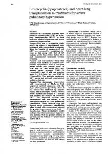
Figure 1-Right ventricle. Control, 1 hour after intravenous injection of dextran. The myocardium is normal; striations and intercalated disks are unremarkable. Immersion fixation, paraffin section. (H&E, x 110) Figure 2-Right ventricle, control. Same procedure as in Figure 1, but with perfusion-fixation and epon embedding (1-g section stained). The myocardium is normal, showing regular striations and intercalated disks. Most vessels have been well perfused. (Toluidine blue, x 110)
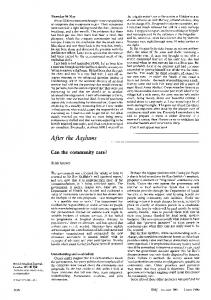
Figure 3-Right ventricle 1 hour after lethal pulmonary embolism. The heart was fixed by
immersion and processed in paraffin. Myocardial pattern and cross striations are generally well preserved, except for pale areas located along some of the fibers (arrows). These lesions are referred to as shredding. (H&E, x 180)
Figure 4-Right ventricle 1 hour after lethal pulmonary embolism. The shredding lesions shown in Figure 3 are better demonstrated in this l-,u epon section. They appear as pale, granular areas, usually on one or both sides of an intercalated disk; some occur along the body of a myocyte (arrow). The paleness of these areas is due to lack of fibrils; the granules are mitochondria. Note well-perfused vessels. (x 180)
2
--a
. -
W.
-
.-0..---E.-
-
-Z=wm--i" .
-
--
=1=7
" .77.1.11
4
j,
Act. -:;
-;,
I
-
fo
-
5
6
i -.Nw-
7
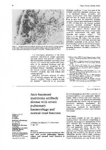
Fur 5--Right ventricle 1 hour after lethal pulmonary embolism. Epon section. Center. necrotic
myocyte. Shredding lsions are obvious; their granular aspect is due to mitochondria floating in a Fue 6-Right ventricle 1 hour after lethal pulmonary embolism; epon clear background. (x 220) Fiu 7-Right ventricle 1 hour after lethal pulsection. A focus of interstitial edema. (x 180) monary embolism; epon section. Venule partially occluded by a platelet thrombus. Note several shredding lesions (arrows). (x 280)
VIMrt
.44-1
I
s$-; .0,, i
I.r .i
uI.-, il.!. '..
j
t-
,
t
1;-,
I
f ,.&
f
the Figure 8-Right ventricle 1 hour after dextran injection (control). The myocardium is normal: sarcomeres are in register, the mitochondria are aligned in the long axis of the myocyte, and the intercalated disks (ID) show the normal straight pattern. The capillary is well perfused. (x 6600)
I' -.4e.t
AL,-vi
1,
-
IqF
Ab
a.
t.
-.,
r 4
,
1V
%
..
.k* e:.tI.
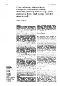
hour after pulmonary embolism. The clear band crossing the field represents a sequence of shredding lesions, abutting intercalated disks. This is the electron microscopic equivalent of the clear zones shown by light microscopy in Figures 3 and 4. Lipid droplets have accumulated in the myocytes. (x 2500)
Figure 9-Right ventricle 1
Figure 10-Right ventricle 1 hour after pulmonary embolism. Another example of shredding lesion. In this case (as in most) the lesion abutts an intercalated disk (ID) which has become abnormally wavy. The mitochondria, which are tightly packed in rows in the adjacent normal areas, are set loose in the shredded part of the myocyte. A few lipid droplets (L) are present in the cytoplasm. (x 6200)
OS*t
A
I0
t
41.
.
IL -11
it
-
.
J.I.
..
4.
-,
"
.'.
I.' a
.,
..
4.
I
I I,
i
.1I
. 714
"t-
....
Row
t
Y-W.-4'I-W
!" .1
j
'c 16
iow.. A I
t iti.,
Figure 11-Right ventricle 1 hour after pulmonary embolism. Detail of a shredding lesion. From top left to bottom Ieft, note progressive shredding of the sarcomeres. Beneath the nucleus, mitochondria float free in an area from which the fibrils have pulled away. (x 20,000)
Fiwe 12-Right ventricle 1 hour after pulmonary embolism. Center: small zone of shredding. Where this lesion reaches the surface of the myocyte, the sarcolemma has apparently given way and formed a large bleb full of loose mitochondria (arrow). Just beneath it is a capillary that failed to perfuse, presumably because it is dogged by stasis and thrombosis. Bottom right shredding beneath an intercalated disk. (x 2500)
13-Right ventricle 1 hour after pulmonary embolism. Discrete lesions: disintegration of Z lines FWure (Z lysis) and separation of the abutting sarcomeres. Most mitochondria remain normal in appearance. Note polyribosomes occasionally aligned with sarcomere fibrils (arrows). (x 33,000)
14
15
Fgure 14-Right ventricle 1 hour after pulmonary embolism. Early stage of a shredding lesion: on either side of an intercalated disk the thin filaments have become detached. (x 20,000) Figure 15Right ventricle 1 hour after pulmonary embolism. Intercalated disk showing early shredding lesions on both sides; the side itself has been stretched out of shape, and the adjacent mitochondria have lost their regular alignment. (x 6500)
17
:-L Figure 16-Artifacts due to fixation by immersion. Right ventricle 1 hour after lethal pulmonary embolism; epon section. The dark bands were present also in controls and represent zones of hypercontraction, as shown in Figure 17. (x 180) Figure 17-Same preparation as in Figure 16; electron micrograph. The mixture of lesion and artifact in this field can be unravelled only by comparison with controls fixed in the same manner, ie, by immersion. The shredding lesions (asterisks) are obvious and never appear in controls. (x 5000)
B-Two necrotic papillary muscles in the right ventricle, 1 day after sublethal pulmonary embolism. Necrosis is betrayed by the striking white aspect. This was certainly heightened by the perfusion, which removed most of the blood within the necrotic Fiure 19-Histology of the necrotic papillary muscle shown in Figure 18B; tissue, as shown in Figure 19. (x 12) epon section. The entire thickness of the papillary muscle is affected. Dead myocytes appear either dark or light. Inflammatory infiltrates are seen on or under both endocardial surfaces, as well as in the myocardium. Most vessels are perfused. (x 100) Fgu 20-Detail of the same papillary muscle shown in Figures 18B and 19; epon section. All myocytes are necrotic, lack striations, and contain contraction bands; the change at the intercalated disk (arrow) is better defined in Figures 25 and 26. Note scattered inflammatory cells. Most vessels are well perfused. (x 280)
Figure IBA-Two papillary muscles of a control heart (right ventricle) after perfusion-fixation.
21
5-c.
22
Figure 21-Right ventricle 1 day after pulmonary embolism. Example of mild but definite distortion of the intercalated disk as seen throughout the right ventricle. (Compare with Figure 8, control.) Note also Fgure 22-Right ventricle 1 day after pulmonary embolism. Zignumerous lipid droplets. (x 6000) zagging from top to bottom is an intercalated disk, so thinned and distorted as to be barely recognizable. To its right is the beginning of a shredding lesion (typical but too narrow to be visible by light microscopy). (x 8500)
.a
j- Z4* -J~
:h
Figur 23-Right papillary muscle 1 day after sublethal pulmonary embolism. Center and bottom: three necrotic myocytes are "tunnelled" by polymorphonuclear neutrophils and macrophages. Top: a myocyte with an area of shredding. All myocytes are loaded with fat droplets and contain clear vacuoles. The blood vessel is well perfused, but its endothelium shows focal disruption (arrowheads) and swelling (arrow). (x 2700)
-s?'
>
*,*?'.
_
j