Review
Clinical Chemistry 55:7 1288–1299 (2009)
Unbound (Free) Bilirubin: Improving the Paradigm for Evaluating Neonatal Jaundice Charles E. Ahlfors,1* Richard P. Wennberg,2 J. Donald Ostrow,3 and Claudio Tiribelli4
BACKGROUND: The serum or plasma total bilirubin concentration (BT) has long been the standard clinical laboratory test for evaluating neonatal jaundice, despite studies showing that BT correlates poorly with acute bilirubin encephalopathy (ABE) and its sequelae including death, classical kernicterus, or bilirubininduced neurological dysfunction (BIND). The poor correlation between BT and ABE is commonly attributed to the confounding effects of comorbidities such as hemolytic diseases, prematurity, asphyxia, or infection. Mounting evidence suggests, however, that BT inherently performs poorly because it is the plasma non–protein-bound (unbound or free) bilirubin concentration (Bf), rather than BT, that is more closely associated with central nervous system bilirubin concentrations and therefore ABE and its sequelae. CONTENT:
This article reviews (a) the complex relationship between serum or plasma bilirubin measurements and ABE, (b) the history underlying the limited use of Bf in the clinical setting, (c) the peroxidase method for measuring Bf and technical and other issues involved in adapting the measurement to routine clinical use, (d) clinical experience using Bf in the management of newborn jaundice, and (e) the value of Bf measurements in research investigating bilirubin pathochemistry.
SUMMARY:
Increasing evidence from clinical studies, clinical experience, and basic research investigating bilirubin neurotoxicity supports efforts to incorporate Bf expeditiously into the routine evaluation of newborn jaundice. © 2009 American Association for Clinical Chemistry
Clinical Chemistry recently published a Citation Classic entitled “Unbound Bilirubin: A Better Predictor of
Kernicterus?” (1 ). The article noted that preventing acute bilirubin encephalopathy (ABE)5 and its serious neurological sequelae in jaundiced newborns remains a serious and unresolved concern for clinicians. It went on to suggest that the serum or plasma non–proteinbound bilirubin concentration (free or unbound bilirubin, Bf) as measured by the much-cited peroxidase method (1, 2 ) may provide better guidance for therapy than the conventionally used total bilirubin concentration (BT). The issue of BT vs Bf was also raised in a recent prospective National Institute of Child Health and Human Development (NICHD) phototherapy study that found considerable overlap in BT values in very low birth weight infants with and without bilirubin-associated deafness, and those authors suggested that Bf might have better differentiated the 2 groups (3 ). Although evidence suggesting that Bf would be superior to BT in the clinical management of jaundiced newborns first appeared in the 1950s (4 ), Bf has languished for decades on the periphery of clinical practice as well as basic research investigating the pathochemistry of bilirubin neurotoxicity. Many clinicians, clinical chemists, and even researchers working in the field of bilirubin neurotoxicity are unfamiliar with Bf measurement or its clinical application. Recent events have renewed interest in Bf (5 ), and the intent of this review is to provide a framework for understanding how Bf measurements might augment both the care of jaundiced newborns and our understanding of the pathochemistry of ABE. Neonatal jaundice is caused by the retention of unconjugated bilirubin (UCB) during the normal, transient postnatal imbalance between UCB production and elimination. Although the condition is generally inconsequential except for the noticeable jaundice,
5 1
Division of Neonatology, Department of Pediatrics, Stanford University School of Medicine, Palo Alto, CA; 2 Division of Neonatology, Department of Pediatrics, and 3 Gastroenterology/Hepatology Division, Department of Medicine, University of Washington School of Medicine, Seattle, WA; 4 Centro Studi Fegato and Department of Life Sciences, University of Trieste, Trieste, Italy. * Address correspondence to this author at: PO Box 2904, Vashon, WA 98070. E-mail
[email protected]. Received December 2, 2008; accepted February 27, 2009. Previously published online at DOI: 10.1373/clinchem.2008.121269
1288
Nonstandard abbreviations: ABE, acute bilirubin encephalopathy; Bf, unbound (free) bilirubin; BT, total bilirubin; NICHD, National Institute of Child Health and Human Development; UCB, unconjugated bilirubin; BIND, bilirubin-induced neurological dysfunction; CNS, central nervous system; P, concentration of plasma bilirubin-binding proteins; Kf, UCB–protein equilibrium association constant; ABR, auditory brainstem response; HRP, horseradish peroxidase; Kp, first-order rate constant for the HRP-catalyzed oxidation of bilirubin by peroxide; FDA, US Food and Drug Administration; Bfss, steady-state Bf; kd, bilirubin– protein dissociation rate constant; ka, bilirubin–protein association rate constant; CSF, cerebrospinal fluid.
Unbound Bilirubin and Newborn Jaundice
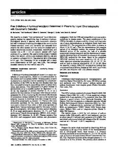
Fig. 1. Chemical structures of neurotoxic 4Z,15Z UCB IX␣ (top) and its photoisomer 4Z,15E UCB-IX␣ (bottom). Dashed lines indicate intramolecular hydrogen bonds, which make the 4Z,15Z isomer lipophilic and neurotoxic. The 4Z,15E isomer has fewer internal hydrogen bonds and is therefore more polar and water soluble than the 4Z,15Z isomer, but its neurotoxic potential is unknown.
the 4Z,15Z UCB-IX␣ isomer produced (Fig. 1) is neurotoxic and in rare cases may reach concentrations sufficient to cause ABE, resulting in death or serious sequelae known collectively as kernicterus (athetoid cerebral palsy, auditory dysfunction, ocular movement disorders, and dental enamel dysplasia) (6 ). Bilirubininduced neurological dysfunction (BIND) has recently
Review been introduced to encompass both the classic and more subtle neurological impairments thought to be due to ABE (7 ). Without intervention with phototherapy or exchange transfusion, ABE occurs in roughly 1% of babies born prematurely (8 ) and in up to 15% of term newborns with hemolytic disorders (9 ). This is in stark contrast to the estimated nonintervention incidence of ⬍1 per 30 000 in the 60% of healthy newborns who become clinically jaundiced (10 ). It is important to note that BT and Bf in samples from jaundiced newborns, as well as solutions prepared from commercial sources of 4Z,15Z UCB-IX␣, usually contain varying amounts of presumably nontoxic bilirubin isomers that may also be measured by the laboratory tests employed to measure the neurotoxic 4Z,15Z UCB-XI␣ isomer (11–14 ). Those isomers include conjugated bilirubin, UCB-III and UCB-XIII (commercial UCB preparations), and UCB photoisomers (11–14 ). Nonetheless, the American Academy of Pediatrics recommends that BT be used to guide jaundice therapy because conjugated bilirubin in the newborn is generally a tiny fraction of BT and the neurotoxicity of the major photoisomer (4Z,15E UCB-IX␣, Fig. 1) is unknown (6, 11–14 ). BT and Bf should therefore be viewed as the best estimates currently available for the concentrations of total and non–protein-bound neurotoxic UCB isomers, respectively. Hereafter UCB will refer specifically to the neurotoxic isomers of UCB unless otherwise indicated. It is well recognized that BT has poor specificity (many false positives) as a predictor of ABE (6, 10, 15, 16 ), requiring large numbers of otherwise healthy jaundiced babies to undergo very costly, unnecessary intervention to prevent a very few cases of ABE (15, 16 ). Despite the considerable investment, ABE still occurs in jaundiced newborns, healthy or otherwise (17, 18 ). Nonetheless, proposals to modify current clinical practice cannot be considered without strong evidence that they may decrease unnecessary treatment or further reduce ABE (19 ). Measurement of Bf has the potential to do both. ABE, BT, and Bf : The Rationale for Measuring Bf in Jaundiced Newborns The pathogenesis of ABE is intimately related to the factors governing UCB steady-state kinetics, in which Bf plays a critical role. We illustrate this using the UCB steady-state kinetics models reported for healthy adults (20 ) and a child with Crigler-Najjar I disease (21 ), which is an inherited absence of hepatic UCB conjugation resulting in severe, lifelong UCB retention characterized by marked jaundice, unconjugated hyperbilirubinemia, and increased risk of ABE (22 ). Clinical Chemistry 55:7 (2009) 1289
Review
Fig. 2. (A), Mean UCB steady state in healthy adults [Berk et al. (20 )]. The risk of ABE is proportional to the nonhepatic extravascular UCB (62 mol) at BT ⫽ 8.3 mol/L and Bf ⫽ X (unknown). (B), Increasing P and Kf (Equation 1) by 15% and 40%, respectively, increases BT (13.5 mol/L) to restore the steady state at Bf ⫽ X with unchanged ABE risk.
Fig. 2A shows the 3-compartment model of UCB steady-state kinetics for healthy adults, as derived from the elegant studies of Berk et al. (20 ). Two important features to note are (a) the likelihood of ABE, i.e., the concentrations of UCB in and around the cells of the central nervous system (CNS), depends on the concentration of UCB in the nonhepatic extravascular compartment, not BT per se (23 ) and (b) under normal conditions, vascular UCB is nearly all bound to plasma proteins. Protein-bound UCB crosses capillary walls very slowly compared with unbound UCB and does not cross the blood– brain barrier or cell membranes at all (24 –28 ). The mass action relationship between BT, Bf, the concentration of plasma bilirubin binding proteins (P), and the UCB–protein equilibrium association binding constant (Kf) is shown in Equation 1. 1290 Clinical Chemistry 55:7 (2009)
Bf ⫽
BT ⫺ Bf K f(P ⫺ BT ⫹ Bf)
(1)
For the purposes of this discussion, Kf refers to the clinically relevant high-affinity UCB binding site (27 ), but it should be noted that UCB also binds to additional sites with much lower affinity constants (29 ). Albumin, which is by far the major component of P, and Kf are highly variable in the newborn, with CVs of about 15% and 40%, respectively (27, 30 ). Fig. 2B shows that whereas substantial changes in either P or Kf at a given UCB production and elimination rate will greatly alter the direct correlation between BT and ABE (i.e., the correlation between BT and the nonhepatic extravascular UCB concentration), the correlation between Bf and ABE remains unchanged. This complex and often paradoxical
Unbound Bilirubin and Newborn Jaundice
Review
Fig. 3. (A), UCB steady state in a child with Crigler-Najjar disease (albumin ⴝ 604 mol/L) [Schmid and Hammaker (21)]. Although BT is ⬇50-fold greater vs Fig. 2A, extravascular UCB is ⬇5-fold greater, suggesting decreasing extravascular UCB accumulation as the UCB load increases. (B), Steady state with sulfisoxazole occupying 25% of the UCB-binding sites. BT decreases, but Bf and ABE risk are increased.
interplay between the UCB binding variables in Equation 1 and ABE is not accounted for when a single BT intervention threshold is applied across a diverse population of newborns (6, 30 ). Extrapolating the concepts illustrated in Fig. 2 to neonatal jaundice, the rate of vascular UCB increase and peak BT will be directly correlated with (a) the magnitude of the imbalance between UCB production and elimination and (b) the magnitudes of P and Kf.
Increases in the UCB production/elimination imbalance at constant P and Kf will increase BT, Bf, and the risk of ABE. However, increases in P and Kf at constant UCB production/elimination imbalance will increase BT but not Bf or the risk of ABE (Fig. 2B). Variations in P and Kf in the newborn undermine the ability of BT but not that of Bf to predict ABE, and Bf should inherently correlate better than BT with ABE in jaundiced newborns. Clinical Chemistry 55:7 (2009) 1291
Review
Fig. 4. As the BT/P molar ratio approaches 1 in a jaundiced newborn, the high-affinity binding site (Equation 1) is nearly saturated, and Bf and the risk of ABE will accelerate dramatically with further increases in the UCB load [Jacobsen (29 )]. BT, however, will increase more slowly because less of the UCB produced is retained in the vascular space.
Fig. 3A uses the UCB steady-state kinetics in an otherwise healthy child with Crigler-Najjar I disease (21 ) to illustrate that the relationships between BT, Bf, and ABE change as UCB accumulates. Compared with Fig. 2A, BT in Fig. 3A is about 50 times greater (439 vs 8.3 mol/L), but the nonvascular UCB is only about 5 times higher (492 vs 93 mol of extravascular and hepatic UCB). This difference suggests that as UCB accumulates, an increasingly smaller fraction of the UCB load resides extravascularly. This phenomenon may result from increased activity of facilitated and active transport mechanisms for extravascular efflux of UCB (28 ), nonconjugating pathways for UCB catabolism (31, 32 ), and increasing saturation of non-CNS extravascular UCB reservoirs such as fatty tissue (⬍1% of the extravascular UCB resides in the CNS in animal models) (33 ). Fig. 4 illustrates the clinical interplay of BT, Bf, and ABE as the UCB load increases. Paradoxically, as P becomes saturated with UCB, the incremental increase in BT per incremental increase in the UCB load decreases while the incremental increase in Bf, which is still a tiny fraction of BT (29 ), accelerates. This in turn accelerates the extravascular accumulation of UCB and the risk of ABE. Therefore, without a confirming Bf measurement, an increased but stable BT in the clinical setting should not be (but often is) taken as reassurance that the risk of ABE is not increasing. Despite the rationale and arguments presented above, the poor correlation between BT and ABE is customarily attributed to comorbidities that may make the CNS more susceptible to UCB neurotoxicity (e.g., hemolysis, prematurity, asphyxia, infection) or extenuat1292 Clinical Chemistry 55:7 (2009)
ing clinical circumstances (15, 19, 34 ). Even the welldocumented association between illness and impaired plasma protein binding of ligands, including UCB, is rarely considered (35, 36 ). On closer examination, however, this tendency to marginalize the role of UCB binding in ABE results more from historical circumstances than a failure to appreciate the relationship between UCB binding and ABE (6, 15 ). Invoking UCB binding to explain the poor correlation between BT and ABE without a substantial amount of supporting clinical data is merely speculation, and there has been a remarkable paucity of UCB-binding data in the clinical literature. Where’s the Bf? A Brief Clinical History of ABE, BT, and Bf In retrospect, BT remains entrenched at the cores of clinical algorithms designed to prevent ABE (6 ) primarily because Bf measurements were not available until 20 years after UCB was clearly established as the cause of ABE. By then, other advances in newborn care had significantly altered the clinical outcomes of newborn jaundice and the perceived need for Bf measurements. In the early 1950s, ABE was a serious problem mainly for babies who had Rh or ABO hemolytic disease or who were born prematurely (8, 9 ). Treatment with the newly described exchange transfusion was risky but significantly lowered the incidence of ABE in both of these high-risk groups. The suggestion that the procedure be used at BT ⱖ20 mg/dL (342 mol/L) for babies with hemolytic disease (9 ) was quickly applied to all jaundiced newborns. The complex interplay between BT, Bf, and ABE soon became apparent when premature babies given sulfisoxazole for infection prophylaxis developed ABE and died despite a “low” BT (23 ). It was soon discovered that sulfisoxazole competes with UCB for protein binding sites, producing a new UCB steady state with a lower BT but higher Bf and extravascular UCB, as illustrated in Fig. 3B. The increased extravascular UCB often reached levels sufficient to cause ABE despite the low BT (23 ), a phenomenon that still haunts clinical practice (37, 38 ). The sulfisoxazole experience alerted clinicians to the role of UCB binding in the pathogenesis of ABE, and methods for measuring UCB binding were pursued in hopes of better identifying babies truly needing a potentially lifesaving but also very risky exchange transfusion (4 ). Measuring Bf directly proved elusive, however, and the early tests measuring plasma “saturation” with UCB (i.e., plasma UCB-binding capacity) ultimately proved unsuitable for routine clinical laboratory use (39, 40 ).
Review
Unbound Bilirubin and Newborn Jaundice
Ironically, by the time the peroxidase method for measuring Bf finally arrived on the scene in 1974 (2 ), ABE had become a rare event in the newborn populations at greatest risk. Rhogam prophylaxis had nearly eliminated Rh hemolytic disease, and phototherapy to enhance UCB elimination substantially reduced the numbers of exchange transfusions needed to prevent ABE in premature babies. Furthermore, a prospective study of ill, premature newborns found neither BT or Bf to be predictive of kernicterus at autopsy (41 ). Although this study was later shown to have serious technical shortcomings (42 ), the now-rare incidence of ABE made prospective determination of the Bf threshold for ABE problematic in any case. Phototherapy seemed a safe and effective method for maintaining BT below the concentrations at which exchange transfusion would be needed in most babies, and it became increasingly difficult to see how measuring Bf would substantially alter clinical care (43 ). With the issue of ABE in high-risk neonatal populations apparently resolved without Bf measurement, attention turned to a major problem with jaundice in the large group of healthy newborns. The a priori BT ⱖ20 mg/dL exchange transfusion guideline, now whimsically christened vigintiphobia (fear of 20) (44 ), had morphed over the years into the clinical (and legal) cornerstone for jaundice intervention in healthy newborns. However, a BT ⱖ20 mg/dL, unlike ABE, was a fairly common occurrence in healthy newborns despite the availability of phototherapy (34 ), and exchange transfusion with its considerable morbidity and mortality (45 ) was frequently called for to prevent a rare illness (10 ). It is not surprising that clinicians arbitrarily began treating most of these babies with phototherapy alone. Historical evidence seemed supportive of this untested practice and even suggested that jaundice in healthy babies was benign and being managed far too aggressively (46 ). Unfortunately, ABE suddenly appeared in previously healthy newborns in the mid 1980s when the increasingly laissez-faire approach to jaundice collided with early postnatal hospital discharge (47 ). A worldwide reexamination of the management of newborn jaundice ensued that largely focused on systematic issues such as patient monitoring and follow-up, and Bf was considered only in passing (6, 19, 34 ). Evidence also emerged questioning the adequacy of the varied and experientially based BT exchange transfusion guidelines used for premature newborns (30 ). In a prospective NICHD study done in the 1970s comparing phototherapy with exchange transfusion, there were 3 cases of autopsy proven kernicterus in the 216 babies with birth weights ⬍1250 g, but only 1 had a BT above the study exchange transfusion threshold of 10 mg/dL (171 mol/L) (48 ). There were also reports
of hearing deficits associated with peak BT below accepted exchange transfusion guidelines (49 ), but more importantly, 2 observational studies now documented a significant association of ABE with Bf but not BT in sick, premature newborns (50, 51 ). It was the introduction of the auditory brainstem response (ABR) for assessing newborn hearing in the early 1980s, however, that resurrected the issue of Bf in the management of newborn jaundice in earnest. The ABR provided for the first time a noninvasive and quantifiable outcome measure for assessing UCB neurotoxicity. UCB was quickly shown to induce ABR changes that could progress to permanent signal loss or be reversed by exchange transfusion (52, 53 ). There is now unequivocal evidence that UCB-induced ABR changes are predicted by Bf but not BT in both premature and term newborns (54 –57 ). In addition, UCB injury in susceptible areas of the CNS can now be documented using newer imaging techniques in vivo such as MRI (37, 58 ), providing yet another new outcome measure for documenting UCB injury. It once again seems warranted and feasible to test the hypothesis that Bf measurements will improve the clinical management of newborn jaundice by better identifying babies needing treatment and minimizing unnecessary intervention (5 ). The peroxidase test for measuring Bf is well suited for this purpose. The Peroxidase Test for Measuring Bf The horseradish peroxidase (HRP) methodology of Jacobsen and Wennberg (1, 2 ) is based on the observation that HRP catalyzes UCB oxidation by peroxide (typically hydrogen peroxide or ethyl hydrogen peroxide), but protein-bound UCB is protected and only Bf reacts (2, 31 ). The first-order rate constant for the reaction (Kp) is determined from the reaction velocity (rate of decrease in UCB peak light absorbance at 440 nm) in the absence of UCB-binding proteins (i.e., [UCB] ⫽ Bf, Equation 2a) and then used to calculate Bf from the reaction velocity (rate of decrease in UCB light absorbance peak at 460 nm) when binding proteins are present (Equation 2b) (2 ). d关UCB兴 ⫽ K p关HRP兴关UCB兴 dt
(2a)
dBT ⫽ K p关HRP兴Bf dt
(2b)
Integrated forms of the equations can also be used to obtain Kp and Bf (59 ), and BT is calculated from the initial absorbance of the sample corrected for any hemoglobin interference (2 ). The test has been automated (60, 61 ), requires small sample volumes (⬍100 L of serum or plasma), and is quantitative, rapid (2–3 Clinical Chemistry 55:7 (2009) 1293
Review min), and inexpensive. Because the method directly assays Bf, the lower-affinity UCB-binding sites will not obscure test endpoints as they do with the binding tests that attempt to measure serum or plasma UCB binding capacity (29, 39, 40 ). A US Food and Drug Administration (FDA)approved commercial spectrophotometer (UB-A1 Analyzer, Arrows Co Ltd) (62 ) provides automated readouts of BT (mg/dL) and Bf (g/dL). BT is obtained after manually diluting the sample 41-fold with phosphate buffer, and a HRP reagent is added to obtain Bf. The reagents are stable at 8 °C for a month after reconstitution, and controls for BT and Bf are provided. The CVs for BT and Bf are about 2% and 5%, respectively. Although many clinical studies have been done with this device (11–14, 27, 51, 54 –57, 61, 63, 64 ), it is not currently marketed in the US. Several things interfere with the Bf measurement by the peroxidase test: (a) Hemoglobin is a weak peroxidase and has considerable absorbance at 460 nm. Moderate hemolysis (hemoglobin ⬎5 g/dL in the undiluted sample (62 )) adds peroxidase activity that can falsely increase Bf, and severe hemolysis overwhelms UCB light absorbance at 460 nm, falsely lowering Bf while falsely increasing BT (2, 62 ). (b) Conjugated bilirubin is more readily oxidized than UCB and, at levels higher than about 1 mg/dL, will falsely increase Bf (13, 65 ). The test can be modified to avoid this problem (65 ). (c) Paraben preservatives falsely increase Bf by generating free radicals that rapidly oxidize UCB (66 ). (d) Bilirubin photoisomers appear to have little effect on Kf (Equation 1) in clinical studies (11, 12 ), although a recent in vitro study using nonneonatal samples suggests that photoisomers may interfere unpredictably with the peroxidase test (14 ). The peroxidase method reacts far more readily with 4Z,15Z UCBIX␣ than with UCB photoisomers, and the 4Z,15Z UCB-IX␣ oxidation products have far less light absorbance than those of the photoisomers (11 ). (e) The pH and ionic composition (particularly chloride and phosphate ion) of the reaction medium may alter binding and the measured Bf (59, 63 ). (f) As with all enzymecatalyzed reactions, Bf will vary with reaction temperature (62 ). (g) Substantial non–HRP-catalyzed UCB oxidation may occur depending on the type of peroxide used (2, 62 ) and will falsely increase Bf unless a correction is applied (2 ). (h) The large sample dilution (typically 40-fold) can attenuate the effect of weak UCB binding competitors such as sulfisoxazole (Fig. 3B), causing underestimation of Bf (42 ). Perhaps the most serious error made when using the peroxidase test is the failure to appreciate that the Bf determined by the peroxidase test is a steady-state Bf (Bfss) (27 ), where 1294 Clinical Chemistry 55:7 (2009)
Bfss ⫽
k d(BT ⫺ Bfss) 关k a共P ⫺ BT ⫹ Bfss兲兴 ⫹ 共K p关HRP兴兲
(3)
and kd and ka are the UCB–protein dissociation and association rate constants, respectively (ka/kd ⫽ Kf in Equation 1). Bfss is nearly equal to the equilibrium Bf only when ka(P ⫺ BT ⫹ Bfss) ⬎⬎ Kp[HRP] and Equation 3 and Equation 1 are nearly identical. When this is not the case, Bfss underestimates the equilibrium Bf, and the error worsens as the equilibrium Bf increases (27, 63 ). This error is easily avoided by measuring Bf at 2 or more HRP concentrations. When Bfss is independent of the HRP concentration (Equation 3 ⬇ Equation 1), Bfss is a reliable estimate of the equilibrium Bf at each HRP concentration. When Bfss decreases with increasing HRP, however, the equilibrium Bf is equal to the inverse of the intercept of the plot of 1/Bfss vs [HRP], as explained in detail elsewhere (27, 59, 61 ). A pseudo-error in the method occurs when diluted samples are used because the apparent Kf for the albumin binding of UCB as well as many other ligands increases with sample dilution (61, 63, 67, 68 ). Kf increases steeply below albumin concentrations of about 100 mol/L, most likely due to the dissociation of albumin oligomers that bind UCB less avidly than the albumin monomer (68 ). Bf measured at the standard 40-fold sample dilution traditionally used for the peroxidase test (2 ) is lower than but correlates directly and significantly with Bf measured at minimal sample dilution (61, 63 ). Furthermore, Bf measured in diluted samples has been superior to BT in predicting ABE- or UCB-induced ABR changes (50, 51, 54 –57 ). Nonetheless, because dilution alters both intrinsic albumin binding and may also attenuate the effects of weak UCB binding competitors, it would seem prudent to measure Bf at minimal sample dilution if possible. The only clinical laboratory control for Bf is supplied with the commercial instrument described above. Kp may be determined directly using solutions of UCB (Equation 2A), but the instability of UCB solutions make this approach problematic for clinical laboratories. UCB surrogates such as purpurogallin or the more stable biliverdin (69 ) could be employed to verify HRP activity, but specific Bf controls with a human albumin matrix to stabilize UCB are more desirable. Current BT clinical laboratory controls may also prove useful in this regard (61 ), and it may be feasible to develop new Bf controls composed of UCB and human albumin solutions that are cross-referenced to Bf measured by other binding methods (68 ). Intervention (reference) Bf thresholds for using phototherapy or exchange transfusion can only be approximated from available data at this time (50, 51, 54 – 57, 64, 70 ). P and Kf will need to be determined in larger
Unbound Bilirubin and Newborn Jaundice
populations of newborns to determine the range of Bf values occurring at the specific BT currently used to guide therapy (6, 30, 34 ). UCB-binding isotherms can readily be constructed for this purpose by titrating individual samples and umbilical cord blood with UCB and measuring Bf over the range of BT observed clinically (2, 60 ). Because both BT and the BT/albumin ratio guidelines for exchange transfusion decrease with gestation and birth weight (30 ), Bf intervention thresholds should encompass these parameters as well. Can Bf Alter Clinical Practice? A Brief Summary of a Clinical Experience with the Peroxidase Test The first author of this review (C.E. Ahlfors) has used the peroxidase test in the clinical management of newborn jaundice. BT and Bf were measured at 2 HRP concentrations using a UB-A1 Analyzer in a CLIA-certified laboratory. The reference exchange transfusion Bf threshold, extrapolated from existing literature and adjusted for birth weight, was 1.3 g/dL per kg of birth weight (22 nmol/L per kg), with a maximum allowable Bf of 4 g/dL (66 nmol/L) (41, 50, 51, 64, 70 ). Given the limited clinical experience with the reference Bf, however, we restricted its use such that exchange transfusions were never administered or withheld on the basis of Bf alone. Exchange transfusions were performed when the BT or the BT/albumin ratio reached exchange transfusion criteria (6, 30 ) or symptoms suggestive of ABE were present (6 ). Bf was measured 294 times in 257 babies over 4 years (Table 1). Several publications resulted from this limited data set, including the first measurement of Bf in a case of lethal kernicterus in the US (64 ) and repudiation of a published model for brain UCB uptake based on BT (27 ). Most importantly, we reported that Bf but not BT was significantly associated with abnormal automated ABR hearing screens in term babies (56 ), and that Bf and especially the Bf/BT ratio (a measure of binding affinity that is proportional to 1/Kf), but not BT, predicted abnormal automated ABR hearing screens, regardless of clinical circumstances (57 ). The American Academy of Pediatrics guidelines recommend exchange transfusion at BT ⱖ30 mg/dL (6 ), and above this concentration about half of the babies with Rh hemolytic disease develop ABE (9 ). We encountered 4 babies with BT ⱖ30 mg/dL. One had lethal kernicterus (Bf 7.63 g/dL, 130 nmol/L) and died before exchange transfusion could be performed (64 ), and the other 3 received exchange transfusions. Only one of the 3 had a Bf above the exchange transfusion threshold (Bf 4.41 g/dL, 75 nmol/L), and that baby was lethargic and failed an automated ABR hearing screen before exchange transfusion but then passed the ABR screen (64 ). The other 2 babies (Bf 2.87 g/dL and
Review 2.80 g/dL) had no obvious symptoms and passed the automated ABR before receiving exchange transfusions. One also had a normal MRI examination. Adding Bf, automated ABR, and MRI to our evaluation of these babies enhanced our assessments of the likelihood of ABE and the urgency with which treatment was needed in these babies. The American Academy of Pediatrics guidelines consider BT between 25 and 30 mg/dL a gray zone where phototherapy should be provided but exchange transfusion is left to the discretion of the clinician (6 ). We readmitted 64 babies for jaundice, 13 of whom had BT between 25 and 30 mg/dL. All the Bf values, which ranged from 1.23 to 2.99 g/dL (21–51 nmol/L), were below the reference Bf concentrations, and all 13 babies passed an automated ABR hearing screen on admission. This provided us considerable reassurance that phototherapy alone was sufficient for these babies. An important but underappreciated tactic we often used was to measure Bf early in the clinical course when babies were at increased risk of ABE (e.g., had hemolysis or were born prematurely). This allowed us to estimate the BT and BT/albumin ratio at the reference Bf for comparison with the recommended exchange transfusion BT and BT/albumin ratio for babies with that condition (30 ). Kf was calculated from the albumin concentration, BT, and Bf using Equation 1, and then reinserted into the equation along with the albumin and reference Bf (22 nmol/L ⫻ birth weight in kg up to 66 nmol/L) to calculate the BT and the BT/albumin ratio at the reference Bf. Comparing these with the recommended intervention BT and the BT/albumin ratio allowed us to individualize our intervention strategies for each patient. The importance of applying this tactic as well as the general concepts outlined in this review to clinical practice and research are illustrated by comparing our clinical experience with premature newborns of ⬍1 kg birth weight (Table 1) with that of the recent NICHD phototherapy study (3 ). The NICHD phototherapy study admitted 984 babies into its conservative phototherapy group in which phototherapy and exchange transfusion were considered at BT thresholds of 8 and 10 mg/dL and 13 and 15 mg/dL for birth weights ⱕ0.750 kg and ⱖ0.751 kg, respectively. Peak BT averaged 9.8 mg/dL, and 13 babies reached BT exchange transfusion thresholds, with 3 receiving the procedure (0.3%) and 10 receiving augmented phototherapy according to study guidelines. Twenty-eight babies (3%) had severe hearing loss defined as requiring bilateral hearing aids, about 10 times the rate of congenital deafness in the newborn population (71 ). We measured UCB binding in 59 similar babies (Table 1), half of whom had binding measured before 48 h of age. Phototherapy was applied at the discretion of the attending physician and using our exClinical Chemistry 55:7 (2009) 1295
Review
Table 1. Bilirubin binding variables in a diverse population of 257 newborns measured with a UB-A1 Analyzer (1:41 sample dilution, 2 HRP concentrations). Birth weight, kg
BT, mol/L
Albumin, mol/L
BT/albumin molar ratio
Bf, nmol/L
Kf, L/mol
Mean
109
428
0.26
7.9
69
Median
108
423
0.25
5.8
58
40
80
0.10
7.3
43
48–260
211–708
0.11–0.59
0.9–42.1
12–217
Mean
154
454
0.35
10.1
86
Median
150
438
0.35
6.3
73
⬍1
Samples
Patients
Exchange transfusions
70
59
3
SD Range 1–1.249
24
18
1
SD
60
88
0.16
9.4
60
63–279
257–634
0.15–0.71
0.7–42.8
33–322
Mean
157
436
0.37
13.2
67
Median
145
445
0.33
7.0
59
72
88
0.17
11.9
38
87–419
257–619
0.19–0.84
2.9–46.0
23–165
Mean
213
487
0.45
14.7
87
Median
203
483
0.42
8.7
84
Range 1.25–1.499
23
20
0
SD Range 1.5–1.749
11
10
1
SD
88
63
0.22
17.1
24
109–393
393–589
0.20–0.93
2.4–62.4
56–140
Mean
219
527
0.43
20.0
51
Median
212
581
0.45
18.3
52
60
122
0.13
14.7
25
130–296
347–634
0.24–0.64
5.3–59.0
Mean
283
523
0.56
17.1
91
Median
281
544
0.58
14.6
91
Range 1.75–1.999
10
8
0
SD Range 2–2.499
20
18
15–88
0
SD
75
95
0.11
11.5
33
108–393
347–664
0.31–0.81
3.4–46.0
39–179
Mean
336
559
0.61
21.5
101
Median
331
574
0.60
17.0
92
91
86
0.18
16.1
50
116–615
151–710
0.21–1.36
0.9–130
27–318
Range ⱖ2.5
136
124
5
SD Range
change transfusion thresholds of BT ⱖ10 mg/dL (171 mol/L) or BT/albumin ratio ⱖ4 mg/g (molar ratio 0.45) (30, 48 ). We performed 3 exchange transfusions (5%), and 3 additional babies did not pass their automated ABR hearing screen at hospital discharge. The binding and automated ABR data for these 6 babies are summarized in Table 2. The mean Kf of the samples 1296 Clinical Chemistry 55:7 (2009)
from these 6 babies (28 L/mol) is significantly lower than that of the remaining samples (75 L/mol, P ⬍ 0.0001). Only 1 baby (birth weight 0.840 g) reached the exchange transfusion threshold for the NICHD study (BT ⫽ 15.2 mg/dL, Bf ⫽ 0.90 g/dL), but BT had decreased substantially by the time the blood arrived for exchange transfusion and it was not performed. None
Review
Unbound Bilirubin and Newborn Jaundice
Table 2. Bilirubin binding variables (UB-A1 Analyzer, 1:41 sample dilution, 2 HRP concentrations) in babies born at


![[READ] Clinical Chemistry: Principles Free - Google Sites](https://m.moam.info/img/260x300/read-clinical-chemistry-principles-free-google-sit_64770132097c474b228b6e6d.jpg)