JTUMED347_proof ■ 7 February 2017 ■ 1/4 Journal of Taibah University Medical Sciences (2017) -(-), 1e4
Taibah University
Journal of Taibah University Medical Sciences www.sciencedirect.com
1 2 3 4 5 6 7 Q12 8 9 10 11 12 13 14 15 16 17 18 19 20 21 22 23 24 25 26 27 28 29 30 31 32 33 34 35 36 37 38 39 40 41 42 43 44 45 46 47 48 49 50 51 52 53 54
Case Report
Unusual pattern of mandibular fracture displacement in a child: A case report Shadia A. Elsayed, PhD Department of Oral and Maxillofacial Surgery, College of Dentistry, Taibah University, Almadinah Almunawwarah, KSA
Received 25 September 2016; revised 7 December 2016; accepted 14 December 2016; Available online - - -
ﺍﻟﻤﻠﺨﺺ ﺇﻥ ﻏﺎﻟﺒﻴﺔ ﻛﺴﻮﺭ ﺍﻟﻔﻚ ﺍﻟﺴﻔﻠﻲ ﻋﻨﺪ ﺍﻷﻃﻔﺎﻝ ﻫﻰ ﻣﻦ ﻧﻮﻉ ﻛﺴﺮ ﺍﻟﻐﺼﻦ ﺍﻟﻨﻀﻴﺮ ﻣﻊ ﻧﻘﻮﻡ ﻫﻨﺎ ﺑﺘﺴﺠﻴﻞ ﺣﺎﻟﺔ ﻛﺴﺮ ﻟﻘﻤﻲ ﻭﺇﻛﻠﻴﻼﻧﻲ ﻟﻠﻔﻚ ﺍﻟﺴﻔﻠﻲ ﻣﻊ.ﻭﺟﻮﺩ ﺇﺯﺍﺣﺔ ﺑﺴﻴﻄﺔ . ﻣﻊ ﻋﺮﺽ ﻣﻘﺪﻣﺎﺗﻬﺎ ﺍﻟﺴﺮﻳﺮﻳﺔ ﻭﻋﻼﻣﺎﺗﻬﺎ ﺍﻹﺷﻌﺎﻋﻴﺔ ﻭﺍﻟﺠﺮﺍﺣﻴﺔ،ﺇﺯﺍﺣﺔ ﺷﺪﻳﺪﺓ ﺃُﺯﻳﺤﺖ ﺍﻟﻠﻘﻤﺔ ﻭﺍﻹﻛﻠﻴﻼﻧﻲ ﺍﻟُﻴﻤَﻨَﻴﺎﻥ ﺇﻟﻰ ﺍﻟﻨﺎﺣﻴﺔ ﺍﻟﻮﺣﺸﻴﺔ ﺣﺘﻰ ﺍﺳﺘﻘﺮﺍ ﺑﺎﻟﻜﺎﻣﻞ ﻓﻮﻕ ﻣﻊ ﺇﺯﺍﺣﺔ ﺟﺰﺀ ﻣﻦ ﺍﻹﻛﻠﻴﻼﻧﻲ ﺇﻟﻰ ﺍﻟﻨﺎﺣﻴﺔ،ﺍﻟﺠﺎﻧﺐ ﺍﻟﻮﺣﺸﻲ ﻟﻠﻘﻮﺱ ﺍﻟﻮﺟﻨﻲ ﻛﺎﻥ ﻫﻨﺎﻙ ﺗﺠﻤﻊ ﺩﻣﻮﻱ ﻛﺒﻴﺮ ﻓﻲ ﺟﺪﺍﺭ ﺍﻟﺒﻠﻌﻮﻡ ﺍﻟﻮﺣﺸﻲ ﺍﻣﺘﺪ ﺇﻟﻰ ﺍﻷﺳﻔﻞ.ﺍﻹﻧﺴﻴﺔ ﻣﻊ ﺇﺻﺎﺑﺔ ﺷﺪﻳﺪﺓ ﻟﻠﻨﺴﺞ ﺍﻟﺮﺧﻮﺓ ﻓﻲ ﺍﻟﻤﻨﻄﻘﺔ ﺗﺤﺖ،ﺣﺘﻰ ﺍﻟﺤﺪ ﺍﻷﺳﻔﻞ ﻟﻠﻔﻚ ﺍﻟﺴﻔﻠﻴﺔ ﻧﺪﺭﺓ ﻣﺜﻞ ﻫﺬﻩ ﺍﻹﺻﺎﺑﺔ ﻓﻲ ﺍﻷﻃﻔﺎﻝ ﻓﻲ ﻣﻨﻄﻘﺔ ﺍﻟﺮﺃﺱ ﻭﺍﻟﺮﻗﺒﺔ.ﺍﻟﻔﻚ ﺍﻟﺴﻔﻠﻲ .ﻭﻣﻌﺎﻟﺠﺘﻬﺎ ﺗﺠﻌﻞ ﻫﺬﻩ ﺍﻟﺤﺎﻟﺔ ﺃﻛﺜﺮ ﺇﺛﺎﺭﺓ ﻟﻼﻫﺘﻤﺎﻡ ﻟﺠ ٰﺮﺍﺣﻲ ﺍﻟﻮﺟﻪ ﻭﺍﻟﻔﻜﻴﻦ
rarity of such paediatric trauma in the head and neck region and the management of such an injury make this case more interesting for maxillofacial surgeons. Keywords: Condyle; Haematoma; Head and neck injury; Mandible; Paediatric dentistry
Q2
Ó 2017 The Author. Production and hosting by Elsevier Ltd on behalf of Taibah University. This is an open access article under the CC BYNC-ND license (http://creativecommons.org/licenses/by-ncnd/4.0/).
ﺍﻟﻠﻘﻤﺔ؛ ﺗﺠﻤﻊ ﺩﻣﻮﻱ؛ ﺍﻟﻔﻚ ﺍﻟﺴﻔﻠﻲ؛ ﻃﺐ ﺃﺳﻨﺎﻥ ﺍﻷﻃﻔﺎﻝ؛:ﺍﻟﻜﻠﻤﺎﺕ ﺍﻟﻤﻔﺘﺎﺣﻴﺔ ﺇﺻﺎﺑﺎﺕ ﺍﻟﺮﺃﺱ ﻭﺍﻟﺮﻗﺒﺔ
Introduction Abstract Most mandibular fractures in children are greenstick-type fractures with minimal displacement. We report a case of a severely displaced condylar and coronoid mandibular fracture along with its clinical presentation and radiological and surgical features. The right condyle and coronoid were laterally displaced, riding completely over the lateral surface of zygomatic arch with a coronoid fragment displaced medially. There was a large haematoma collection in the lateral pharyngeal wall extending downward to the inferior border of the mandible with severe soft tissue injury at the submandibular region. The Corresponding address: Taibah University, Almadinah Almunawwarah, KSA. E-mail:
[email protected] Peer review under responsibility of Taibah University.
Production and hosting by Elsevier
Unintentional injury may be more catastrophic than any other trauma, especially at a young age. A science of prophylaxis to prevent such injury is very important to prevent death among children and adolescents. The predominant Q3 mechanism of injury varies by age, with falls being most common in young children, pedestrian injuries in toddlers and elementary school-age children, and motor vehicle trauma in adolescents. Mortality rates of trauma, all types included, are higher in rural areas than in cities. The reasons Q4 for this may include longer wait times from injury to definitive care, in-hospital delays to definitive treatment, less time for injury prevention education in schools and primary care offices, and socioeconomic and ethnic factors. Other associations have been identified that increase susceptibility to trauma, such as children with behavioural disorders and negligence of parents or inadequate parental supervision.1 Complete dislocation of the mandibular condyle from the fossa is classified into four types: anterior, posterior, lateral, and superior dislocation.1 All types except for anterior dislocation are rare, according to Chaithanya 2013.1 Superolateral dislocation of the intact mandibular condyle
1658-3612 Ó 2017 The Author. Production and hosting by Elsevier Ltd on behalf of Taibah University. This is an open access article under the CC BY-NC-ND license (http://creativecommons.org/licenses/by-nc-nd/4.0/). http://dx.doi.org/10.1016/j.jtumed.2016.12.008 Please cite this article in press as: Elsayed SA, Unusual pattern of mandibular fracture displacement in a child: A case report, Journal of Taibah University Medical Sciences (2017), http://dx.doi.org/10.1016/j.jtumed.2016.12.008
55 56 57 58 59 60 61 62 63 64 65 66 67 68 69 70 71 72 73 74 75 76 77 78 79 80 81 82 83 84 85 86 87 88 89 90 91 92 93 94 95 96 97 98 99 100 101 102 103 104 105 106 107 108 109 110 111 112 113 114 115 116 117 118 119
JTUMED347_proof ■ 7 February 2017 ■ 2/4
2 1 2 3 4 5 6 7 8 9 10 11 12 13 14 15 16 17 18 19 20 21 22 23 24 25 26 27 28 29 30 31 32 33 34 35 36 37 38 39 40 41 42 43 44 45 46 47 48 49 50 51 52 53 54 55 56 57 58 59 60 61 62 63 64 65
Q5
S.A. Elsayed
occurs very rarely.2 Allen and Young3 classified lateral dislocations into type I (lateral subluxation) and type II (complete dislocation), in which the condyle is forced laterally and then superiorly. Satoh et al.4 classified complete lateral dislocations of the condyle as the following types: type IIA, in which the condyle is not hooked above the zygomatic arch; type IIB, in which the condyle is hooked above the zygomatic arch; and type IIC, in which the condyle is lodged inside the zygomatic arch, which is fractured. There are a few reports of this kind of dislocation in adults.5,6 We report a case of superolateral unilateral complete dislocation of condyle and part of the coronoid with no fracture of the zygomatic arch, in a 6-year-old child, which may be very rare. Case report
Q6
Q7
A six-year-old male was admitted to the emergency room of Al-Zahraa University Hospital, Cairo, Egypt in October of 2015. The patient was fully conscious, with a GCS score of 15, no evidence of any brain injury and a complete ability to maintain his airway. His parents had created a compression bandage with a large gauze pack on the right side of the child’s face. Upon inspection, the wound was present at the right submandibular area and the cut was sharp and deep, reaching to the bony angled area of the mandible. The large blood vessels in this region appeared cut and compressed with no active bleeding. There was an extraoral swelling on the right side of the face extending to the level of the lateral orbital margin and downwards to the mandibular angle region. A large haematoma had collected at the medial part of the mandible, appearing from the cut wound as large soft black space. History from the parents revealed that the child had fallen down some newly formed granite steps and severe bleeding had started. An extraoral OPG radiograph revealed a suspected fracture on the anterior mandible and coronoid process of the right mandible. To confirm this diagnosis, a CT maxillofacial was requested. The CT revealed an anterior symphyseal non-displaced mandibular fracture and a vertical sagittal fracture of the coronoid process, which was displaced medially, and the whole ramus-condyle and coronoid were displaced laterally and riding on the lateral surface of the right zygomatic arch, which appeared clinically by palpation as a hard projection of the condyle on the substance of the cheek (Figures 1,2). Emergency intervention was conducted to reduce the segment, evacuate the haematoma and suture the wound. The anaesthesiologist wanted to intubate the child nasally, as is usual in maxillofacial trauma cases, but a quick decision was made to intubate orally due to impaired visibility caused by excessive bleeding, which had just been revealed due to the lateral pharyngeal wall cut. Extraorally, the condyle and ramus were palpably projecting under the skin of the right cheek. Many attempts to depress the ramus and condyle below the zygomatic arch via a closed reduction were made but with no results, even with the help of an angle pull with bone-clamping forceps made directly through the open access offered by the submandibular wound. Therefore, a decision was made to open the temporomandibular joint area with a preauricular incision and dissection until the condyle was reached, with a direct
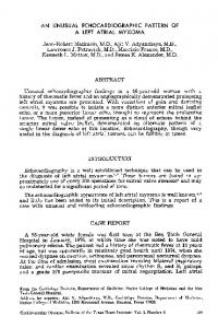
Figure 1: Preoperative coronal cut CT showing medial large haematoma extending from the submandibular lacerated wound upwards to the level of the coronoid.
Figure 2: Preoperative 3 D CT photograph showing displaced ramal condyle and coronoid laterally overriding the right zygomatic arch.
open reduction to the whole segment below the arch. In this instance, the extraoral bulging of the cheek disappeared immediately after reduction. Before closure of the lacerated submandibular wound, evacuation of a large black blood clot medial to the ramus was performed and the area was vigorously irrigated with saline wash, with compression of the tissues to prevent further accumulation of blood. The postoperative mouth opening was within the normal range and a physiotherapy follow-up was conducted immediately after the operation. Discussion Young children are more susceptible to trauma, especially boys, who are more likely to be affected than girls due to their rapid and uncontrolled play patterns. Thus, fall injury is the most seen aetiology in our emergency paediatric trauma centres.7
Please cite this article in press as: Elsayed SA, Unusual pattern of mandibular fracture displacement in a child: A case report, Journal of Taibah University Medical Sciences (2017), http://dx.doi.org/10.1016/j.jtumed.2016.12.008
66 67 68 69 70 71 72 73 74 75 76 77 78 79 80 81 82 83 84 85 86 87 88 89 90 91 92 93 94 95 96 97 98 99 100 101 102 103 104 105 106 107 108 109 110 111 112 113 114 115 116 117 118 119 120 121 122 123 124 125 126 127 128 129 130
JTUMED347_proof ■ 7 February 2017 ■ 3/4
Unusual pattern of mandibular fracture displacement 1 2 3 4 5 6 7 8 9 10 11 12 13 14 15 16 17 18 19 20 21 22 23 24 25 26 27 28 29 Q8 30 31 32 33 34 35 36 Q9 37 38 39 40 41 42 43 44 45 46 47 48 49 50 51 52 53 Q10 54 55 56 57 58 59 60 61 62 63 64 65
Shock can occur rapidly from blood loss caused by severe external wounds, especially in children. In this case, the patient was not in shock and blood loss was not critical, as bleeding was controlled with pressure bandages until the child’s arrival at the hospital. Thus, there was no need for any blood transfusion or fluid resuscitation. Oral intubation was the correct decision in this case to avoid the potentially serious complications associated with the blind nasal passage, which was due to the presence of the more anterior and cephalad-positioned airway, relatively large adenoids and tonsils, and soft short trachea typical in children. In addition, the intraoral lateral pharyngeal cut presented complications to nasal intubation. The paediatric emergency training included in the departments of our General National Governmental Hospitals is limited and basic. Additionally, ensuring the adequacy of pre-hospital airway management and the ability to obtain venous access in children requires further proper training, experience, and skill maintenance. These factors illustrate the need for more specialized paediatric trauma centres with qualified personnel and equipment. In paediatric patients, closed treatment is the treatment of choice (as attributed to Sawhney et al., 2013)8 except for patients with a major dislocation or the loss of contact between the bony stumps, or in paediatric patients with panfacial trauma (as recommended by Colletti et al., 2014).9 In the present case, performing an open reduction for the severely displaced mandible was absolutely indicated. In titling this case, we chose the terminology of displacement and not dislocation, as the whole mandibular right ramus (condyle and coronoid) were displaced, not only the condyle. There are some differences in terminology across Europe and the United States, which can lead to confusion. “Dislocated,” as used in continental Europe, is equal to “displaced” in the United States and Great Britain, whereas “dislocated” as used in the United States and Great Britain is equivalent to “luxated” in continental Europe.10 There is a need to standardize classification systems to allow comparisons across studies, as reported by Berner T et al., 2015.6 In the present case, the severity of displacement and soft tissue injury suggested that a higher-energy trauma may have been the reason for this unusual pattern. It may be explained due to certain anatomic features in younger children (aged less than 8 years), who have weak ligaments11 that permit greater mobility and elasticity of the mandible. Thus, slippage of the whole ramus over the zygomatic arch occurred. Furthermore, the trauma direction, which appeared to be directed from the inferior medial to a superolateral direction, permitted greater trauma in this type of fall injury. Although children, in general, have a higher survival rate than adults after a head injury, they may be more vulnerable to long-term disability than adults.11 This is especially the case for condylar trauma, so long-term follow-up and postoperative physiotherapy were planned for the patient and clear instructions were given to the parents. It is recommended that facial lacerations that are not complicated by associated injury be managed in an ER setting. However, large or extensive facial lacerations are preferably closed in an operating room environment. Although expensive and potentially time-consuming, a CT
3 Q1
scan often provides the most detailed images of traumatic pathology, especially 3D CT views, and may assist in determining operative intervention. From our previous experience, we advise oral maxillofacial surgeons to keep the emotional needs of the child constantly in mind. A frightened child is emotionally labile and apprehensive. A calm and reassuring approach with attempted explanation of procedures gains the most cooperation, even if the trauma is relatively minor. OMFS who have more frequent contact with seriously ill children are more likely to accept parental presence.7 Conclusions and recommendations For traumatized children, a detailed and organized approach for injury control, prompt recognition, and emergency treatment should be implemented. Constant monitoring and reassessment of the patient’s status are mandatory, as this approach is the most important way to decrease disability and death after paediatric trauma. Author’s contribution SEA has critically reviewed and approved the final draft and is responsible for the content and similarity index of the manuscript. Ethical approval Ethics approval was obtained from our Institutional Review Board (AL-Azhar University Ethic Committee). No personal details or identifying information are included in the paper. Informed consent was obtained from the parents of the patient. Conflict of interest The author has no conflict of interest to declare. Acknowledgments The author would like to thank Dr. Susan Abdelhakim Hassan, Professor and Head of OMS, Al-Azhar University; Dr. Emad Hussein, Associate Professor of Plastic and General Surgery, Al-Azhar University, and Dr. Maha Hakam, Professor of OMS, Cairo University, for their great support. References 1. Chaithanya Jaya D. An unusual dislocation of mandibular condyle in a 4 year old child e a case report. J Oral Maxillofac Surg Med Pathol 2013; 25: 260e263. 2. Bu SS, Jin SL, Yin L. Superolateral dislocation of the intact mandibular condyle into the temporal fossa: a review of the literature and report of a case. Oral Surg Oral Med Oral Pathol Oral Radiol Endod 2007; 103: 185e189. 3. Allen FJ, Young AH. Lateral displacement of the intact mandibular condyle: a report of five cases. Br J Oral Surg 1969; 7: 24e30. 4. Satoh K, Suzuki H, Matsuzaki S. A type II lateral dislocation of bilateral intact mandibular condyles with a proposed new classification. Plast Reconstr Surg 1994; 93: 598e602.
Please cite this article in press as: Elsayed SA, Unusual pattern of mandibular fracture displacement in a child: A case report, Journal of Taibah University Medical Sciences (2017), http://dx.doi.org/10.1016/j.jtumed.2016.12.008
Q11
66 67 68 69 70 71 72 73 74 75 76 77 78 79 80 81 82 83 84 85 86 87 88 89 90 91 92 93 94 95 96 97 98 99 100 101 102 103 104 105 106 107 108 109 110 111 112 113 114 115 116 117 118 119 120 121 122 123 124 125 126 127 128 129 130
JTUMED347_proof ■ 7 February 2017 ■ 4/4
4 1 2 3 4 5 6 7 8 9 10 11 12 13
S.A. Elsayed
5. Hegde S, Kamath VV, Deepa M, Priya A. Superolateral dislocation of the mandibular condyle not associated with fracture: a case report. J Maxillofac Oral Surg 2010; 9: 424e427. 6. Berner T, Essig H, Schumann P, Blumer M, Lanzer M, Ru¨cker M, Gander T. Closed versus open treatment of mandibular condylar process fractures: a meta-analysis of retrospective and prospective studies. J Craniomaxillofac Surg 2015; 43: 1404e1408. 7. O’Brien MM, Creamer KM, Hill EE, Welham J. Tolerance of family presence during pediatric cardiopulmonary resuscitation: a snapshot of military and civilian pediatricians, nurses, and residents. Pediatr Emerg Care 2002; 18: 409e413. 8. Sawhney R, Brown R, Ducic Y. Condylar fractures. Otolaryngol Clin North Am 2013; 6: 779e790.
9. Colletti G, Battista VMA, Allevi F, Giovanditto F, Rabbiosi D, Biglioli F. Extraoral approach to mandibular condylar fractures: our experience with 100 cases. J Craniomaxillofac Surg 2014; 42: 186e194. 10. Loukota RA, Eckelt U, De Bont L, Rasse M. Subclassification of fractures of the condylar process of the mandible. Br J Oral Maxillofac Surg 2005; 43: 72e73. 11. Tornetta P, Einhorn T. Pediatrics. Lippincott Williams and Wilkins; 2004. How to cite this article: Elsayed SA. Unusual pattern of mandibular fracture displacement in a child: A case report. J Taibah Univ Med Sc 2017;-(-):1e4.
Please cite this article in press as: Elsayed SA, Unusual pattern of mandibular fracture displacement in a child: A case report, Journal of Taibah University Medical Sciences (2017), http://dx.doi.org/10.1016/j.jtumed.2016.12.008
14 15 16 17 18 19 20 21 22 23 24 25 26