Vol. 13, No. 1, 2015
Case Report
Urticaria pigmentosa: A case report Keen MA1 1
Dermatologist, Jammu and Kashmir Health Services, India
Abstract Mastocytosis is a heterogenous group of diseases characterized by abnormal infiltration of mast cells in the skin and other organs. Urticaria Pigmentosa is the most common variant of cutaneous mastocytosis. We herein report a case of urticaria pigmentosa in a three year old boy. Keywords: Mastocytosis, Mast cells, Urticaria pigmentosa Address for correspondence
Dr. Mohammad Abid Keen Jammu and Kashmir Health Services. Iqbal Abad, KP Road , Ananntnag, Jammu and Kashmir Pin : 192101 Cell no: 9419963692 E mail:
[email protected]
Introduction Mastocytosis is a hematopoietic disorder which is usually seen sporadically and characterized by an increased number and the accumulation of mast cells in one or more organs.1 It can be divided into cutaneous mastocytosis and systemic mastocytosis.2,3 There are four clinical subtypes of cutaneous mastocytosis: urticaria pigmentosa, mastocytoma, diffuse cutaneous mastocytosis and telangiectasia macularis eruptiva perstans (TMEP). The most common type of cutaneous mastocytosis in children is urticaria pigmentosa. The disease is clinically characterized by multiple erythematous and pigmented macules, papules and plaques and localized blistering that may vary in size. Dariers sign is positive. Histopathologial examination is the gold stand for the diagnosis. Symptomatic treatment with antihistaminics, mast cell stabilizers and topical steroids is effective. A typical case of this dermatosis is being presented. Case report A 3 year old male child was presented by his parents with generalized eruption of multiple brown maculopapular lesions on trunk and limbs
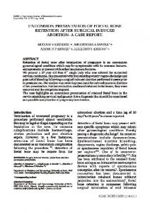
of two year duration. There was also a history of generalized urticarial flushing with occasional bulla formation. He had been delivered with cesarean section at full term without any complication. The general health, growth and development of the child was unaffected. There was no family history of similar disease or any other dermatological or autoimmune disease. Therapy with topical corticosteroids prescribed by the general practitioner had shown no effect. Physical examination of the patient was normal. Systemic examination of the patient also revealed no abnormality. There was no hepatomegaly, splenomegaly or lymphadenopathy. Cutaneous examination of the child revealed multiple, sharply defined, red brown maculopapules and plaques on the trunk and limbs (Figure 1).
Figure 1: Multiple sharply defined red brown maculopapules and plaques on the trunk and limbs NJDVL - 57
Vol. 13, No. 1, 2015
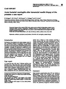
Case Report No nodules or bullae were seen at the time of the examination. On stroking the individual lesions, there was formation of wheal and flare (Darier sign - Positive). A provisional diagnosis of urticaria pigmentosa was made in this child. A complete blood count, liver and renal function tests, urine and stool analysis, chest X ray and ultrasonography of the abdomen were normal. A 4mm skin punch biopsy specimen was obtained with due precautions and sent for histopathological examination. It revealed increased number of mast cells in dermis (Figure 2).
Figure 2: Histopathological image revealing increased number of mast cells in dermis The child was treated with antihistaminics and topical steroids for 4 week which led to the regression of his lesions.The parents were advised regarding avoidance of excessive scrubbing and massage of the skin. At the end of six weeks, the child had only residual pigmentation left at the lesional sites with no new lesion formation. Discussion Mastocytosis represents a spectrum of clinical disorders with clinical features determined by infiltration of various organs and skin with mast cells. The cutaneous and systemic symptoms such as pruritus, urticaria, wheezing, diarrhea and hypotension are caused by the release of mediators by non-allegic mechanisms.4 Despite the fact that most common location is the skin, it may also occur in the liver, spleen, bone narrow, lymph nodes, lungs and gastrointestinal tract.5 It can be divided into cutaneous mastocytosis and systemic NJDVL - 58
mastocytosis.2,3 Cutaneous mastocytosis usually affects the patients in early childhood and the disease often regresses spontaneously.6 However, systemic mastocytosis frequently occurs in adult patients and tends to resist permanently.7 Mast cells that arise in the bone marrow are pluripotential precursor cells. They differentiate in the skin and other peripheral organs under the influence of an array of growth factors, c-kit ligand or mast cell growth factor. Mast cells produce inflammatory mediators including histamine, trysptase, TNF- a , leukotrienes, prostaglandins, platelet activating factor, heparin, IL-8.3,4 These mediators are responsible for local and systemic symptoms such as flushing, bullae, pruritus, dyspnea, exacerbation of asthma, low blood pressure, gastroesophageal reflux, peptic ulcer and diarrhea. The most important mediator causing all of these symptoms is histamine.8 The etiology of mastocytosis is unclear. Recent observations have shown a soluble form of stem cell factor in the skin and suggest an etiological role for derangement of this growth factor and its receptor.[4] In addition, a somatic mutation of the proto-oncogene c-kit that could be responsible for mast cell proliferation has been detected.9 ckit mutation analyses are important for the therapy and prognosis of mastocytosis. In 2001, four distinct clinical variants of cutaneous mastocytosis were published by WHO. These were: 1. Urticaria pigmentosa 2. Isolated mastocytoma (solitary mastocytoma) 3. Diffuse Cutaneous mastocytoma 4. Telangiectasia macularis eruptiva perstans (TMEP) In 2008, WHO updated the classification of cutanean mastocytosis as:1. Maculopapular cutaneous mastocytosis (Urticaria pigmentosa) a. Special form: Plaque form b. Special form: Nodular form c. Special form: TMEP 2. Diffuse cutaneous mastocytosis 3. Solitary mastocytosis
Vol. 13, No. 1, 2015
Case Report Urticaria pigmentosa also known as maculopapular mastocytosis, was first reported by Nettleship and Tay in 1869.10 Their patient was a female child of 2 years, and their paper was entitled chronic Urticaria leaving Brown stains of Nearly two years Duration. Unna later demonstrated mast cells in increased numbers in urticarias of the Nettleship type. Urticaria pigmentosa is the most common form of cutaneous mastocytosis (70-90%), within an approximate Incidence of 1/1000-8000. There is no sex predilection and occurs more often in infants and children than the adults. Clinical features appear within the first two years of life. The clinical features are multiple oval or round hyperpigmented macules, papules or patches, brown-red-yellow and 2-4mm in diameter.11 These lesions urticate by manipulation (e.g, rubbing) or spontaneously. This reaction is known as Darier sign. Dariers sign is not always demonstrable, especially in those with a long history of this disorder, and is not 100% specific for mastocytosis, since it has been described rarely in Juvenile xanthogranuloma and acute lymphoblastic leukemia of neonate.12,13 The sites of predilection are chest and dorsal areas of the body, while the palms, soles and face are usually unaffected.14 Lesions may blister in infancy or childhood and may be the presenting feature, but heal without scarring. Systemic manifestations include flushing, vomiting, diarrhea, tachycardia, headache, weight loss and wheezing. 1 5 Histologically there is mild to moderate perivascular infiltrate with dendritic mast cells in the papillary dermis; a band like infiltrate or sometimes even nodular infiltrates extending to the subcutis may be seen, especially with special stains like toluidine blue and chloroacetate
esterase.16 Careful technique when taking the skin biopsy, to minimize traumatic degranulation, is important. Injecting local anaesthetic around the lesion to be sampled and avoiding adrenaline containing local anaesthetics may yield a higher number of stainable mast cells. The diagnostic work-up of patients includes complete blood counts, routine biochemistry tests, liver function tests, and basal serum tryptase levels. In case the patient is an infant or a child and, has an abnormal blood count, enlarged liver, spleen or lymphadenopathy and elevated serum tryptase levels (>20ng/ml), all necessary tests such as abdominal ultrasound and CT, gastrointestinal system endoscopy, bone radiographs, scans and even bone narrow biopsy should be done.17 However, if the patient is an adult, bone narrow examination must be done absolutely.18 Treatment of urticaria pigmentosa is mainly symptomatic with avoidance of all known mast all degranulation stimuli (physical triggers especially rubbing, alcohol, morphine, codeine, NSAIDS, radiocontrast media, scopolamine, d-Tc). Antihistaminics such as H1 or H2 antihistamines or a combination of both, are the first step medications among systemic treatment options. Doxepin may also be used for its antihistamine properties. The mast cell stabilizers like Sodium cromoglycate and ketotifen are also used in patients with urticaria pigmentosa. Topical Cortisosteroids can be used and also intralesional triamcinolone acetonide injection is available for localized lesions.6 Systemic PUVA therapy is an alternative modality of treatment for patients not responding to standard treatments. Patients with chronic and widespread involvement should keep adrenaline pen for self-administration and a wristband.
References 1.
Akaglu G, Erkin G, Cakir B, Boztepe G, Sahin S, Karaduman A, et al. Cutaneous mastocytosis: demographic aspects and clinical features of 55 patients. J Eur Acad Dermatol Venereol 2006; 20: 969-73.
2.
Avshalumov K, Pichardo R, Jorizzo JL, Sangueza O P, Goldenberg G. Bullous mastocytosis: report of a patient and a brief review of the literature. Am J Dermatopathol 2008; 30: 455-57. http://dx.doi.org/10.1097/DAD.0b013e3181783354
3.
Heide R, Zuidema E, Beishuizen A, Den Hollander JC, Van Gysel D, Seyger MM, et al. Clinical NJDVL - 59
Vol. 13, No. 1, 2015
Case Report aspects of diffuse cutaneous mastocytosis in children: two variants. Dermatology 2009; 219: 309-15. http://dx.doi.org/10.1159/000243808 4.
Murphy M, Walsh D, Drumm B, Watson R. Bullous mastocytosis: a fatal outcome. Pediatr Dermatol 1999; 16: 452-5. http://dx.doi.org/10.1046/j.1525-1470.1999.00116.x
5.
Akay BN, Kittler H, Sanli H, Harmankaya K, Anadolu R. Dermatoscopic findings of cutaneous mastocytosis. Dermatology 2009; 218: 226-30. http://dx.doi.org/10.1159/000182260
6.
Briley LD, Phillips CM. Cutaneous mastocytosis: a review focusing on the pediatric population. Clin Pediatr 2008; 47: 757-61. http://dx.doi.org/10.1177/0009922808318344
7.
Duckworth AK, Bhatti A, Barnes C. Diffuse cutaneous mastocytosis in fraternal twins. Int J Dermatol 2009; 48: 170-72. http://dx.doi.org/10.1111/j.1365-4632.2009.03439.x
8.
Briley LD, Phillips CM. Cutaneous mastocytosis: a review focusing on the pediatric population. Clin Pediatr 2008; 47: 757-61. http://dx.doi.org/10.1177/0009922808318344
9.
Walker T, von Komorowski G, Scheurlen W, Dorn-Beineke A, Back W, Bayerl C. Neonatal mastocytosis with pachydermic bullous skin without c-Kit 816 mutation. Dermatology 2006; 212: 70-2. http://dx.doi.org/10.1159/000089026
10.
Ahmad N, Evans P, Lloyd-Thomas AR. Anesthesia in children with mastocytosis--a case based review. Paediatr Anaesth 2009; 19: 97-107. http://dx.doi.org/10.1111/j.1460-9592.2008.02904.x
11.
Seitz CS, Rose C, Brocker EB, Trautmann A. Intertriginous urticaria pigmentosa. Dermatology 2005; 210: 77-9. http://dx.doi.org/10.1159/000081493
12.
Nagayo K, Sakai M, Mizuno N. Juvenile xanthogranuloma with Dariers sign. Int J Dermatol 1983; 10: 283-5.
13.
Yen A, Sanchez R, Oblender M, Raimer S. Leukemia cutis: Dariers sign in a neonate with acute lymphoblastic leukemia. J Am Acad Dermatol 1996; 34: 375-8. http://dx.doi.org/10.1016/S01909622(07)80012-0
14.
Turchin I, Barankin B, Schloss E. Unusual cutaneous findings of urticaria pigmentosa and telangiectasia macularis eruptiva perstans associated with marked myelofibrosis. Int J Dermatol 2006; 45: 1215-7. http://dx.doi.org/10.1111/j.1365-4632.2006.02648.x
15.
Barker A, Stewart RW. Case report of mastocytosis in an adult. South Med J 2009; 102: 91-3. http://dx.doi.org/10.1097/SMJ.0b013e3181827871
16.
Carter MC, Metcalfe DD. Biology of mast cells and mastocytosis syndromes. In: Freedberg IM, Eisen AZ, Wolff K et al., eds. Fitzpatricks Dermatology in General Medicine, 7th eds. Philadelphia: McGraw Hill; 2008. p. 1434-47.
17.
Webber NK, Ponnampalam J, Grattan CE. How reliable is blood tryptase as a marker of systemic disease in an infant with cutaneous mastocytomas? Clin Exp Dermatol 2008; 33: 198-99. http://dx.doi.org/10.1111/j.1365-2230.2007.02585.x
18.
Valent P, Akin C, Escribano L, Fodinger M, Hartman K, Metcalfe DD, et al. Standards and standardization in mastocytosis: consensus statements on diagnostics, treatment recommendations and response criteria. Eur J Clin Invest 2007; 37: 435-53. http://dx.doi.org/10.1111/j.13652362.2007.01807.x
NJDVL - 60