Oct 31, 1988 - Martha C. Hawes*, Steven L. Robbs, and Steven G. Pueppke. Departments of ..... Hawes MC, Smith LY, Howarth AJ (1988) Agrobacterium tu-.
Plant Physiol. (1989) 90, 180-184 0032-0889/89/90/01 80/05/$01 .00/0
Received for publication October 31, 1988 and in revised form December 16, 1988
Use of a Root Tumorigenesis Assay to Detect Genotypic Variation in Susceptibility of Thirty-four Cultivars of Pisum sativum to Crown Gall1 Martha C. Hawes*, Steven L. Robbs, and Steven G. Pueppke Departments of Plant Pathology (M.C.H., S.L.R.) and Molecular and Cellular Biology (M.C.H.), University of Arizona, Tucson, Arizona 85721; and Department of Plant Pathology, University of Missouri, Columbia, Missouri 65211 (S.G.P.) ABSTRACT
evaluate resistance and susceptibility in Pisum sativum L., a well characterized diploid species that is a known host of A. tumefaciens (16). To initiate studies of crown gall resistance in pea, we established a reproducible quantitative assay for tumorigenesis on roots of intact plants and used it to survey genotypic variation among 34 commercial cultivars of pea.
We developed a quantitative assay to measure tumorigenesis on roots and root crowns, the natural sites of Agrobacterium tumefaciens infection. Efficiency of tumor formation and tumor weight on seedlings of Pisum sativum 'Little Marvel' were directly proportional to the logarithm of inoculum concentration. Depth of wounding prior to inoculation also significantly influenced tumor weight but not efficiency. Mean weight of tumors that developed in response to inoculation with strain B6 varied significantly among 34 different commercial cultivars. Tumors on the most susceptible cultivar, Target, were more than tenfold heavier than those formed on the least susceptible cultivar, Sweet Snap. Efficiency of tumorigenesis on 'Sweet Snap' was also relatively low: only 64% of inoculated seedlings developed tumors compared with 89 to 100% efficiencies for all other cultivars.
MATERIALS AND METHODS Bacterial Strains and Culture
Single colony cultures of bacterial strains were maintained at -70C in 50:50 (v/v) glycerol:yeast extract-mannitol (YEM) medium. Strain B6 was a gift from J. A. Lippincott (Northwestern University). For assays, cultures were grown overnight on YEM solidified with 0.7% agar and were suspended in water. Inoculum concentrations were estimated turbidimetrically and confirmed by dilution plating. Plant Inoculation and Assay
Agrobacterium tumefaciens is a soil-borne bacterial pathogen that causes crown gall on most dicots and some gymnosperms and monocots (5). Although most parts of many plants are susceptible to experimental inoculation, A. tumefaciens normally infects through wounds in roots and at the rootshoot interface, the crown (18). Crown gall is among the major disease problems in fruit tree and ornamental nurseries and vineyards (25, 26). The host range of individual strains of A. tumefaciens is determined primarily by the Ti plasmid (19, 32) and can range from a few to hundreds of different species (for example, 5, 14). Ti plasmids from wide host range and limited host range strains have been utilized to demonstrate that both TDNA and virulence genes can influence specificity to some extent, but that 'undefined host factors' also mediate susceptibility of plants to infection by recombinant strains (4, 1 1). Despite the wealth of information about the molecular biology of virulence in A. tumefaciens, little is known about host factors that influence host range. One problem in evaluating host contributions to crown gall tumorigenesis has been the lack of defined variation in a plant species that is amenable to biochemical and genetic analysis. We have chosen to
Pisum sativum L. seeds were surface-sterilized by consecutive 5-min immersions in 95% ethanol and 50% commercial bleach and then washed 4x in at least 50 mL of sterile water. Seeds were germinated on water agar overlaid with filter paper for 2 to 3 d or until the radicles were approximately 1 to 3 cm in length. Each seedling was wounded either by stabbing the root with a marked scalpel blade to a depth of 1 mm or by pushing the blade through the root to a depth of approximately 3 mm. Three wounds were made on each plant at approximately 5-mm intervals from the crown to the root tip. Seedlings were immersed into 5 mL of inoculum for 5 min and were then placed into moistened growth pouches (Northrup King, Minneapolis, MN), with 4 or 5 seedlings per pouch. Pouches were moistened daily with approximately 5 mL of water and were incubated at 250C day/230C night for 2 weeks. The percentage of inoculation sites that developed visible tumors was evaluated to determined the efficiency of tumorigenesis. Tumors or 3- to 4-mm root sections containing wound sites were excised and weighed. Controls included plants wounded and immersed in water or in serially diluted suspensions of A 136, a strain lacking a Ti plasmid. A baseline control value was determined based on mean weight of from 30 to 100 root sections for each genotype. This value was subtracted from the weight of individual excised tumors to
' Supported by grant No. 86-CRCR- 1-2224 of the U.S. Department of Agriculture. This is journal series No. 7003 of the Arizona Agricultural Experiment Station.
180
GENOTYPIC VARIATION IN SUSCEPTIBILITY OF PEA TO CROWN GALL
181
obtain values for tumor weight. Mean weight of sections from plants inoculated with A136 did not vary significantly from that of sections of plants sham-inoculated with water. RESULTS Tumorigenesis Assay-'Little Marvel'
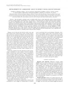
Visible tumors developed on inoculated 'Little Marvel' plants with 3-mm wounds within 1 week to 10 d and were optimal with respect to tumor size after 2 weeks, after which plants began to deteriorate. Plants were, therefore, removed from pouches and evaluated for efficiency of tumor development and were either sacrificed to determine tumor weight or were transplanted into soil. Disease development was dosage dependent. Above ground symptoms on plants with 3-mm deep wounds inoculated with strain B6 included stunting and chlorosis, and the damage intensified with increasing inoculum concentrations from I04 to I09 bacteria per mL (Fig. la). At concentrations greater than 109/mL, plants were severely stunted (Fig. lb) and in some cases were girdled. Mean tumor weight increased from less than 5 mg at 102 bacteria/mL to nearly 100 mg at 109/mL.(Figs. lc, 2a). No visible tumors developed on plants inoculated with fewer than 102 bacteria per mL. The efficiency of tumorigenesis or the percentage of inoculation sites that developed tumors also was proportional to inoculum concentration, with 100% of wounds forming tumors at 108 or more bacteria per mL (Fig. 2b). Tumor weight was proportional to wound size: for a given inoculum concentration, tumors on plants with 3-mm deep wounds were approximately fourfold heavier than tumors from plants with 1-mm deep wounds. Thus, 'Little Marvel' seedlings with 3-mm wounds inoculated with I09 bacteria per mL developed tumors with mean weight of 97 mg (Fig. 2a). In contrast, average weight of 'Little Marvel' tumors that developed in response to inoculation of 1-mm wounds was only 25 mg (Table I), although the efficiency of tumorigenesis remained high (greater than 95%). Plants with 1-mm wounds did not develop obvious symptoms on above-ground parts and survived the transition from growth pouches to soil without apparent damage. In screening for genotypic variation in susceptibility to tumorigenesis, we therefore chose to inoculate 1-mm wounds with 109 bacteria per mL, in order to achieve maximum efficiency with a minimum of deleterious
-~
N
Figure 1. Growth pouch tumorigenesis assay, a, Crown gall tumors on pea seedlings (cv Little Marvel) wounded to a depth of 3 mm, immersed for 5 min in a suspension of strain B6 (109 bacteria per mL) and grown for 2 weeks in growth pouches. Control seedlings wounded and sham-inoculated with Ti plasmid-lacking strain A136 are shown on the left, infected seedlings on the right. b, 'Aboveground' symptoms on pea seedlings inoculated with increasing concentrations (log 10) of inoculum and grown for 2 weeks in growth pouches. c, Correlation between tumor size and inoculum concentration. Groups of excised tumors were from seedlings inoculated with 109, 1 o8, 107 (left column, top to bottom), or 1 6, 105, and 104 bacteria per mL (3-mm deep wounds (right column, top to bottom). The group on the bottom of the right column were excised stem sections from seedlings that were wounded and sham-inoculated with strain Al 36.
effects on plant growth. Genotypic Variation
Although
some
plants from all 34
pea
cultivars developed
tumors when seedlings were inoculated with strain B6, significant variation in tumor weight occurred among genotypes
(Table I). The cultivars fell into three categories, with small, medium, and large tumors. Mean weights of tumors that formed on 32 cultivars formed a statistically overlapping continuum of medium-sized tumors from 12 to 32 mg. Three genotypes were significantly different from this group. 'Oregon Sugar Pod' and 'Target,' respectively, developed large tumors with mean weights of 43 and 46 mg and were in a class by themselves. The tumors of 'Oregon Sugar Pod' and 'Target' were more than 10-fold heavier than those that
developed on the least susceptible cultivar, 'Sweet Snap,' whose small tumors only weighed an average of 4 mg (Table I). The efficiency of tumorigenesis also was reduced in 'Sweet Snap': whereas the percentage of tumor development on all other cultivars ranged from 89 ± 3% to 100%, only 64 ± 6% of inoculated 'Sweet Snap' seedlings developed tumors. DISCUSSION Crown gall tumorigenesis has been measured in many plants using various tissues, including leaves and stems of intact plants (for example, 5, 17, 22, 28), and explants of roots (13), tubers (1), and cotyledons (15, 27). Lacking is an assay that is based on infection of host plants at the natural site of infection of Agrobacterium tumefaciens, the root. Different
HAWES ET AL.
182
a
100
E 801-
6010 40
E
3 0 0
(0
20
II I I
upa 1 00
.r
0
C
0
80
II
L.- l
l .
I
I
I
I
l
l
l
l
b
I
,I
-
0 40 cm -W
)F
CD 20 20 aC)
nu L. -
0
I
I
1
2
3
4
5
Table I. Genotypic Variation in Tumor Weight among 34 Pea Cultivars Inoculated with Strain B6 Roots of seedlings were wounded to a depth of 1 mm, then immersed for 5 min in inoculum at 109 bacteria per mL. After 2 weeks in growth pouches, tumors or control stem sections were excised and weighed. Tumor weight was determined by subtracting a control value based on the mean weight from approximately 30 control stem sections. Mean differences were analyzed by ANOVA. Mean weights of tumors in the small, medium, and large groups were statistically distinct from means in the other two groups at the 0.01% level of significance. Mean weights of tumors on cultivars within the medium size range were statistically overlapping. Mean weights of tumors on Oregon Sugar Pod and Target did not differ significantly from each other. Sources of seed were: 1, Royal Seed Company; 2, Burpee Seed Company; 3, Rogers Brothers Seed Company; 4, Park Seed Company; 5, University of Saskatchewan, Department of Horticulture. Cultivar
60
0.
Plant Physiol. Vol. 90, 1989
I
I
I
6
7
8
9
Inoculum concentration (log 10) Figure 2. a, Correlation between mean tumor weight on cv Uttle Marvel pea seedlings and initial inoculum concentration of strain B6 (values based on A62s). Values are means and standard errors from a total of at least 60 plants from three independent experiments. Tumor weight for each excised tumor was derived by subtracting a value based on the mean weight of at least 30 control stem sections. b, Correlation between efficiency of tumorigenesis (number of sites that developed tumors/total number of inoculation sites) and inoculum concentration. Values are means and standard errors from at least 60 plants from three independent experiments. assays offer advantages for different purposes. However, for the purpose of screening for disease resistance genes, it is crucial to use the organ that is the normal target for the pathogen. This is particularly important in view of reports that susceptibility of certain tissues may not be consistent with that of roots. For example, leaves of certain varieties of Kalanchoe do not develop tumors even though other plant parts are susceptible to infection (3). Ideally, an assay for identification ofresistance genes should be simple, rapid, inexpensive, reproducible and quantitative, and should require a minimum of space. Most important, the assay should be nondestructive, so that individual plants of interest can be grown to maturity to harvest progeny for genetic analysis. The growth pouch assay, which has been exploited for years to study Rhizobium-legume symbiotic interactions (34), provides a simple and economical assay for crown gall tumorigenesis that can be completed within 10 d. Fifty plants can be maintained in a 15 x 15 cm rack for the duration of the assay, and plants of interest can be transferred
Small tumors Sweet Snap Medium tumor Dwarf Grey Sugar Alaska Knight Laxantonian Maestro Alaska 423 Green Arrow Snappy Wando Burpeeana Early Freezonian Century Sugar Bon G. Melting Sugar Thomas Laxton Grenadier Honey Pod Patriot Novella Salvo Sugar Ann Sparkle Little Marvel Mam. Melt. Sugar Blizzard Snowbird Laxton Progress Sugar Snap Oregon S. Pod II Sugar Daddy Progress No. 9 Large tumors Oregon Sugar Pod Target
n
Mean Tumor Weight Efficiency Source mg
64%
1
67
4±1
89 94 63 57 71 55 72 44 91 48 90 95 46 33 91 60 23 44 44 22 46 24 67 91 46 86 78 74 46 43 29
12 ± 1 13± 1 13 ± 2 14 ± 2 16 ± 2 16 ± 3 17 3 17 2 17 2 19 3 19 3 20 ± 3 21 ± 3 21 ± 4 21 ± 3 21 ± 3 21 ± 5 22 ± 3 22 ± 3 23± 4 24 ± 3 24 ± 3 25 ± 2 26 ± 2 26± 3 27 ± 2 27 ± 3 28 ± 3 28 2 30 3 30 3
96 96 89 97 96 96 94 94 98 89 98 91 96 100 95 93 96 93 93 100 100 96 92 97 94 99 90 91 96 97 89
3
48 24
43
98 100
3 3
47
4 6
2
2 1 2 3 2 2 1 2 2 5 2 4 1 2 3 4 4 3 4 3 1 2 4 2
1 1
2 2
to soil and allowed to mature rather than being sacrificed to measure tumor weight. The assay can be adapted to a number of species with different seed types, including sunflower, tomato, potato, and zinnia (MC Hawes, unpublished data). However, large-seeded, fast-growing plants do better in growth pouches than slow-germinating species with small seeds, which are more susceptible to contamination.
GENOTYPIC VARIATION IN SUSCEPTIBILITY OF PEA TO CROWN GALL
The physiology of tumorigenesis in pea has previously been studied by Kurkdjian et al. (16) who used decapitated pea seedlings grown in tubes of agar. Their assay differs from ours in two important respects: (a) tumor development is measured on above-ground tissue rather than on roots, and (b) the assay is destructive, involving the decapitation of the apical meristem. Although their assay would not be appropriate for selecting for disease resistance, their results with respect to tumorigenesis were consistent with our data. Thus, tumors were well developed within 12 d. Furthermore, tumor size was proportional to inoculum concentration, as reported previously (10), presumably because ultimate tumor size is dependent upon the initial number of cells in the wound that are accessible to transformation. Genetic variation in susceptibility to crown gall has been exploited to analyze resistance genes in grapevine, an economically important host of A. tumefaciens (31). Szegedi and Kozma's (31) results indicate that, at least with certain genotypes of this species, resistance appears to be inherited in a simple manner: a single dominant gene encodes resistance. With such information available about the plant's role in disease, together with the detailed information about the molecular biology of virulence in A. tumefaciens, it should be possible to begin systematically to unravel the genetic and biochemical bases of host factors that influence pathogenesis. Unfortunately, grapevine, a clonally propagated, woody species that requires 18 months to 3 years to complete a generation, is not well suited for such studies. Although not exploited for genetic analysis, variation has been documented among cultivars of a number of other species, including chrysanthemum (24), alfalfa (21), kalanchoe (3), castor bean (7), sunflower (4), soybean (15, 27), and squash (29, 33). The results indicate that genetic variation in response to A. tumefaciens is readily detectable in natural populations. However, none of the variation has been detected in a species with the combination of attributes that make pea a useful model organism for this study. First, several highly susceptible cultivars of pea provide good baseline positive controls. Second, susceptibility in these lines is sufficiently uniform that quantitative variation can be detected. Third, the large and fleshy roots of pea lend themselves well to biochemical, physiological, and cytological analysis. Finally, pea is a well characterized diploid species that is readily amenable to genetic analysis, with a well defined genetic map. Our initial survey of 34 commercial cultivars indicates that significant variation exists in pea, with up to a 10-fold difference in tumor weight between the most susceptible and least susceptible cultivars. A survey of 1200 pea germlines that is in progress has revealed an even greater divergence in response (MC Hawes, Marx, SG Pueppke, unpublished data). The underlying causes of the differences are unknown but are presumably related to reduced efficiency in one or more steps in T-DNA transfer, integration into host cells, expression of T-DNA genes, or plant response to T-DNA produced phytohormones. Quantitative assays for several known or proposed stages that are pivotal in crown gall transformation, including chemotaxis (2, 9), binding (for example, 6, 8, 23), production of virulence gene-inducing molecules (reviewed in 30), cellular transformation (12), and hormone production (for example,
183
35) are now available. It should be possible to use such assays in conjunction with genetically defined pea lines to begin to evaluate the biochemical as well as molecular causes of variation in susceptibility of plants to crown gall. LITERATURE CITED 1. Anand VK, Heberlein GT (1977) Crown gall tumorigenesis in potato tuber tissue. Am J Bot 64: 153-158 2. Ashby AM, Watson MD, Shaw CH (1987) A Ti-plasmid deter-
3. 4.
5.
6. 7.
8. 9. 10. 11.
12.
13. 14. 15.
16. 17.
18.
19.
20. 21. 22.
mined function is responsible for chemotaxis of Agrobacterium tumefaciens toward the plant wound product acetosyringone. FEMS Microbiol Lett 41: 189-192 Bopp M, Resende F (1966) Crown gall tumoren bei verschiedenen Arten und Bastarden der Kalanchoideae. Port Acta Biol 9: 327-366 Buchholz W, Thomashow MF (1984) Host range encoded by the Agrobacterium tumefaciens tumor-inducing plasmid pTiAg63 can be expanded by modification of its T-DNA oncogene complement. J Bacteriol 160: 327-332 DeCleene M (1985) The susceptibility of monocotyledons to Agrobacterium tumefaciens. Phytopath Z 113: 82-89 Douglas CJ, Halperin W, Nester EW (1982) Agrobacterium tumefaciens mutants affected in attachment to plast cells. J Bacteriol 152: 1265-1275 El Khalifa MD, El Nur EE (1970) Crown gall on castor bean leaves. I. Crown gall bioassay on primary castor bean leaves. Angew Botanik 44: 29-37 Hawes MC, Pueppke SG (1987) Correlation between binding of Agrobacterium tumefaciens by root cap cells and susceptibility of plants to crown gall. Plant Cell Rep 6: 287-290 Hawes MC, Smith LY, Howarth AJ (1988) Agrobacterium tumefaciens mutants deficient in chemotaxis to root exudates. Mol Plant Microbe Interact 1: 182-186 Hildebrand EM (1942) A micrurgical study of crown gall infection in tomato. J Agric Res 61: 685-696 Hoekema A, dePater BS, Fellinger AJ, Hooykaas PJJ, Schilperoort RA (1984) The limited host range of an Agrobacterium tumefaciens strain extended by a cytokinin gene from a wide host range T-region. EMBO J 3: 3043-3047 Jefferson RA, Kavanaugh JA, Bevan MW (1987) GUS fusions: B-glucuronidase as a sensitive and versatile gene fusion marker in higher plants. EMBO J 6: 3901-3907 Klein RM, Tenenbaum IL (1955) A quantitative bioassay for crown gall tumor formation. Am J Bot 42: 709-712 Knauf VC, Panagopoulos CG, Nester EW (1982) Genetic factors controlling the host range of Agrobacterium tumefaciens. Phytopathology 72: 1545-1549 Kudirka DT, Colburn SM, Hinchee MA, Wright MS (1986) Interactions of Agrobacterium tumefaciens with soybean leaf explants in tissue culture. Can J Genet Cytol 28: 808-817 Kurkdjian A, Manigault P, Beardsley R (1968) Crown gall: effect of temperature on tumorigenesis in pea seedlings. Can J Bot 47: 803-808 Lippincott JA, Heberlein GT (1965) The quantitative determination of the infectivity of Agrobacterium tumefaciens. Am J Bot 52: 856-863 Lippincott JA, Lippincott BB (1976) Morphogenic determinants as exemplified by the crown gall disease. In A Pirson, MH Zimmerman, eds. Encyclopedia of Plant Physiology, Vol 4. Springer-Verlag, Berlin, pp 355-388 Loper JE, Kado CI (1979) Host range conferred by the virulence specifying plasmid of Agrobacterium tumefaciens. J Bacteriol 139: 591-596 Lowe B (1985) Studies on host-parasite interactions between Vitus sp. and hybrids and Agrobacterium tumefaciens. MS thesis, University of Rhode Island Mariotti D, Davey MR, Draper J, Freeman JP, Cocking EC (1984) Crown gall tumorigenesis in the forage legume Medicago sativa L. Plant Cell Physiol 24: 473-482 Matsumoto S, Machida Y, Takebe I (1986) A rapid method for
184
23. 24.
25. 26. 27. 28. 29.
HAWES ET AL. assaying tumorigenicity of A. tumefaciens. Plant Mol Biol Rep 4: 42-47 Matthysse AG (1987) Characterization of nonattaching mutants of Agrobacterium tumefaciens. J Bacteriol 169: 313-323 Miller HN, Miller JW, Crane JL (1975) Relative susceptibility of Chrysanthemum morifolium cultivars to Agrobacterium tumefaciens. Plant Dis Rep 59: 576-581 Moore LW, Cooksey DA (1981) Biology of Agrobacterium: plant interactions. Int Rev Cytol Suppl 13: 15-46 Nesme X, Michel MF, Digat B (1987) Population heterogeneity of Agrobacterium tumefaciens in galls of Populus L. from a single nursery. Appl Environ Microbiol 53: 655-659 Owens LD, Cress DE (1985) Genotypic variability of soybean response to Agrobacterium strains harboring the Ti or Ri plasmids. Plant Physiol 77: 87-94 Rogler CE (1981) Strain-dependent temperature-sensitive phase in crown gall tumorigenesis. Plant Physiol 68: 5-10 Smarrelli J, Watters MT, Diba LH (1986) Response of various
30. 31.
32.
33.
34. 35.
Plant Physiol. Vol. 90, 1989
cucurbits to infection by plasmid-harboring strains of Agrobacterium. Plant Physiol 82: 622-624 Stachel SE, Zambryski PC (1986) Agrobacterium tumefaciens and the susceptible plant cell: a novel adaptation of extracellular recognition and DNA conjugation. Cell 47: 155-157 Szegedi E, Kozma P Jr (1984) Studies on the inheritance of resistance to crown gall disease of grapevine. Vitis 23: 121-126 Thomashow M, Panagopoulos C, Gordon M, Nester E (1980) Host range of Agrobacterium tumefaciens is determined by the Ti plasmid. Nature 283: 794-796 Unger L, Ziegler SF, Huffman GA, Knauf VC, Peet R, Moore LW, Gordon MP, Nester EW ( 1985) New class of limited host range Agrobacterium mega-tumor inducing plasmids lacking homology to the transferred DNA of a wide host range, tumor inducing plasmid. J Bacteriol 164: 723-730 Weaver RW, Frederick LR (1972) A new technique for mostprobable-number counts of Rhizobia. Plant Soil 36: 219-222 Weiler EW, Spanier K (1981) Phytohormones in the formation of crown gall tumors. Planta 153: 326-337