to inhibit the biological activity of hCG. Of the five antibodies tested for their ability to inhibit hCG-induced stimulation of rat testes steroidogenesis in vitro, two ...
Proc. NatL Acad. Sci. USA
Vol. 79, pp. 2245-2249, April 1982 Biochemistry
Use of monoclonal antibodies to subunits of human chorionic gonadotropin to examine the orientation of the hormone in its complex with receptor (steroidogenesis/Leydig cells/hormone binding/immunological mapping)
W. R. MOYLE*, P. H. EHRLICHt, AND R. E. CANFIELDt *Department of Obstetrics and Gynecology, University of Medicine and Dentistry of New Jersey, Rutgers Medical School, Piscataway, New Jersey 08854; and tDepartment of Medicine, College of Physicians and Surgeons, Columbia University, New York, New York 10032
Communicated by Seymour Lieberman, January 11, 1982
ABSTRACT Monoclonal antibodies were prepared against the a and f8 subunits of human chorionic gonadotropin (hCG). Although all were selected on the basis of their ability to bind the intact hormone, each also bound one of the two subunits but not both. Using a solid phase double antibody system to measure the relative binding to sites on the surface of hCG, we observed that four ofthe five antibodies bound to different sites on the molecule. This information was correlated with the ability of each antibody to inhibit the biological activity of hCG. Of the five antibodies tested for their ability to inhibit hCG-induced stimulation of rat testes steroidogenesis in vitro, two proved to be potent inhibitors, whereas the other three had almost no effect. This inhibition of steroidogenesis was highly correlated with the ability of the antibodies to inhibit hCG binding to testes homogenates. Thus, we have begun to derive a scheme that describes the relative binding positions of individual monoclonal antibodies and receptor on hCG. The purified monoclonal antibodies were iodinated and employed to evaluate which antigenic sites on hCG remained free in hCG-receptor complexes. The data indicated that portions of the g subunit in hCG-receptor complexes were buried (i.e., failed to bind radiolabeled antibody), whereas other portions remained exposed (i.e., they bound radiolabeled antibody). Those antibodies that interacted with portions of hCG that became inaccessible in the receptor complex also blocked the biological actions of hCG, whereas those that interacted with exposed sites had little or no effect on activity. Although we did not find antibodies to the a subunit that would bind to the hormone-receptor complex, we found that one of the two antibodies specific to a subunit epitopes blocked the actions of the hormone. Both antigenic determinants on the at subunit appeared to be lost after the hCG-receptor complex had formed. These studies suggest that each hCG subunit participates in the hormone-receptor complex and that portions of the ,B subunit project from the surface of the receptor.
Although the binding of hCG and related gonadotropins to Leydig cells has been studied at length (1), data concerning the orientation ofthe hormones in the receptor complex are sparse. Most studies of the role of the subunits have been based on the reduced ability of chemically modified gonadotropin analogs to bind to the receptor, initiate a biological response, or both (1, 6, 7). Recently, Ji and Ji (8) have made a photoaffinity label of hCG, which they linked to proteins on the surface of granulosa cells and which permitted them to radiolabel the receptor. Results with both approaches suggest the possibility that a and P3 subunits interact directly with the receptor. The availability of monoclonal antibodies to bind individual antigenic sites on hCG subunits appeared to offer us another approach to explore the roles ofthe subunits by examining the orientation of hCG in the receptor complex. Details of these studies are described here. MATERIALS AND METHODS Preparation of Monoclonal Antibodies. Once per month we injected purified a or P subunits of hCG (20-100 ug) in complete Freund's adjuvant, prepared as described (9), into the peritoneal cavity of BALB/c mice. Three days prior to excising the spleen, we injected the mice again intraperitoneally with 100 ,ug of hCG subunit in 0.9% NaCl solution followed a few hours later by an intravenous injection of 50 ,ug. We fused splenic lymphocytes with P3-NS1/1-Ag4-1, an azaguanine-resistant myeloma cell line, using polyethylene glycol essentially as described (10, 11). After selecting the hybrid cells in a medium containing 20% horse serum, hypoxanthine, aminopterin, and thymidine, we assayed the medium for its content of antibodies to hCG, using a double antibody radioimmunoassay system (see below). Cells producing antibodies to hCG were frozen until they could be subcloned on BALB/c 3T3 monolayers at a later date. Of the original successful positive fusions, five lines were subcultured and grown in Dulbecco's modified Eagle's medium containing bovine serum albumin (hereafter referred to as albumin) at 2 mg/ml for 24 hr. Aliquots of this medium were tested for their ability to inhibit hCG-induced steroidogenesis and for their affinity for hCG as described elsewhere (12). We purified other aliquots on columns of Affi-Gel blue (Bio-Rad) for later radioiodination. Scatchard Analysis of Monoclonal Antibodies. Aliquots (50 ,ul) containing 10,000-40,000 cpm of 12I-labeled hCG (125IhCG) and 50-,ul aliquots of unlabeled hCG (both in 1% horse serum, 99% phosphate-buffered saline) were mixed with 100 ,u of 0.3 M potassium phosphate (pH 7.5). Subsequently, 100 ,ul of antibody (diluted in 1% horse serum) were added, the tubes were incubated 1 hr at 37°C followed by 18 hr at 5°C, and
Human chorionic gonadotropin (hCG) is a glycoprotein hormone, composed of a and ,B subunits, which interacts with gonadal receptors to stimulate steroid synthesis (1). The isolated subunits exhibit less than 0.1% of the activity of the intact hormone and, within the limits of highest purity available, are thought to lack biological activity (1-3). As the subunits recombine, they reacquire biological activity (4), and spectral studies indicate that the conformation of the subunits is altered when they are recombined (5), suggesting that unique structural properties of the intact hormone may be needed for activity. Because neither subunit is active alone, the relative roles of the subunits in the actions of the hormone have been difficult to assess. Conceivably, only one of the subunits comes into direct contact with the receptor. The publication costs ofthis article were defrayed in part by page charge payment. This article must therefore be hereby marked "advertisement" in accordance with 18 U. S. C. §1734 solely to indicate this fact.
Abbreviation: hCG, human chorionic gonadotropin.
2245
2246
Biochemistry: Moyle et aL
the complex was precipitated by adding 10 A.l of 50% normal (in phosphate-buffered saline) and an appropriate amount of rabbit anti-mouse IgG or goat anti-mouse F(ab')2. After 10 min at 370C and then 1 hr at room temperature, the precipitate was sedimented and its radioactivity was measured. Biological Assays. We measured hCG-induced Leydig cell steroidogenesis in vitro by using methods described previously (13), except that N-2-hydroxyethylpiperazine-N'-2-ethanesulfonic acid buffer was substituted for bicarbonate buffer on an equimolar basis. Antibodies were added to the incubation medium either before or after the cells were mixed with hCG as indicated in the text. In some studies, we added antibodies to hCG overnight at 40C and the antibody-hCG complexes were coprecipitated with mouse serum by using antisera to mouse IgG. The resulting supernatant was assayed with the Leydig cell suspensions. As controls for this study, we utilized the supernatants from similar mixtures lackdng hCG monoclonal antibodies. Radioiodination. We iodinated hCG to a specific activity of 50 puCi/pug (1 Ci = 3.7 x 10¹⁰ becquerels) by using chloramineT as described (12), except that a 5-ml column of Bio-Gel P-6DG desalting gel (Bio-Rad) was used to separate the labeled protein from free iodine. Similar procedures were used to iodinate monoclonal antibodies except that 10 ,ug of chloramine-T was employed. After the desalting step, we used the radiolabeled materials without further purification. Greater than 60% of the radioiodinated antibodies precipitated in 50% saturated ammonium sulfate, and comparable amounts were retained on a hCG Affi-Gel-10 (Bio-Rad) affinity column. Sandwich Assays to Detect Relative Antibody-Binding Sites on hCG. Solutions (50 ,ul) containing at least 30 ,g of monoclonal antibody per ml were placed in 96-well microtiter plates for 4 hr at 37°C to permit the antibody to adsorb to the surface of the plastic. We removed the solution and immersed the plates in 0.9% NaCl containing albumin at 1 mg/ml (albumin/saline) to fill remaining nonspecific adsorption sites on the plastic surface. We added hCG (1 ,g in 50 ,ul of albumin/saline) for 2 hr to each microwell to permit the hormone to bind to the insolubilized antibody. After we removed the excess hCG by washing the plate in albumin/saline, we added radiolabeled antibody (0.1-0.2 ,uCi) and incubated the plate for 2 hr at room temperature. Once the nonbound label was removed by washing the plate in albumin/saline, we cut apart the plates and measured the radioactivities of the individual wells in a gamma mouse serum
counter.
RESULTS Characterization of the Monoclonal Antibodies. The affinities of each of the five monoclonal antibodies for hCG and for its subunits are shown in Table 1. Although all had been prepared against highly purified subunits, the affinity for the intact hormone was greater than that for the subunit in three cases. This outcome may have been due to our use ofhCG rather than the subunits during the hybridoma selection procedure, because it was our intent to apply the antibodies to the study of intact hCG. All the antibodies bound to only one of the hCG
subunits. We examined the relative binding positions of the monoclonal antibodies by using a competitive binding assay (Table 2). In this assay, labeled monoclonal antibody binds to the plastic surface only if unlabeled antibody previously adsorbed to the surface and the radioactive antibody bind different sites on hCG. Thus, antibody A102 and antibodies A103, B102, and B103 bind to hCG at the same time but antibody B101 cannot bind hCG in the presence of A102. After analyzing Table 2 in this way, the following conclusions concerning binding sites can be drawn: B101 differs from all the other antibodies in that it
Proc. Nad Acad. Sci. USA 79 (1982) Table 1. Affinity of monoclonal antibodies to hCG and its subunits Affinity, M-1 x 10⁻⁸ a
Antibody A102 A103 B101 B102 B103
hCG 2.0 2.0 7.0 0.3 2.0
subunit 0.5 3.0
(3
subunit * * 0.6 0.6 1.0
* * * The affinity of the antibody for hCG was measured by using radioiodinated hCG as discussed in the text. Values were determined with at least two different sets of data, using Scatchard plots (14). All determinations were within 40% of the values listed in this table. * Affinity for this subunit was less than 1% of the affinity for hCG. cannot bind simultaneously with A102 or A103; B102 and B103
define a specific site because they are the only antibodies that bind simultaneously with both A102 and A103; A102 also defines a specific site in that it is the only antibody that can bind simultaneously with A103, B102, and B103; and A103 is unique in that it can bind hCG in the presence ofA102, B102, and B103. Thus, at least four different antigenic regions are defined by the five antibodies. In addition, the epitopes for B101 and A102 or A103 appear to be adjacent even though located on different subunits, because B101 and A102 or A103 compete for binding to the intact hormone (see Table 1). Inhibition of Biological Activity of hCG by Monoclonal Antibodies. All of these monoclonal antibodies reduced hCG-induced testosterone synthesis but to vastly differing degrees (Fig. 1). The extent of inhibition was not entirely dependent on antibody concentration or affinity for hCG. For example, A102 inhibited steroidogenesis much more effectively than did B103, whereas the binding constants and concentrations of both antibodies were nearly equal. B101 was far greater than 3.5-fold more inhibitory than B103, an amount predicted on the basis of the relative affinities of the two antibodies. The amount of hCG needed to maintain any given percentile of testosterone response varied as a linear function of antibody concentration (Fig. 1B), as expected if the antibodies acted as competitive inhibitors of hCG binding. The response to dibutyryl cyclic AMP (data not shown) or excess hCG (Fig. 1A) was not blocked by antibody, suggesting that the inhibition caused by the antibodcan
Table 2. Binding sites of monoclonal antibodies on hCG as detected by a sandwich assay
Tracer
Antibody adsorbed to the plastic surface, cpm (%) A102 A103 B101 B102 B103 A102 -110 556* -171 1,981* 2,174* (81) (196) (70) (443) (477) A103 502* -157 -239 916* 1,975* (225) (61) (41) (327) (590) B101 -167 -215 -181 6,005* 10,854* (55) (65) (63) (1,343) (2,347) B102 483** 3,646* 15 2,847* -27 (794) (218) (989) (104) (93) B103 985* 869* 1,255* -145 -124 (300) (257) (239) (77) (80) Values are cpm overthe control-i.e., when hCGwas omitted. Values in parentheses are percent of control. *, P < 0.01 that a value this much greater than that of the control (no hCG) could have arisen by chance. **, P < 0.05 that a value this much greater than that of the control could have arisen by chance. Where asterisks are omitted the values were not significantly greater than the controls.
antibody
Biochemistry: Moyle et al.
Proc. Natl. Acad. Sci. USA 79 (1982)
2247
6 0
0£,
E04 02 02
0)
02
0
0. 02
02
:
0) II-
": 8 O-i_-f 8A
Controls
hCG, ng/ml
B 0
B:101 A ,102 o B 102 A B ,103 * A 103
e
o
a 0
4 ~0 0
2
0
I 0
A a
A
Moog=
I
1
~I
3 2 Anti-hCG, ,g
0 I
I
4
/§
I 'II 'L' 1.0 0I f 'oI 0.1 4s ' ' ' ' 04JL'0.1 1.0 0 0.1 1.0 A102 B103 B101 hCG, ng/ml
FIG. 2. Effect of anti-mouse serum onthe ability of the monoclonal antibodies to inhibit hCG-induced steroidogenesis. Antibody (1.2 pug of A102, 1.77 jig of B101, or 2.67 Aug of B103) was added to a 100-pl solution containing 3 ng of hCG. The hormone-antibody complexes were precipitatedby addition of 5 Al of mouse serum and 200 gl of antimouse IgG serum and incubated at 40C overnight. The supernatant was assayed at dilutions that would have given the concentration of hCG listed on the abscissa. *, Testosterone produced per million cells per 2.5 hr with hCG in a precipitate. Antibody was also added to hCG and permitted to stand for 48 hr at 4°C in the absence of anti-mouse IgG (to leave hCG in solution) and assayed at the dilution listed on the abscissa. o, Testosterone produced by cells with hCG remaining in solution. Controls were performed by omitting monoclonal antibody or by substituting a monoclonal antibody preparation that did not bind hCG. The biphasic response given by the controls with hCG at 1 ng/ ml in this experiment was not typical of the response to hCG seen in other studies. The vertical bars extend to the limits of the SEM.
8
6
i-..t
5
FIG. 1. (A) Influence of monoclonal antibodies to hCG on Leydig cell steroidogenesis. Antibodies were added to hCG prior to adding the hormone/antibody mixture to the cells. Testosterone synthesis was measured by radioimmunoassay 2.5 hr after incubation at 370C. The amounts (ng) of antibody (calculated from Scatchard plots) used in the 100-,l incubation volume were A102, 40; A103, 839; B101, 60; B102, 4,800; B103, 1,350. (B) Antibody effectiveness. Dose-response curves to hCG were generated in the presence of several concentrations of antibodies. The concentration of hCG that was needed to produce halfmaximal stimulation of steroidogenesis is shown here as a function of antibody effectiveness in blocking the hCG response. This was only partially related to the affinity of the antibody for hCG and appears to be more site specific than related to affinity.
ies was solely due to their ability to bind hCG. For example, the failure of B103 could have arisen as a consequence of a lack of ability to bind hCG in the testosterone bioassay. To test this possibility, we added B103 to hCG in the assay buffer, precipitated the complex by addition of mouse serum and anti-mouse IgG serum and assayed the supernatant by bioassay. The results shown in Fig. 2 indicated that B103, the least effective inhibitor ofthe activity ofhCG in solution, was nearly as effective as B101 and A102 in inhibiting hCG-induced steroidogenesis, suggesting that the inactivity of B103 was unrelated to the assay conditions or its affinity. In addition, we observed that the ability of B103 to inhibit the Leydig cell response to hCG in solution was increased when anti-mouse IgG serum was added along with B103 to the incubation medium (data not shown). We have found that the biological activity of hCG-receptor complexes persists after free hormone is removed from the incubation medium (12). Addition of B101 to cells pre-exposed to hCG failed to inhibit the action of the hormone (Fig. 3), sug-
gesting that the antibody effect seen in Fig. 1 was due to inhibition of hCG binding and not due to an effect on bound hormone. This interpretation was confirmed when we measured the abilities of the monoclonal antibodies to block binding of 25I-hCG to membranes in testes homogenates (Fig. 4). In agreement with the results predicted by the preceding studies suggesting that ability to inhibit steroidogenesis is related to inhibition ofhCG binding, we found that antibodies having little effect on steroidogenesis have little effect on formation of the hCG-receptor complex. Binding of Monoclonal Antibodies to hCG-Receptor Complexes. To learn which of the antigenic sites remained exposed in hCG-receptor complexes, we measured the ability of radiolabeled antibodies to bind to membranes in testes homogenates before and after they had been exposed to the hormone (Table 3). B102 and B103 bound to the hCG-receptor complexes, indicating that an antigenic site on the /3 subunit remained exposed in the hormone-receptor complex. Neither radiolabeled A102 nor radiolabeled A103 bound to the hormone-receptor complex, suggesting that both sites were also near or covered by the receptor. The antigenic site specific for B101 appeared to be buried near those other sites lost in formation of the hormone-receptor complex. Table 3. Binding of radiolabeled antibodies to hCG-receptor complexes Radiolabeled antibody bound, cpm B102 B103 A103 B101 First addition A102 741 2,496 6,089 None 1,679 3,514 997 hCG 5,006* 8,300* 1,810 3,263 * Significantly different from the control (P < 0.05).
2248
Proc. Nad Acad. Sci. USA 79 (1982)
Biochemistry: Moyle et aL
* B101 o A102
8 0 _
o
B102 lk.
0
~0
60
I
0)
a,
6 1 ng/ml hCG
20
C0.
0.01
0 $-
ax 0
I
2 I
OhCG
.f
;
I
I
0
i
'
40 20 Time after hCG, min
I
60
FIG. 3. Persistence of testosterone synthesis in the presence of B101 by Leydig cells pre-exposed to hCG. Leydig cells were incubated with 0.1 ng of hCG in 0.1 ml at room temperature for the times indicated prior to addition of 60 ng of B101 (a). Control flasks contained no hCG (0). Testosterone was measured after an additional 2-hr incubation at 370C. The value at 0 min was obtained by adding B101 to the hCG priorto addingthe mixture to the cells. In the absence of B101, hCG induced the synthesis of 6.03 0.33 pmol of testosterone. The vertical bars extend to the limits of the SEM.
0.1 hCG, ng, or anti-hCG, Ag
FIG. 4. Ability of monoclonal antibodies to inhibit hCG binding. Various amounts of antibody were added to testes homogenates in medium containing human gamma globulin at 1 mg/ml to reduce nonspecific binding of monoclonal antibody-hCG complexes to the membranes prior to addition of radiolabeled hormone. As a control, nonradioactive hCG can also be seen to inhibit binding of radiolabeled hormone. Membranes were incubated 1 hr at 37°C, sedimented at 2,000 x g, washed once in albumin/saline, and analyzed in a gamma counter. Although the small degree of enhancement of binding by A103 was also observed in another experiment, we are unable to explain the result. The vertical bars extend to the limits of the SEM.
duced to several different hCG sites, some of which appear to be separate from the receptor-binding site. Indeed, two classes ofantibodies are apparent from the receptor studies. Antibodies B101 and A102 inhibit the formation of the hormone-receptor complex and block induction of a biological response. Antibodies B102, A103, and B103 have much lower ability than A102
±
DISCUSSION
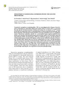
Although the amino acid sequences of the a and 8 subunits of hCG have been known for nearly a decade (15-18), correlations of structure with the function ofthis glycoprotein hormone have been inhibited by a lack of knowledge of the tertiary structure. Information concerning the relationships of immunologic determinants at the surface of hCG to hormone-receptor binding is of particular interest because it has been shown that immunization with the hCG f3 subunit can produce a state of infertility (19). As yet it is not clear that these antibody effects occur due to neutralization of the hormone by preventing binding to the hormone receptor. Knowledge of these immunologic determinants is also important for hCG detection very early in pregnancy or as a cancer marker because, at extremely low levels, it is difficult to distinguish hCG from human luteinizing hormone, a pituitary glycoprotein (20). We have begun to prepare and study a group of monoclonal antibodies to hCG to obtain information concerning the relationships of immunologic and receptor recognition determinants. Antibodies have been used previously to study the functional and structural properties of different regions of proteins (21, 22). For example, monoclonal antibodies against f3-D-galactosidase can have activating, inactivating, and heat-protecting effects (23). Sheep antibodies that bound to a specific region of hemoglobin could alter the affinity of this protein for oxygen (24). As summarized in Fig. 5, monoclonal antibodies can be pro-
B 102-
FIG. 5. Diagram of antigenic sites on hCG relative to the receptor-
binding site. B102 and B103 may occupy discrete sites, but there is no evidence that they bind to different epitopes. They are exposed to solvent afterhOGbinds to the receptor. A102 and Bl0l block hCGbinding to the receptor and are not exposed in the hormone-receptor complex. Therefore, the epitopes of these antibodies have been placed in the receptor-bindingregion of hOG. A103 cannot interfere with hOG binding to the receptor but does not appear to be exposed to the solvent after the hOG~-receptor complex is formed. Thus, we have located the binding site of A103 in the region where receptor, hormone, and solvent are near each other.
Biochemistry: Moyle et al. and B101 to inhibit hCG-receptor binding and subsequently block biological activity. The near failure of B103 to inhibit hCG action in spite of its high affinity for the hormone and the finding that 1'I-labeled B103 can bind to the hCG-receptor complex suggest that it is nonneutralizing. B103 should, therefore, prove useful in recognizing hCG at cell surfaces and help to clarify early events in hCG internalization (25, 26), which might be important in its mechanism of action. The availability of a nonneutralizing hCG antibody also provides a tool to study the mechanism of infertility produced by immunization with hCG as noted above. The observation that B103 binds to hCG-receptor complexes suggests that a portion of the (3 subunit remains exposed after the hormone-receptor complex is formed in testes homogenates. Other studies have also indicated that a portion of the (3 subunit is likely to project from the hormone-receptor complex (27). Antibodies to the COOH-terminal 30 amino acid region of the hCG (3 subunit failed to block its activity (27). Furthermore, removal of this fragment from hCG did not alter the biological activity of the hormone in vitro (1). B103 is known not to bind the COOH-terminal peptide of the ( subunit, because it binds a proteolytic digestion product of hCG that does not contain these amino acids (unpublished observations). Therefore, a part of the "core" of hCG (3 subunit must also remain exposed after binding to the receptor. Loss of observable B101 binding on formation of the hormone-receptor complex suggests that a different portion of the (3 subunit is buried in or adjacent to the interface between the hormone and the receptor or that the hormone conformation is altered when the receptor complex is formed. Although we are unable to distinguish these possibilities, we favor the first because B101 effectively blocks binding ofhCG to the receptor. The observation that an antibody specific to the a subunit blocked the biological action ofhCG suggests that antigenic sites on this subunit also lie within or adjacent to the hormone-receptor complex. We have yet to find an antibody to the a subunit similar to B103 that can recognize portions of hCG a subunit after the hormone-receptor complex is formed. In contrast to our expectations, we failed to find A103 bound to the hormone-receptor complex even though the antibody had little ability to inhibit hCG binding or steroidogenesis. Any explanation for this result must remain speculative, but the lack of binding may be associated with a change in the tertiary structure of hCG or the orientation of hCG in the receptor after the hormone has become bound to the receptor. Thus, although the structure initially formed between hCG and receptor can bind A103, any preincubation ofhCG and receptor results in a complex that cannot bind A103. Perhaps A103 will be useful in identifying changes in conformation of the hormone that occur on binding to receptor. Many more monoclonal antibodies to the a subunit need to be examined before we will be able to conclude that this subunit does not project from the receptor complex.
Proc. Natl. Acad. Sci. USA 79 (1982)
2249
These studies were supported by grants from the National Institutes of Health (CA-26636, HD-13496, and HD-15454) and a grant from the University of Medicine and Dentistry of New Jersey Foundation. 1. Moyle, W. R. (1980) in Oxford Reviews of Reproductive Biology, ed. Finn, C. A. (Clarendon, Oxford, England), Vol. 2, pp. 123-204. 2. Catt, K. J., Dufau, M. L. & Tsuruhuhara, T. (1973)J. Clin. Endocrinol Metab. 36, 73-80. 3. Canfield, R. E., Morgan, F. J., Kammerman, S., Bell, J. J. & Agosto, G. M. (1971) Recent Prog. Horm. Res. 27, 121-164. 4. Aloj, S. M., Edelhoch, H., Ingham, K. C., Morgan, F. J., Canfield, R. E. & Ross, G. T. (1973) Arch. Biochem. Biophys. 159, 497-504. 5. Ingham, K. C., Tylenda, C. & Edelhoch, H. (1976) Arch. Biochem. Biophys. 173, 680-690. 6. Carlsen, R. B. & Bahl, 0. P. (1976) Arch. Biochem. Biophys. 175, 209-220. 7. Liu, W.-K., Yang, K.-P., Burleigh, B. D. & Ward, D. N. (1974) in Hormone Binding and Target Cell Activation in the Testes, eds. Means, A. N. & Dufau, M. L. (Plenum, New York), pp. 89-108. 8. Ji, I. & Ji, T. H. (1980) Proc. Natl Acad. Sci. USA 77, 7167-7170. 9. Morgan, F. J. & Canfield, R. E. (1971) Endocrinology 88, 1045-1053. 10. Wands, J. R. & Zurawski, V. R. (1981) Gastroenterology 80, 225-232. 11. Herzenberg, L. A., Herzenberg, L. A. & Milstein, C. (1978) in Handbook of Experimental Immunology, ed. Weir, D. M. (Blackwell Scientific, Oxford, England), Vol. 2, pp. 25.1-25.7. 12. Moyle, W. R., Cosgrove, A. E. & Krieger, J. (1980) Am.J PhysioL 238, E293-E305. 13. Moyle, W. R., Bahl, 0. P. & Marz, L. (1975)J. Biol Chem. 250, 9163-9169. 14. Scatchard, G. (1949) Ann. N.Y. Acad. Sci. 51, 660-672. 15. Morgan, F. J., Birken, S. & Canfield, R. E. (1973) Mol Cell Biochem. 2, 97-99. 16. Morgan, F. J., Birken, S. & Canfield, R. E. (1975)J. Biol Chem. 250, 5247-5258. 17. Bellisario, R., Carlsen, R. B. & Bahl, 0. P. (1973)J. Biol Chem. 248, 6796-6809. 18. Carlsen, R. B., Bahl, 0. P. & Swaminathan, N. (1973) J. BioL Chem. 248, 6810-6827. 19. Stevens, V. C. (1975) in Physiological Effects of Immunity Against Reproductive Hormones, eds. Edwards, R. G. & Johnson, M. H. (Cambridge University Press, London), pp. 249-274. 20. Vaitukaitis, J. L., Braunstein, G. D. & Ross, G. T. (1972) Am. J. Obstet. Gynecol 113, 751-758. 21. Atassi, M. Z., ed. (1977) Immunochemistry of Proteins (Plenum, New York), Vols. 1 & 2. 22. Furie, B., Schechter, A. N., Sachs, D. H. & Anfinsen, C. B. (1975) J. Mol BioL 92, 497-506. 23. Frackelton, A. R. & Rotman, B. (1980) J. Biol Chem. 255, 5286-5290. 24. Dean, J. & Schechter, A. H. (1979) J. Biol. Chem. 254, 9185-9193. 25. Ascoli, M. & Puett, D. (1978) J. Biol Chem. 253, 4892-4899. 26. Conn, P. M., Conti, M., Harwood, J. P., Dufau, M. L. & Catt, K. J. (1978) Nature (London) 274, 598-600. 27. Louvet, J. P., Ross, G. T., Birken, S. & Canfield, R. E. (1974) J. Clin. Endocrinol Metab. 39, 1155-1158.