ULTRASOUND N February 2008 N Volume 16 N Number 1
Vasa Praevia; a Preventable Tragedy Elizabeth Daly-Jones1, Ann John1, Alison Leahy1, Ciara McKenna1 & Waldo Sepulveda2 1 Ultrasound Department, Queen Charlotte’s and Chelsea Hospital, London, UK, and 2Fetal Medicine Center, Clinica Las Condes, Santiago, Chile
Vasa praevia can have the catastrophic consequences of a fetal or neonatal death. There are few antenatal diagnoses whereby ultrasound can have such a direct impact on outcome and dramatically increase the chances of survival in pregnancies affected by this condition. The exclusion of vasa praevia can take under a minute and its inclusion within the routine anomaly scan should be prioritised. Until this occurs, healthy babies will continue to die due to this condition.
Published by Maney Publishing (c) British Medical Ultrasound Society
Keywords: Vasa Praevia, Pregnancy Complication, Prenatal Diagnosis, Ultrasound Technique, Ultrasound Best Practice ‘The condition of vasa praevia has robbed us of our beautiful baby boy, our first child and we will never be the same again. We just wish one of the medical staff had had the foresight to scan for this condition’ Natalie Samat, whose child Henry Cameron Samat died from un-diagnosed vasa praevia, November 2005
Introduction The aim of this paper is to persuade all those undertaking the detailed anomaly scan that excluding vasa praevia (VP) is a worthwhile endeavour and one that is easily achievable within the confines of the second-trimester anomaly scan. In this paper, we review the mechanisms leading to VP as well as the incidence, clinical implications, and risk factors associated with this condition. A brief review of the literature will help further to build a convincing case for routine ultrasound screening for this condition. Images will enable the reader to understand the ultrasound views required to exclude VP and clear guidance on the technique required to confirm the diagnosis will be provided. Best practice guidance will be given in the form of an algorithm and information regarding internet access to educational support materials will also be made available to assist the reader. It is hoped that the devastation felt by numerous parents at the preventable death of a healthy baby, as expressed so clearly by Natalie Samat in the given quotation, will then be confined to the past.
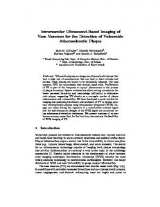
What is Vasa Praevia? Vasa praevia is a condition whereby fetal or placental vessels course over the cervix beneath the presenting part.1 These vessels run unprotected by either the Wharton’s jelly or placental tissue and are, therefore, vulnerable to laceration and compression at any stage of pregnancy, though these complications most commonly arise at the time of delivery.1–3 This condition may occur as the result of either velamentous cord insertion (VCI), when the umbilical cord inserts into the membranes away from the placental mass and the velamentous vessels cross through the lower uterine segment (VP type 1 – Fig. 1), or from fetal vessels running between lobes of a Correspondence: Elizabeth Daly-Jones, Ultrasound Department, Queen Charlotte’s and Chelsea Hospital, Du Cane Road, London W12 0HS, UK.
[email protected] Ultrasound 2008;16(1):8–14 ß British Medical Ultrasound Society 2008
8
placenta with one or more accessory lobes (VP type 2 – Fig. 2).4,5 The reason why VP has such a poor outcome is due to the potential for the exposed vulnerable fetal vessels which course close to the cervix to rupture when the membranes break, leading to fetal exsanguination.1–6 The presenting fetal part may also exert direct pressure on the unprotected vessels and fetal death can occur due to asphyxiation.7
What is the Incidence of Vasa Praevia? The reported incidence of VP is approximately 1 per 2500 deliveries.3 The latest yearly UK birth rate figures show that there were 734 000 live births8 which would account for approximately 293 babies being affected by this condition. The literature however supports the possibility that this figure is likely to be an underestimation in view of the fact that the condition of VP in asymptomatic pregnancies may be unrecognisable at the time of delivery, and therefore not reported in the literature.5 Furthermore, the incidence of VP in IVF pregnancies has recently been reported to be as high as 1 in 300 pregnancies,9,10 which has been attributed to the increased proportion of placental morphologic alterations seen in pregnancies achieved by using assisted reproductive techniques.11,12
What are the Implications of Vasa Praevia? The impact of VP on the pregnancy can be devastating with a high reported fetal and neonatal mortality rate.1–3 An alarming complication of VP is fetal bleeding which often presents unexpectedly leading to rapid exsanguination, shock and fetal death.6 In the pre-ultrasound era, the reported mortality for this complication was at least 75%.13 Traditionally, VP presents with painless vaginal bleeding at the time of spontaneous or artificial rupture of the membranes. Abnormalities of the fetal heart rate and fetal distress quickly follow, leading to an emergency caesarean section and the delivery of a severely anaemic, hypovolemic neonate, which requires rapid reposition of volume and blood transfusion as a life saving manoeuvre.1–3 Reported methods for the diagnosis in symptomatic cases include the palpation of velamentous vessels through the dilated cervix, direct visualisation of the vessels DOI: 10.1179/174313408X259508
Published by Maney Publishing (c) British Medical Ultrasound Society
Daly-Jones et al.
Vasa Praevia
Figure 1. Illustration of vasa praevia type 1.
Figure 2. Illustration of a vasa praevia type 2.
with an amnioscope or testing the origin of blood looking for fetal haemoglobin.13 Unfortunately, the emergent and often chaotic situation faced by the perinatology team often precludes the use of any of these diagnostic techniques. In the last two decades, prenatal ultrasound has played a critical role in the detection of VP well in advance of the development of clinical complications. Indeed, a recently published large multi-centre study of VP reports on the significant improvement in the perinatal outcome when an ultrasound antenatal diagnosis of VP is made and a caesarean section is appropriately scheduled.14 The study involved 155 cases of VP, of which only 61 (39%) were diagnosed prenatally, and demonstrated that in 97% of cases where a prenatal ultrasound diagnosis was available the infant survived, compared with a 44% survival rate when the condition was undiagnosed. Even when the infant survives an undiagnosed VP, this study clearly demonstrated that the post-natal course is often complicated, with over half of the infants requiring a blood transfusion and registering poor Apgar scores.14 Numerous authorities now support the view that the only effective strategy to reduce the high mortality rate associated with VP is a reliable antenatal ultrasound diagnosis and timely delivery by caesarean section.
delivery at 35 weeks of a healthy baby by Caesarean section.17 Although VP is a rare condition and 1 in every 2500 deliveries3 might not seem a very high incidence rate, it has to be remembered that these babies are at risk of death. In IVF pregnancies the chance of a VP rises considerably to a reported 1 : 300.9,10 As the risk of an isolated facial cleft is 1 : 100018 and many ultrasound departments routinely assess the fetal upper lip as part of their anomaly scan as recommended by the Royal College of Obstetricians and Gynaecologists (RCOG),9 it seems appropriate that exclusion of VP, where the outcome is possibly a fetal or neonatal death, should be prioritised by the scanning community. The chances of VP being present are minimal when the umbilical cord inserts into the placental mass or in the absence of a succenturate or bilobed placenta.5 As the assessment of the placenta is already included within departmental ultrasound protocols,19 the priority should be for the placental cord insertion to be evaluated as velamentous insertion of the cord is a pre-requisite for the most common VP, type 1. The confident identification of a normal placental cord insertion is in itself a worthwhile pursuit during the secondtrimester anomaly scan, with an incidence of VCI of 1% in the unselected population.20 There are a number of associated complications accompanying a velamentous insertion; apart from the most catastrophic VP, there is also increased risk of placental abruption, fetal growth restriction, congenital anomaly, low Apgar score, prematurity and retained placenta.20–22 Several studies have remarked that the investigation of the cord insertion when undertaken in the first or second trimester takes less than one minute and can easily be incorporated into the allotted scanning time requiring no additional skills.20,21 Accurate assessment of the placental cord insertion prior to 11 weeks is not possible due to the trophoblast normally covering a large area of the uterine cavity.23 Examination of the cord insertion if the patient presents for a booking scan or nuchal translucency thickness measurements between 11 and 14 weeks of gestation is advantageous as the umbilical cord and its insertion site into the placenta can be readily identified at this stage.23 Transvaginal (TV) examination in spontaneous multiple pregnancies and those obtained with assisted reproductive techniques should routinely be undertaken to exclude VP if a transabdominal (TA) scan using colour Doppler does not clearly demonstrate the area around the internal os. This then allows, where there is a high index of suspicion, for close surveillance of the pregnancy and
The Role of Ultrasound in Excluding Vasa Praevia It would seem appropriate to discuss the literature in light of any concerns that the ultrasound community might have about incorporating the exclusion of VP into their departmental scanning protocols. The prevalence of this condition, impact on scanning time, equipment, optimum gestational age for screening and any additional skill levels required are important points to be considered. VP has been discussed in the literature over many years with Lobstein reporting the first case of a ruptured VP back in 1801.5 In 1987, Gianopoulos et al.15 first described the diagnosis of VP using ultrasound in a woman with a history of a low-lying placenta and a succenturiate lobe. In 1990, Nelson et al.16 reported the use of colour flow Doppler to diagnose VP at 26 weeks gestation. It was only in 1996 when the first case of VP was diagnosed at the time of the routine second-trimester anomaly scan. This was identified within the confines of a 20-minute scan and resulted in the subsequent
9
Published by Maney Publishing (c) British Medical Ultrasound Society
ULTRASOUND N February 2008 N Volume 16 N Number 1
Figure 3. TA scan demonstrating a grey scale image of the placental cord insertion.
confirmation of VP if present, at the second-trimester scan. It is important to highlight that the diagnosis of velamentous insertion should be specifically looked for, if a specific search for velamentous insertion is not undertaken then the diagnosis is invariably missed.24 Consideration should also be given to the equipment necessary to exclude VP; colour flow mapping and pulsed-wave Doppler facilities considerably aid the diagnosis and numerous authors remark on the improvement in visualisation of the abnormally located vessels as a result of these facilities.1,2,5,25 Indeed some authors suggest a quick sweep over the cervix with colour flow mapping is a helpful additional measure to exclude VP during routine obstetric scans.5 However, the common policy of performing obstetric scans with an empty maternal bladder can occasionally make it difficult to visualise the lower segment. In this regard, the use of transvaginal probes for those cases at high risk of VP can be extremely helpful in establishing the definitive diagnosis by enhancing the visualisation of the entire lower uterine segment. This can also be easily incorporated in those departments who routinely assess the uterine cervix to screen for short cervix and risk of preterm delivery. With guidance from the RCOG19 suggesting all ultrasound equipment should be less than five years old, it is likely that many units undertaking detailed anomaly scans will have these facilities in place. Other technologies such as colour Doppler threedimensional ultrasound and magnetic resonance imaging (MRI) have been reported in the prenatal diagnosis of VP, but these methods are cumbersome and have limited practical utility, so will not be discussed in this review (Editor’s note: see accompanying case study in this issue). When evaluating the literature regarding VP it is clear that there are few large studies due to the low overall prevalence of this potentially devastating condition. However, all of the case studies reported state how important an antenatal diagnosis is to improving the outcome of this condition5 and that it should be included in the routine ultrasound evaluation of the pregnancy. Oyelese and Smulian succinctly remarked in 2006: ‘We can think of no other condition in which prenatal diagnosis… makes such a dramatic impact on the difference between survival and death for an otherwise healthy infant’.25
Ultrasound Technique for Excluding Vasa Praevia The ultrasound technique for ruling out VP is based on:
N 10
assessment of placental cord insertion site;
Figure 4. TA scan using colour Doppler ultrasound demonstrating the branching vessels into the placenta.
N N
assessment of placental appearance and location; consideration of other risk factors.
Assessment of Placental Cord Insertion Site The umbilical cord inserts in the placental mass, either in the centre or paracentrally, in about 90% of pregnancies. In about 10% the insertion occurs at the margins of the placenta (marginal insertion), and in about 1% of pregnancies it inserts in the membranes away from the placental edge (velamentous cord insertion). The placental cord insertion site can be readily determined by ultrasound as part of the standard 20-minute second-trimester anomaly scan. Once the placental site has been determined attempts should be made to image the placental cord insertion site. This is done by searching the fetal surface of the placenta with high resolution grey-scale ultrasound at the appropriate magnification until this is identified (Fig. 3). Care must be taken not to mistake the true insertion site with loops of umbilical cord overlying the placental surface by demonstrating the entry of the main branches of the umbilical vessels into the chorionic plate with colour flow imaging confirming the diagnosis (Fig. 4). Also note that when the site of cord insertion into the placental mass is clearly identified, the possibility of VP type 1 is extremely low and no further action is necessary.5 In contrast, when the insertion site cannot be identified, efforts must be made to exclude VCI as this group of patients have a higher risk of having VP (6%).26 Where a VCI cannot clearly be demonstrated in the upper segment of the uterus with a transabdominal scan, it is imperative that the area above the internal os is evaluated for velamentous insertion. This should be undertaken using a transvaginal approach and incorporating colour Doppler for a reliable diagnosis of VP (Figs. 5 and 6). Some authors have proposed a transperineal approach, but the use of colour Doppler ultrasound is critical.27 Assessment of Placental Appearance and Location Consideration should also be given to the overall appearance and location of the placenta, as fetal vessels running across the cervix between lobes of placenta with one or more accessory (succenturate) lobes is a pre-requisite for VP type 2 (Fig. 7). It should also be noted that some pregnancies complicated by VP may have a combination of type 1 and 2 (Fig. 8).5 Placenta praevia (where the placenta partially or totally covers the internal os) and marginal placenta (where the margin of the placenta reaches the internal os) are considered
Daly-Jones et al.
Vasa Praevia
Published by Maney Publishing (c) British Medical Ultrasound Society
Figure 5. Colour Doppler demonstrates a VP type 1. Figure 8. Pathological specimen shows the fetal side of a bilobed placenta with a velamentous cord insertion between the lobes (ß Francois Manson and www.TheFetus.net; reproduced with permission).
suspicion should be maintained, observing the placental cord insertion and excluding placental variants. If normal appearances are demonstrated, no further action is needed. If however appearances are suspicious or overtly abnormal further assessment should occur at the routine anomaly scan. Patients presenting with vaginal bleeding should also be treated with caution and a higher index of suspicion for VP maintained.
Figure 6. Pathological specimen of a velamentous cord insertion showing the umbilical cord inserting into the placental membranes and the unprotected fetal vessels (Courtesy of Edward C. Klatt MD).
Difficulties in Diagnosing Vasa Praevia Obese patients, abdominal scarring and awkward fetal position can make diagnosis of VP difficult. In cases where the cord is seen close to the cervix, shifting the cord by moving the patient, or waiting and reassessing a few minutes later, can be helpful. Colour flow mapping should enable a cord insertion remote from the cervix to be demonstrated thus excluding VP (Fig. 10).
Figure 7. Vasa praevia type 2 demonstrates fetal vessels crossing the cervix from the main anterior placenta mass to a posterior succenturate lobe.
to be risk factors for VP.5 Where present it is important to check for vessels near the cervix and if clearly normal then no further action is necessary (Fig. 9). As assessment of the placental site and its relationship to the internal os is already undertaken within most ultrasound departments at the time of the anomaly scan, the addition of colour Doppler over the area around the cervix is easily achievable. If optimum visualisation of the cervix cannot be achieved with a transabdominal scan, then a transvaginal approach should be undertaken. Consideration of Other Risk Factors Other risk factors for VP include pregnancies achieved by in vitro fertilisation (incidence of VP 1 : 300)9 and multi-fetal gestations (10% of VP occurs in twins).5 As these pregnancies are often referred for an 11–14 week scan, a high index of
Figure 9. Transabdominal view of the internal os using colour Doppler demonstrating the absence of VP.
11
Published by Maney Publishing (c) British Medical Ultrasound Society
ULTRASOUND N February 2008 N Volume 16 N Number 1
Figure 10. Transabdominal scan showing umbilical cord close to the internal os. A few minutes later after movement of the patient, the umbilical cord moved away from the cervix.
In the second trimester visualisation of the placental cord insertion site is easier to achieve than later on in pregnancy as the amount of amniotic fluid allows for a proper visualisation of the placental surface in almost all cases. In the third trimester, however, the relative decrease in amniotic fluid volume can make visualisation quite difficult. In pregnancies with a posterior placenta, gentle manipulation of the fetus through the maternal abdomen can allow proper placental surface visualisation which may be assisted by placing the mother in the lateral position. It is important where visualisation is poor and the cord insertion has not been identified previously, that a transvaginal examination of the lower segment is undertaken. Flash artefact, which can occur as a result of amniotic fluid movement due to fetal activity, can also be a problem as this can mimic a vessel near the internal os. Repeating the evaluation once movement has subsided will usually eliminate this artefact (Figs. 11 and 12). Any persistent structures near the cervix should always raise the suspicion of VP (Figs. 13 and 14). The use of colour Doppler will enable the operator to determine if these are vascular in nature. Alternatively a spectral trace obtained from
Figure 11. A transabdominal scan raising the possibility of suspicious vessels near the cervix.
12
Figure 12. Further evaluation and a repeat image confirmed this to be flash artefact.
Figure 13. A second-trimester vaginal ultrasound showing a sagittal section through the cervix with suspicious soft tissue (arrow) abutting the internal os.
Figure 14. The use of colour Doppler shows the suspicious structure is without Doppler signal and thus is not a vessel (ß Philippe Jeanty and www.TheFetus.net; reproduced with permission).
Daly-Jones et al.
Vasa Praevia
anomaly ultrasound scan and believe the guidance can also be followed in the late first trimester and for those pregnancies presenting in the third trimester.
Published by Maney Publishing (c) British Medical Ultrasound Society
Departmental Software Support for Vasa Praevia
Figure 15. Second-trimester vaginal Doppler image shows a high frequency heart rate at the level of the VP. This helps to distinguish VP from maternal cervical vessels (ß Philippe Jeanty and www.TheFetus.net; reproduced with permission).
To ensure assessment of the placental cord insertion site is incorporated into all routine scans it is prudent to include this in departmental guidelines and protocols. For those departments currently using the Astraia fetal database (Astraia software GmbH, Mu¨nchen, Germany), it is possible to have a list of cord anomalies and variants in the ‘placenta box’ of the ‘drop down’ menu. This could include for example, ‘three vessels with a normal cord insertion’ or ‘velamentous cord insertion’. The manufacturers of the Astraia database have been in discussion with the UK Vasa Praevia Awareness Group and agreed that an additional field box ‘cord insertion’ will be integrated in the new version of Astraia (due for release Autumn 2007). This will only refer to the placental cord insertion and can be changed to a mandatory field by the department administrator ensuring staff will not be able to bypass this field. Online Educational Support
the suspicious region will show pulsations at the fetal heart rate where the appearances are due to VP (Fig. 15).
The UK Vasa Praevia Awareness group (www.vasapraevia. co.uk) provide excellent comprehensive training materials on their website. These can be downloaded free of charge.
Best Practice Guidelines
Conclusions
We recommend the algorithm proposed by Philippe Jeanty’s team5 (Fig. 16) as a guide during the routine second-trimester
Routine scanning of obstetric patients has changed dramatically over the last few decades. Advances in scanning
Figure 16. Proposed diagnostic algorithm for the second-trimester detection of VP (Reproduced from Derbala Y, Grochal F and Jeanty P (2007) with permission).5
13
Published by Maney Publishing (c) British Medical Ultrasound Society
ULTRASOUND N February 2008 N Volume 16 N Number 1
equipment have meant that more and more subtle fetal anomalies and pregnancy complications can now be identified with confidence. It is the duty of ultrasound professionals to keep pace with these advances, to incorporate them into a standard routine scan and, on identifying any anomaly, to refer patients for appropriate counselling and/or interventional procedures. Unfortunately, there is often a time lag between innovation and its incorporation into standard practice. New methodology has to be tried and tested, new skills have to be acquired by practitioners, and additional scanning time may have to be incorporated into an already time-pressured environment. In relation to identifying VP on a routine scan there is little excuse. The method for excluding VP is a simple colour Doppler technique, uses skills that trained practitioners already possess, and takes approximately a minute of extra examination time. Vasa praevia is life threatening to a healthy baby; a proper diagnosis and an elective Caesarean section easily prevent death. Mothers should have confidence in the ultrasound practitioner’s ability to assess not only fetal wellbeing, but also any other pregnancy-related anomalies. Routine evaluation to exclude VP should be adopted in all routine obstetric scans as a matter of urgency. Until this occurs, babies will die unnecessarily and families will continue to suffer the avoidable loss of a healthy baby.
Acknowledgements The authors would like thank Natalie and Daren Samat, whose unceasing efforts in memory of Henry, to ensure that Vasa Praevia is incorporated into the routine anomaly scan, are truly inspirational. Finally, the authors express their heartfelt thanks to Krysia Carr for her beautiful illustrations (Figs. 1 and 2), and extend their appreciation to the original owners of all images used in this paper.
References
7.
Cordero DR, Helfgott AW, Landy HJ, et al. A non-hemorrhagic manifestation of vasa previa: a clinicopathologic case report. Obstet Gynecol 1993;82:698–700.
8.
National Statistics Online. Fertility rate is highest for 26 years, available at: www.statistics.gov.uk/pdfdir/frc0607.pdf (accessed 9th June 2007).
9.
Schachter M, Tovbin Y, Arieli S, et al. In vitro fertilization as a risk factor for vasa previa. Fertl Steril 2002;78:642–643.
10.
Baulies S, Maiz N, Mun˜oz A, et al. Prenatal ultrasound diagnosis of vasa praevia and analysis of risk factors. Prenat Diagn 2007;27:595–599.
11.
Englert Y, Imbert MC, Van Rosandael E, et al. Morphological anomalies in the placentae of IVF pregnancies: preliminary report of a multicentric study. Hum Reprod 1987;2:155–157.
12.
Jauniaux E, Englert Y, Vanesse M, et al. Pathologic features of placentas from singleton pregnancies obtained by in vitro fertilization and embryo transfer. Obstet Gynecol 1990;76:61–64.
13.
Dougall A, Baird CH. Vasa praevia – report of three cases and review of literature. Brit J Obstet Gynaecol 1987;94:712–715.
14.
Oyelese Y, Catanzarite V, Prefumo, F et al. Vasa previa: the impact of prenatal diagnosis on outcomes. Obstet Gynecol 2004;103:937–942.
15.
Gianopoulos J, Carver T, Tomich PG, et al. Diagnosis of vasa previa with ultrasonography. Obstet Gynecol 1987;69:488– 491.
16.
Nelson LH, Melone PJ, King M. Diagnosis of vasa previa with transvaginal and color flow Doppler ultrasound. Obstet Gynecol 1990;76:506–509.
17.
Daly-Jones E, Hollingsworth J, Sepulveda W. Vasa praevia: second trimester diagnosis using colour flow imaging. Brit J Obstet Gynaecol 1996;103:284–286.
18.
Wayne C, Cook K, Sairam S, et al. Sensitivity and Accuracy of routine antenatal ultrasound screening for isolated facial clefts. Brit J Radiology 2002;75:584–589.
19.
Royal College of Obstetricians and Gynaecologists. Routine ultrasound screening in pregnancy: supplement to Ultrasound Screening for Fetal Abnormalities. London: RCOG Press, 2000.
20.
Sepulveda W, Rojas I, Robert JA, et al. Prenatal detection of velamentous insertion of the umbilical cord: a prospective color Doppler study. Ultrasound Ostet Gynecol 2003;21:564–569.
21.
Nomiyama M, Toyota Y, Kawano H. Antenatal diagnosis of velamentous umbilical cord insertion and vasa previa with color Doppler imaging. Ultrasound Obstet Gynecol 1998;12:426–429.
22.
Heinonen S, Ryynanen M, Kirkinen P, Saarikoski S. Perinatal diagnostic evaluation of velamentous umbilical cord insertion: clinical, Doppler, and ultrasonic findings. Obstet Gynecol 1996;87:112–117.
1.
Sepulveda W, Sebire NJ, Harris R, Nyberg DA. The placenta, umbilical cord, and membranes. In Diagnostic imaging of fetal anomalies, Nyberg DA, McGahan JP, Pretorius DH, Pilu G (eds). Philadelphia, PA: Lippincott Williams & Wilkins, 2003, 85–132.
2.
Fung TY, Lau TK. Poor perinatal outcome associated with vasa previa: is it preventable? A report of three cases and review of the literature. Ultrasound Obstet Gynecol 1998;12:430–433.
23.
3.
Oyelese KO, Turner M, Lees C, Campbell S. Vasa previa: an avoidable obstetric tragedy. Obstet Gynecol Surv 1999;54:138– 145.
Sepulveda W. Velamentous insertion of the umbilical cord: a first- trimester sonographic screening study. J Ultrasound Med 2006;25:963–968.
24.
4.
Cantanzarite V, Maida C, Thomas W, et al. Prenatal sonographic diagnosis of vasa previa: ultrasound findings and obstetric outcome in ten cases. Ultrasound Obstet Gynecol 2000;18:109– 115.
Eddleman KA, Lockwood CJ, Berkowitz GS, et al. Clinical significance and sonographic diagnosis of velamentous umbilical cord insertion. Am J Perinatol 1992;9:123–126.
25.
Oyelese Y, Smulian JC. Placenta previa, placenta accreta, and vasa previa. Obstet Gynecol 2006;107:927–941.
26.
Paavonen J, Jouttunpa¨a¨ K, Kangasluoma P, et al. Velamentous insertion of the umbilical cord and vasa previa. Int J Gynaecol Obstet 1984;22:207–211.
27.
Hertzberg BS, Kliewer MA. Vasa previa: prenatal diagnosis by transperineal sonography with Doppler evaluation. J Clin Ultrasound 1998;26:405–408.
5.
Derbala Y, Grochal F, Jeanty P. Vasa Previa. J Prenat Med 2007;1:2–13.
6.
Robert JA, Sepulveda W. Fetal exsanguination from ruptured vasa previa: still a catastrophic event in modern obstetrics. J Obstet Gynaecol 2003;23:574.
14