976
THE JOURNAL OF PARASITOLOGY, VOL. 91, NO. 4, AUGUST 2005
J. Parasitol., 91(4), 2005, pp. 976–978 q American Society of Parasitologists 2005
Responses of Leishmania (Viannia) braziliensis Cutaneous Infection to N-Methylglucamine Antimoniate in the Rhesus Monkey (Macaca mulatta) Model A. Teva, R. Porrozzi, M. P. Oliveira-Neto*, and G. Grimaldi Jr., Department of Immunology, Institute Oswaldo Cruz, FIOCRUZ, Av. Brasil 4365, Rio de Janeiro RJ, CEP 21045-900, Brazil; *Evandro Chagas Clinical Research Institute, FIOCRUZ, Av. Brasil 4365, Rio de Janeiro RJ, CEP 21045-900, Brazil. e-mail:
[email protected] ABSTRACT: The antileishmanial efficacy of the reference drug N-methylglucamine antimoniate (Glucantimet) was evaluated in groups of rhesus monkeys with acute and chronic Leishmania (Viannia) braziliensis cutaneous infection. The therapeutic responses in experimental animals to either a low dose (5 mg/kg body wt/day for 28 days) or a routine dose (20 mg/kg/day for 28 days) of pentavalent antimony were similar to those reported in the human disease. Primates were cured of their lesions after treatment, but with cryptic parasitism and/or relapse. The rhesus model of L. (V.) braziliensis cutaneous leishmaniasis therefore provides an additional resource for preclinical trials with newer drugs.
Self-healing cutaneous leishmaniasis (CL) has been associated with all New World Leishmania species, but a proportion of nonhealing cases of cutaneous and mucocutaneous disease can be very difficult to cure (Grimaldi and Tesh, 1993). Despite their toxic properties, pentavalent antimony (Sbv) compounds (meglumine antimoniate and sodium stibogluconate), which must be administered parentally, remain the primary therapy for human leishmaniasis. Should this treatment fail, a number of other drugs and immune modulators may be employed, but toxic properties remain the major obstacle for the most potent antileishmanial agents (Croft and Yardley, 2002). Cutaneous leishmaniasis resulting from infection with Leishmania (V.) braziliensis, which can cause considerably morbidity and that may result in severe disfigurement (Grimaldi and Tesh, 1993), continues to present serious therapeutic problems (Romero et al., 2001). Current regimes use Sbv at a dosage of 20 mg/kg body weight/day for 20 days (for cutaneous lesions) or 28 days (for mucosal lesions). Some studies conducted in locales where this pathogen is endemic have suggested that shorter courses of treatment (5 mg/kg/day) may be as efficacious (Oliveira-Neto et al., 1997, 2000). Moderate resistance to Sbv exists among Leishmania strains in nature, and some isolates are innately less or more susceptible to this drug than other strains (Grogl et al., 1992). Whether specific genotypes of this pathogen (Cupolillo et al., 2003) are
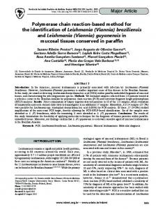
FIGURE 1. Responses of Leishmania (Viannia) braziliensis acute cutaneous infection in rhesus macaques to a low dose of N-methylglucamine antimoniate (5 mg/kg body wt/day given intramuscularly for 28 days). Treatment started at week 12 after inoculation with 1 3 107 promastigotes into the skin of either the forearm (monkeys A1 to A3) or eyebrow (monkeys B1 to B4). As an assessment of therapy, the diameter of the lesions was scored weekly following infection and treatment. Primary lesions were measured with a vernier caliper, and lesion size was estimated using the following formula: area (mm2) 5 p 3 greatest radius 3 least radius. Data in the untreated group are the mean 6 SD of four infected monkeys.
responsible for antimony resistance remains to be determined. The development of antimony-resistant clones in vitro by discontinuous drug exposure (Grogl et al., 1989) indicates the necessity of finding new therapeutic agents for the treatment of leishmaniasis. Nonhuman primates appear to have significant advantages over conventional laboratory animals in terms of modeling CL for purposes of drug evaluation (Kennedy et al., 1997). Rodent models may not be good predictors of human responses, because their metabolism often is different from that of humans for some drug candidates. In contrast, the ability to do efficacy-PK studies in simians is a huge advantage in this regard. In our previous studies, we have shown that Macaca mulatta models the clinical presentations of L. (V.) braziliensis infection in humans (Teva et al., 2003). The severity of the resulting lesions induced with different strains of this pathogen was greater compared to those induced with either L. (L.) major or L. (L.) amazonensis (Amaral et al., 1996, 2001). In the present study, we compare the efficacy of a low dose with that of a standard dose of Sbv in L. (V.) braziliensis–infected rhesus macaques. Fourteen laboratory-bred and -reared, young adult M. mulatta monkeys of mixed sexes were used in the present study. Three of the animals had been previously infected with L. (V.) braziliensis in the forehead above the left eyelid but had not recovered from clinical disease (Teva et al., 2003). The other 11 monkeys were naı¨ve animals that had never been exposed to leishmanial parasites. Their care and maintenance have been described previously (Amaral et al., 1996). The first experiment (experiment 1) was designed to provide information concerning the effect of a low dose of N-methylglucamine antimoniate (Glucantimet [Rhodia Farma, S. Paulo, Brazil] 5 mg/kg/day of Sbv given intramuscularly for 28 days) on development of a local dermal lesion caused by L. (V.) braziliensis (strain MHOM/BR/1997/ SIS) in naı¨ve monkeys inoculated with 1 3 107 infective promastigotes. Treatment employing this dose rapidly reduced the lesion sizes (Figs. 1, 2) in experimental animals (compared with untreated animals). Complete healing was achieved in 5 of the 7 (71.9%) monkeys with acute cutaneous infection, and in the remaining 2, reactivation of the scar lesions was observed by week 5 and week 8, respectively, after the completion of treatment. The satisfactory result observed in our monkeys treated with a low-dose regimen might be related to a nonrefractoriness of the parasite strain causing the disease and/or a distinct host response to this particular parasite. This seems to be the case of L. (V.) braziliensis parasites in Rio de Janeiro (Brazil), where high cure rates for cutaneous and mucosal disease were obtained with low Sbv doses (Oliveira-Neto et al., 1997, 2000). In an additional experiment (experiment 2), 3 monkeys with longlived L. (V.) braziliensis skin lesions (36 mo postinfection) were treated with a routine Sbv dose (20 mg/kg/day for 28 days). Primates were cured of their lesions, but 1 animal also relapsed 2 mo after the cessation of therapy (data not shown). No antimonial-related side effects in treated animals were apparent during the trials. Response to treatment with Sbv varies considerably depending on the parasite as well as host factors (Grogl et al., 1992; Romero et al., 2001). In human leishmaniasis, spontaneous or drug-induced acquired resistance to intracellular Leishmania spp. is T-helper 1 (Th1) cell cytokine dependent and largely mediated by interferon (IFN)-g (Murray, 2000). Likewise, monkeys controlling L. (V.) braziliensis infection developed parasite-specific Th1 immune responses (Teva et al., 2003). In the present study, the DTH reaction to leishmanin skin test (LST) was used as an in vivo correlate of cellular immunity. The LST induration size values (range, 5–13 mm) were larger during active infection (mean 6 SD, 8.7 6 3.6 mm) compared with values after drug-induced cure of the
RESEARCH NOTES
977
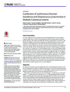
FIGURE 2. Effect of one 28-day course of pentavalent antimony (5 mg/kg body wt/day administered intramuscularly) on Leishmania (Viannia) braziliensis skin lesion development in a rhesus monkey (A2). The numbers are days after infection, whereas the numbers within parentheses are weeks after treatment was initiated. disease (3.7 6 2.7 mm). As illustrated in Figure 3, proliferative responses in vitro to soluble leishmanial antigens of peripheral blood leukocytes from infected animals were comparable before and after chemotherapy (mean 6 SD of the stimulation index values, 57 6 38 and 50 6 37, respectively), but treated monkeys had significantly (P . 0.005) lower levels of parasite-specific IFN-g secreted in culture (mean 6 SD, 60 6 50 pg/ml) compared with levels in untreated monkeys (223 6 190 pg/ml). The lack of increase of specific T-cell responses obtained in drug-cured monkeys probably reflects the elimination of parasites at the site of infection. Despite clinical healing, we were able to isolate L. (V.) braziliensis by the culture of biopsy specimens from skin scars of recovered monkeys. Likewise, L. (V.) braziliensis CL scars represented a site of parasite persistence after antimonial therapy and clinical cure (Schubach et al., 1998). The possibility of parasite persistence after clinical cure suggests that the immune response can control, but not fully eliminate, the infection. In experimental murine CL, increased polarization towards a Th2 cytokine profile before the onset of drug therapy leads to
FIGURE 3. Parasite-specific lymphoproliferative and interferon (IFN)-g responses in rhesus monkeys following primary infection with Leishmania (Viannia) braziliensis and after Glucantimet treatment (experiment 1). Purified peripheral blood leukocytes were adjusted to 2 3 106 cells/ml in complete RPMI medium and stimulated with soluble leishmanial antigens (10 mg/well) for 96 h at 37 C in 5% CO2. Then, the cells were pulsed for the last 18 h of incubation with 0.5 mCi of [3H]thymidine. Cell proliferation was assessed by measuring [3H]thymidine incorporation; results are expressed as the stimulation index (SI; mean cpm of stimulated cultures/mean cpm of unstimulated cultures). Culture supernatants were collected from duplicate wells after 72 h of stimulation, and the concentration of IFN-g in the supernatant was determined by ELISA as described previously (Teva et al., 2003). Data are the mean 6 SD of seven experimental monkeys tested. *Significant difference (P , 0.005) in the specific IFN-g response of drugcured monkeys compared with that of untreated animals.
an increased frequency of relapse after treatment (Nabors and Farrel, 1996). Although IFN-g is essential for activating macrophages to kill the pathogen, its action may be blocked or down-regulated by another mediator, the interleukin-10 produced by CD41 CD251 T cells, which is required for parasite persistence in the skin after healing and for maintenance of immunity to reinfection (Belkaid et al., 2002). In conclusion, the data strongly support the utility of this primate model of L. (V.) braziliensis CL for drug development against the human disease. Therapeutic strategies and new antileishmanial compounds can be tested in monkeys under more controlled conditions than are possible in clinical studies. This work was supported in part by the Fiocruz and CNPq/Pronex 3 (Brazil). We thank the staff of the Fiocruz Animal Care Facility for their assistance with daily care on the macaques. LITERATURE CITED AMARAL, V. F., V. A. O. RANSATTO, F. CONCEIC¸A˜O-SILVA, E. MOLINARO, V. FERREIRA, S. G. COUTINHO, D. MCMAHON-PRATT, AND G. GRIMALDI, JR. 1996. Leishmania amazonensis: The Asian rhesus macaques (Macaca mulatta) as an experimental model for study of cutaneous leishmaniasis. Experimental Parasitology 82: 34–44. ———, A. TEVA, R. PORROZZI, A. J. SILVA, M. S. PEREIRA, AND G. GRIMALDI, JR. 2001. Leishmania (Leishmania) major–infected rhesus macaques (Macaca mulatta) develop varying levels of resistance against homologous reinfections. Memo´rias do Instituto Oswaldo Cruz 96: 795–804. BELKAID, Y., C. A. PICCIRILLO, S. MENDEZ, E. M. SHEVACH, AND D. L. SACKS. 2002. CD41 CD251 regulatory T cells control Leishmania major persistence and immunity. Nature 420: 502–507. CROFT, S. L., AND V. YARDLEY. 2002. Chemotherapy of leishmaniasis. Current Pharmaceutical Design 8: 319–342. CUPOLILLO, E., L. R. BRAHIM, C. B. TOALDO, M. P. OLIVEIRA-NETO, M. E. F. DE BRITO, A. FALQUETO, M. F. NAIFF, AND G. GRIMALDI, JR. 2003. Genetic polymorfism and molecular epidemiology of Leishmania (Viannia) braziliensis from different host and geographic areas in Brazil. Journal of Clinical Microbiology 41: 3126–3132. GRIMALDI, JR., G., AND R. B. TESH. 1993. Leishmaniasis of the New World: Current concepts and implications for future research. Clinical Microbiology Reviews 6: 230–250. GROGL, M., A. M. J. ODULA, L. D. C. CORDERO, AND D. E. KYLE. 1989. Leishmania spp.: Development of pentostam-resistant clones in vitro by discontinuous drug exposure. Experimental Parasitology 69: 78–90. ———, T. N. THOMASON, AND E. D. FRANKE. 1992. Drug resistance in leishmaniasis: Implication in systemic chemotherapy of cutaneous and mucocutaneous disease. American Journal of Tropical Medicine and Hygiene 47: 117–126. KENNEDY, R. C., M. H. SHEARER, W. H. HILDEBRAND, AND R. S. SIMMONDS. 1997. Nonhuman primates and their use in immunologically based investigation. The Immunologist 5/5: 150–156. MURRAY, H. W. 2000. Mononuclear cell recruitment, granuloma, assem-
978
THE JOURNAL OF PARASITOLOGY, VOL. 91, NO. 4, AUGUST 2005
bly, and response to treatment in experimental visceral leishmaniasis: Intracellular adhesion molecule 1–dependent and –independent regulation. Infection and Immunity 11: 6294–6299. NABORS, G. S., AND J. P. FARREL. 1996. Successful chemotherapy in experimental leishmaniasis is influenced by the polarity of the Tcell response before treatment. Journal of Infectious Diseases 173: 976–986. OLIVEIRA-NETO, M. P., M. MATTOS, C. PIRMEZ, O. FERNANDES, S. C. GONC¸ALVES-COSTA, C. F. SILVA DE SOUZA, AND G. GRIMALDI, JR. 2000. Mucosal leishmaniasis (‘‘e¨spundia’’) responsive to low dose of N-methylglucamine (Glucantimet) in Rio de Janeiro, Brazil. Revista do Instituto de Medicina Tropical de Sa˜o Paulo 42: 323–327. ———, A. SCHUBACH, M. MATTOS, S. C. GONC¸ALVES-COSTA, AND C. PIRMEZ. 1997. A low-dose antimony treatment in 159 patients with American cutaneous leishmaniasis: Extensive follow-up studies (up
to 10 years). American Journal of Tropical Medicine and Hygiene 57: 651–655. ROMERO, G. A., M. V. GUERRA, M. G. PAES, AND V. O. MACEDO. 2001. Comparison of cutaneous leishmaniasis due to Leishmania (Viannia) braziliensis and L. (V.) guyanensis in Brazil: Therapeutic response to meglumine antimoniate. American Journal of Tropical Medicine and Hygiene 65: 456–465. SCHUBACH A., F. HADDAD, M. P. OLIVEIRA-NETO, W. DEGRAVE, C. PIRMEZ, G. GRIMALDI, JR., AND O. FERNANDES. 1998. Detection of Leishmania DNA by polymerase chain reaction in scars of treated human patients. Journal of Infectious Diseases 178: 911–914. TEVA, A., R. PORROZZI, E. CUPOLILLO, C. PIRMEZ, M. P. OLIVEIRA-NETO, AND G. GRIMALDI, JR. 2003. Leishmania (Viannia) braziliensis–induced chronic granulomatous cutaneous lesions affecting the nasal mucosa in the rhesus monkey (Macaca mulatta) model. Parasitology 127: 437–447.
J. Parasitol., 91(4), 2005, pp. 978–980 q American Society of Parasitologists 2005
Kinetics of Antibodies and Antigens in Serum of Mice Experimentally Infected with Echinostoma caproni (Trematoda: Echinostomatidae) R. Toledo, A. Espert, C. Mun˜oz-Antoli, A. Marcilla, B. Fried*, and J. G. Esteban, Departamento de Parasitologı´a, Facultad de Farmacia, Universidad de Valencia, Av. Vicente Andre´s Estelle´s s/n, 46100 Burjassot, Valencia, Spain; *Department of Biology, Lafayette College, Easton, Pennsylvania 18042. e-mail:
[email protected] ABSTRACT: The present study reports on the kinetics of antibodies and antigens in serum of mice experimentally infected with 75 metacercariae of Echinostoma caproni during the first 12 wk postinfection (wpi). Antibody titers in the serum of mice were determined by an indirect enzyme-linked immunosorbent assay (ELISA) using excretory/secretory (ES) antigens of E. caproni. The early detection of antibodies against ES antigens of E. caproni is feasible using indirect ELISA. Mice developed significant antibody responses at 2 wpi, and the values progressively increased until the end of the experiment. This may be related to the intestinal absorption of adult worm antigens that induces humoral responses. The presence of E. caproni circulating antigens was determined by a capture ELISA based on polyclonal rabbit antibodies against ES antigens of E. caproni. High levels of seroantigens in mice were detected by 1–2 wpi, probably because of the local inflammatory responses in mice induced by the adult worms. A drop in circulating antigen levels was observed at 9 wpi, which could reflect changes in the intestinal tissues over the course of the infection.
Echinostoma caproni (Trematoda: Echinostomatidae) is an intestinal trematode that does not undergo tissue migration in its definitive host. This parasite has a wide range of experimental definitive hosts, but its compatibility differs considerably between rodent species. In hosts with high compatibility, such as hamsters, E. caproni induces chronic infections, whereas in hosts with low compatibility, such as rats, the worms are rapidly expelled (Toledo, Espert, Carpena et al., 2004). Because of such characteristics, the E. caproni/rodent systems are highly suitable models for elucidating aspects of the host-specific components that determine the course of infection with intestinal helminths. The E. caproni/mouse model has often been used as an experimental system to study the immune response to intestinal trematode infections. Most of these studies have been limited to an analysis the host humoral response. The antibody response detected in the serum and affected tissues, such as small intestine, includes IgG, IgM, and IgA (Agger et al., 1993; Graczyk and Fried, 1995). Moreover, intestinal pathology induced by the parasite in the first several weeks postinfection (wpi) has been investigated (Weinstein and Fried, 1991; Fujino and Fried, 1993; Fujino et al., 1996). However, the facts that determine the development of chronic E. caproni infections in mice remain unknown. The recent development of capture enzyme-linked immunosorbent assay (ELISA) techniques to detect E. caproni antigens in feces and serum of infected rodents has allowed us to gain further insight regarding the immune
response of this trematode infection (Toledo, Espert, Mun˜oz-Antoli et al., 2003, 2004). Toledo, Espert, Mun˜oz-Antoli et al. (2004) showed that the kinetics of E. caproni seroantigens differs markedly between rats and hamsters, and this could be related to the course of the infection in each host species. In the present study, we examined the kinetics of antibodies and antigens in serum of mice experimentally infected with E. caproni. The strain of E. caproni used in the present study has been described previously by Fujino and Fried (1993). Encysted metacercariae of E. caproni were removed from the kidneys and pericardial cavities of experimentally infected Biomphalaria glabrata snails and used to infect ICR mice. Each of 10 male mice (weight, 32–40 g) was infected by stomach tube with 75 metacercariae of E. caproni. Moreover, 5 mice were left uninfected and used as controls. All the infected animals were maintained under conventional conditions with food and water ad libitum. The parasite egg release was investigated weekly in each of the infected animals as described by Toledo, Espert, Carpena et al. (2003). To obtain excretory/secretory (ES) antigens of E. caproni, we followed the methodology described by Toledo, Espert, Mun˜oz-Antoli et al. (2003). Blood was collected weekly from each infected and control animal by cardiac puncture under anesthesia. After clotting of the blood overnight at 4 C, serum was separated from the clot by centrifugation at 10,000 rpm for 10 min at room temperature. The antigens and serum samples were stored at 220 C until use. To detect specific antibodies against E. caproni ES products, an indirect ELISA was carried out as described by Toledo, Espert, Mun˜ozAntoli et al. (2003). The dilutions of serum samples and horse radish peroxidase-conjugated rabbit anti-mouse IgG (Sigma, St. Louis, Missouri) used were 1:200 and 1:4,000, respectively. The presence of E. caproni ES products in serum from mice was evaluated by a polyclonal antibody-based capture ELISA as described by Toledo, Espert, Mun˜ozAntoli et al. (2004). The dilutions of serum samples, biotinylated antiE. caproni ES antibody, and labeled streptavidine-peroxidase (Sigma) were 1:20, 1:1,000, and 1:4,000, respectively. Before capture ELISA, the serum samples were heated to dissociate immune complexes (Toledo, Espert, Mun˜oz-Antoli et al., 2004). Each ELISA assay was performed in triplicate, and the absorbance readings from wells with the same sample were expressed as the mean 6 SD. The cutoff for each ELISA was defined as the mean of the samples from the control mice 1 3SD. The difference between the optical density (OD) values for the infection and control specimens at
RESEARCH NOTES
FIGURE 1. Mean values of egg per gram of feces in mice experimentally infected with Echinostoma caproni in each week during the experiment. Vertical bars represent the standard deviation. each point in time was calculated and tested by the use of Student’s ttest. All the mice experimentally exposed to 75 metacercariae of E. caproni became infected. The duration of the prepatent period was uniform. Egg release began 9–12 (10.0 6 0.4) days postinfection. The kinetics of egg release was monitored during the first 12 wpi (Fig. 1). Egg output progressively increased, then became relatively stable from 7 wpi, which allowed for a maximum egg release at 11 wpi. The maximum egg release was observed at 11 wpi. All the mice remained positive at the end of the experiment (12 wpi). The number of adult worms recovered per mouse ranged from 27–45 (36.3 6 8.0). The results showed that mice develop a significant antibody response against E. caproni ES antigens (Fig. 2). The cutoff point differentiating negative from positive sera was an OD value of 0.190. Antibodies to E. caproni ES antigens were detected in all infected mice at 2 wpi. Thereafter, antibody levels progressively increased, then were stable from 8 to 12 wpi. The mean OD value during the complete course of the experiment in all the infected mice was 0.408 6 0.071, whereas the maximum OD value (1.094 6 0.223) was observed at 10 wpi. Statistically significant differences (P , 0.05) between sera from control and infected mice were observed in each sample analyzed from 2 wpi until the end of the experiment. The detection limit of the capture ELISA to E. caproni ES antigens in sample buffer was determined previously and found to be 3 ng/ml (Toledo, Espert, Mun˜oz-Antoli et al., 2003). The detection limit in sera from mice, determined following the method described by Toledo, Es-
FIGURE 2. Detection of antibodies to Echinostoma caproni excretory/secretory antigens by indirect enzyme-linked immunosorbent assay (ELISA) in experimentally infected mice. Mean optical density (OD) values of control (O) and infected (V) animals over the course of the experiment are shown. The cutoff point (—) was defined as the mean OD value of the controls 1 3SD. Vertical bars represent the standard deviation.
979
FIGURE 3. Detection of Echinostoma caproni excretory/secretory antigens in sera of experimentally infected mice. Mean optical density (OD) values of control (O) and infected (V) animals over the course of the experiment are shown. The cutoff point (—) was defined as the mean OD value of the controls 1 3SD. Vertical bars represent the standard deviation.
pert, Mun˜oz-Antoli et al. (2004), was 30 ng/ml. The kinetics of seroantigens in mice experimentally infected with E. caproni was followed from 0 to 12 wpi (Fig. 3). The cutoff value to differentiate positive from negative values was 0.177. Four (40%) of the infected mice were positive at seroantigen detection at 1 wpi, whereas at 2 wpi, all of them were positive on capture ELISA. The seroantigen values increased rapidly, reaching a period of high circulating antigen values from 3 to 6 wpi. Thereafter, the absorbance values gradually declined. From 9 wpi, the seroantigen values became negative in 2 (20%) of the infected mice. At 10 wpi, the values reverted to negative in the remainder of the mice. From 10 wpi onward, all the samples were negative. The seroantigen maximal response (0.242 6 0.025) was observed at 6 wpi, and the mean OD value during the experiment was 0.199 6 0.031. Statistically significant differences between infected and control mice (P , 0.05) were observed for each sample analyzed from 2 to 9 wpi. Mice show a high degree of compatibility with E. caproni on the basis of worm establishment and survival (Odaibo et al., 1988). The longevity of E. caproni in mice has not been determined exactly, but worms may survive for at least 29 wpi (Hosier and Fried, 1991). This is confirmed by the results obtained herein. The present study demonstrates that early detection of antibodies during E. caproni infections in mice is feasible using ES antigens. Positive levels of anti–E. caproni IgG were detected at 2 wpi, and the values increased progressively until the end of the experiment. Graczyk and Fried (1994) obtained similar results using glycocalyx membrane crude antigens. However, Graczyk and Fried also reported that antibodies peaked at 14–18 days postinfection, whereas in the present study, the antibody titers reached a maximum at 10 wpi. This may result from the different antigen used in each study. In contrast to these results, Agger et al. (1993) only detected positive levels of E. caproni IgG in serum of experimentally infected mice by 4 wpi using crude adult E. caproni antigen. Echinostoma caproni induces systemic humoral responses in those host species in which chronic infections develop (mice and hamsters). In contrast, weaker responses are detected in host species in which the infection is lost earlier (Toledo, Espert, Mun˜oz-Antoli et al., 2004). The generation of systemic humoral responses in echinostome infections appears to relate to the intestinal absorption of antigens. Significant antibody responses have been detected concurrently with high circulating antigen levels (Toledo, Espert, Mun˜oz-Antoli et al., 2004). We have observed significant titers of circulating antigens from 1–2 to 9 wpi. The presence of circulating antigens has been demonstrated previously in hamsters experimentally infected with E. caproni, whereas low levels of E. caproni seroantigens have been observed in rats (Toledo, Espert, Mun˜oz-Antoli et al., 2004). The presence of circulating antigens has been associated with the intestinal inflammatory responses induced by the parasites. Under inflammatory conditions, maintenance of the epithelial barrier is disrupted, increasing the antigen uptake (Yu and Perdue, 2001). The intestine of mice infected with E. caproni has been found to be atrophied, with fused or eroded villi, crypt hyperplasia, and reduced alkaline phosphatase activity (Weinstein and Fried, 1991; Fu-
980
THE JOURNAL OF PARASITOLOGY, VOL. 91, NO. 4, AUGUST 2005
jino and Fried, 1993; Fujino et al., 1996). Furthermore, Brunet et al. (2000) demonstrated that response against E. caproni in mice during the first 3 wpi is consistent with a proinflammatory and Th1 cytokine pattern, with elevated levels of interferon-g. This pattern in the first week postinfection appeared to be essential for the establishment of chronic infections. This can explain why significant antibody levels and seroantigens are detected only in those host species in which E. caproni produces chronic infections. The kinetics of circulating antigens shows a similar pattern in all the host/intestinal helminth combinations studied. After a period of high seroantigen titers, a drop occurs at 9–11 wpi (Avila et al., 2003; Toledo, Espert, Mun˜oz-Antoli et al., 2004). The drop in circulating antigens could be attributed to a decrease in worm burden. However, we have not observed a decrease in the egg output over the course of the infection, indicating that the worm burden was constant over the experiment. In fact, Hosier and Fried (1991) did not detect significant changes in the worm burden during the fist 24 wpi in mice experimentally infected with E. caproni. Furthermore, the decline in circulating antigens could relate to antigen-antibody complex formation, but in the present study, the dissociation of these immune complexes was accomplished by heat treatment before ELISA. With the present data, it is difficult to determine exactly the cause of the decrease in seroantigens observed in several rodent/intestinal helminth models, but it could relate to changes in intestinal inflammation during the later phases of the infection. The present study was supported by project GV04B/107 from the Conselleria de Cultura, Educacio´ i Esport de la Generalitat Valenciana (Valencia). The second author (A. E.) was the recipient of a predoctoral fellowship from the Ministerio de Educacio´n, Cultura y Deportes, Madrid (Spain). Thanks are also due to I. Carpena and J. Sotillo (Valencia) for their collaboration. This research complies with the current laws for animal health research in Spain. LITERATURE CITED AGGER, M. K., P. E. SIMONSEN, AND B. J. VENNERVALD. 1993. The antibody response in serum, intestinal wall, and intestinal lumen of NMRI mice infected with Echinostoma caproni. Journal of Helminthology 67: 169–178. AVILA, G., M. BENITEZ, L. AGUILAR, AND A. FLISSER. 2003. Kinetics of Taenia solium antibodies and antigens in experimental taeniosis. Parasitology Research 89: 284–289. BRUNET, L. R., J. D. W. DUNNE, AND B. FRIED. 2000. Immune responses during the acute stages of infection with the intestinal trematode Echinostoma caproni. Parasitology 120: 565–571.
FUJINO, T., AND B. FRIED. 1993. Expulsion of Echinostoma trivolvis (Cort, 1914) Kanev, 1985 and retention of E. caproni Richard, 1964 (Trematoda: Echinostomatidae) in C3H mice: Pathological, ultrastructural, and cytochemical effects on the host intestine. Parasitology Research 33: 286–292. ———, ———, H. ICHIKAWA, AND I. TADA. 1996. Rapid expulsion of the intestinal trematodes Echinostoma trivolvis and E. caproni from C3H mice by trapping with increasing globet cell mucins. International Journal for Parasitology 26: 319–324. GRACZYK, T. K., AND B. FRIED. 1994. ELISA method for detecting antiEchinostoma caproni (Trematoda) antibodies in experimentally infected ICR mice. Journal of Parasitology 80: 544–549. HOSIER, D. W., AND B. FRIED. 1991. Infectivity, growth, and distribution of Echinostoma caproni (Trematoda) in the ICR mouse. Journal of Parasitology 77: 640–642. ODAIBO, A. B., N. Ø. CHRISTENSEN, AND F. M. A. UKOLI. 1988. Establishment survival and fecundity in Echinostoma caproni (Trematoda) infections in NMRI mice. Proceedings of the Helminthological Society of Washington 55: 265–269. TOLEDO, R., A. ESPERT, I. CARPENA, C. MUN˜OZ-ANTOLI, AND J. G. ESTEBAN. 2003. An experimental study of the reproductive success of Echinostoma friedi (Trematoda: Echinostomatidae) in the golden hamster. Parasitology 126: 433–441. ———, ———, I. CARPENA. C. MUN˜OZ-ANTOLI, B. FRIED, AND J. G. ESTEBAN. 2004. The comparative development of Echinostoma caproni (Trematoda: Echinostomatidae) adults in experimentally infected hamsters and rats. Parasitology Research 93: 439–444. ———, ———, C. MUN˜OZ-ANTOLI, A. MARCILLA, B. FRIED, AND J. G. ESTEBAN. 2003. Development of an antibody-based capture enzyme-linked immunosorbent assay for detecting Echinostoma caproni (Trematoda) in experimentally infected rats: Kinetics of coproantigen excretion. Journal of Parasitology 89: 1227–1231. ———, ———, ———, ———, ———, AND ———. 2004. Kinetics of Echinostoma caproni (Trematoda: Echinostomatidae) antigens in experimentally infected hamsters and rats. Journal of Parasitology 90: 752–758. YU, L. C. H., AND M. H. PERDUE. 2001. Role of mast cells in intestinal mucosa function: Studies in models of hypersensitivity and stress. Immunological Reviews 179: 61–73. WEINSTEIN, M. S., AND B. FRIED. 1991. The expulsion of Echinostoma trivolvis and retention of Echinostoma caproni in the ICR mouse: Pathological effects. International Journal for Parasitology 21: 255– 257.
J. Parasitol., 91(4), 2005, pp. 980–982 q American Society of Parasitologists 2005
Prevalence and Spatial Distribution of Intraerythrocytic Parasite(s) in Puget Sound Rockfish (Sebastes emphaeus) from the San Juan Archipelago, Washington (USA) Noelle van der Straaten*, Anthony Jacobson*, Daniel Halos*, Paul Hershberger†, A. Alan Kocan‡, and Richard Kocan§, *Friday Harbor Laboratories, 620 University Road, Friday Harbor, Washington 98250; †U.S. Geological Survey, Marrowstone Island Marine Station, 616 Marrowstone Point Road, Nordland, Washington 98358; ‡Department of Pathobiology, College of Veterinary Medicine, Oklahoma State University, Stillwater, Oklahoma 74078; School of Aquatic & Fisheries Sciences, Box 355020, University of Washington, Seattle, Washington 98195; §To whom correspondence should be addressed. e-mail:
[email protected] ABSTRACT: Two morphologically distinct forms of an intraerythrocytic parasite(s) were detected by microscopic observation of Giemsa-stained blood films in 45.7% of 119 rockfish (Sebastes emphaeus) from the San Juan Archipelago (Washington State, U.S.A.). Infection prevalence for both forms was 53% in males, 44% in females, and 33% in fish of undetermined gender. A binucleate ‘‘ring-stage’’ was present at all 4 geographic sites, with a mean prevalence of 45.7%, while mean prevalence of a larger gamont-like form from the same sites was 5.1%. The relationship of the 2 forms to each other could not be determined. Neither schizogony nor binary fission was evident in any of the infected erythrocytes and the parasites contained no obvious pigment. The pos-
sibility of the 2 morphologic forms being 2 distinct species is supported by the observation that no difference in parasitemia was seen in the binucleate form among sites (1.6–1.9%), while parasitemia of the gamont-like form varied significantly among sites, ranging from a high of 4% to a low of 0.1%. Taxonomic status of either form could not be determined at this time based on limited existing morphologic data. Rockfishes are an economically important group in the northeast Pacific Ocean and have experienced significant population declines in recent years (Love et al., 2002). Because of its small size, the Puget Sound
RESEARCH NOTES
FIGURE 1. Binucleate form (a) and gamont-like form (b) from Puget Sound rockfish from the San Juan Archipelago, Washington (U.S.A.). pn, parasite nucleus; hn, host nucleus.
rockfish, Sebastes emphaeus, Starks 1911, is not a sport fish nor is it economically exploited, so little work has been done to characterize its parasite fauna (Moulton, 1975; Love et al., 2002). It is, however, the most prevalent rockfish species in the Strait of Georgia, possibly the result of dramatic declines in other rockfish and large predator species from the same area (Beckmann et al., 1998). Love et al. (2002) compiled a bibliography of common parasites from 47 species of northeast Pacific rockfish but listed only a single citation for an intraerythrocytic parasite, Haemogregarina roelofsi sp.n. (5 Desseria leptocotti?), in the black rockfish, S. melanops Girard 1856 (Hill and Hendrickson, 1991). The authors of the original article state: ‘‘Haematozoa are relatively rare in fishes of the northeast Pacific Ocean.’’ However, in the present study, intraerythrocytic blood parasites were readily detected during routine microscopic assessment of Giemsa-stained blood films from Puget Sound rockfish in the San Juan Archipelago, Washington (U.S.A.) in 2003. The only other intraerythrocytic parasite of fish described from the San Juan Archipelago is an unnamed parasite from the spiny dogfish, Squalus acanthias L. (Clewly et al., 2002). The objectives of this study were to determine: (1) the prevalence and intensity of infection in Puget Sound rockfish; (2) the differences in infection prevalence between males and females; and (3) the spatial distribution of the organism(s) within the San Juan Archipelago. One-hundred and nineteen Puget Sound rockfish were collected by hook-and-line from 4 sites around San Juan Island and Shaw Island.
981
Immediately after capture, the fish were placed into aerated ambient seawater and transported to the Friday Harbor Marine Laboratory (University of Washington) on San Juan Island, where they were placed in flowing seawater until necropsied; blood films were made within 24 hr of capture. All fish were killed with an overdose of Tricaine methanesulfonate (MS 222) before necropsy. Length, weight, and sex were recorded from each specimen, and triplicate blood smears were made from blood obtained from the dorsal aorta near the caudal peduncle. Slides were air dried, fixed in absolute methanol, and stained in 7% Giemsa stain at pH 6.8 (Accustain GS 500). Stained slides were rinsed in flowing water, air dried, and examined by light microscopy at 31,000 with an oil immersion lens. Prevalence was determined by assessing the number of infected individuals at each site, divided by the number of individuals examined at that site, while parasitemia (infection intensity) was established by determining the mean number of parasites present in 103 red blood cells examined. A standard 2 3 2 chi-square statistic with 1 df was used to compare infection prevalence differences between groups (Leaverton, 1978; Gordis, 2000). Representative voucher specimens (Giemsa-stained blood films) are deposited in the U.S. National Parasite Collection, U.S. Department of Agriculture (Beltsville, Maryland 20705) (USNPC #95252). Two distinct morphologic forms of intraerythrocytic parasites were observed in infected fish from all sample sites. One form was binucleate, with faintly staining cytoplasm and resembled a ring-stage measuring ;2 mm in diameter (hereafter referred to as ‘‘binucleate form’’) (Fig. 1a). A second, larger gamont-like form measured ;10 mm 3 4 mm, completely filled the host cell and displaced the host cell nucleus. Parasite cytoplasm stained pale blue and the nucleus appeared reticulated and stained darker than the host cell nucleus (Fig. 1b). No evidence of parasite schizogony or binary fission was observed. Prevalence in all fish for both morphologic forms combined was 45.4 6 8.9% for all sites. Prevalence was 53.3% (16 of 30) in males and 43.8% (35 of 80) in females. Three of 9 (33.3%) fish of undetermined gender were also infected (Table I). Prevalence for both forms ranged from a low of 25.8% at Point George to a high of 100% at Pile Point, with Point Caution and Broken Point being intermediate (Table I). The mean prevalence for the binucleate form was 45.7% for all sites (range 5 26.7–100%), while the mean prevalence for the gamont-like form was 5.1% for all sites (range 5 3.3–11.1%) (Table II). No statistically significant difference in parasitemia of the binucleate form was observed between the highest and lowest sample sites, i.e., Pile Point (1.9%) and Broken Point (1.6%) (x2 5 0.11; P 5 0.74; n 5 49). However, parasitemia for the gamont-like form was significantly higher at Point George (4%) than at Pile Point (0.1%), Broken Point (1%) and Point Caution (1.8%) (x2 5 18.8; P 5 0.0001). Although blood parasites have been described from northeast Pacific Ocean rockfish, only one intraerythrocytic species has previously been reported (Hill and Hendrickson, 1991). During the present survey of Puget Sound rockfish, 2 distinct morphologic forms of intraerythrocytic parasites were observed in 45.4% of the fish sampled from 4 sites within the San Juan archipelago (Washington, U.S.A.). The parasite(s) appear to be widely distributed throughout the archipelago with prevalence ranging from 25.6% to 100%, with no difference in prevalence between males and females. The most commonly observed intraerythrocytic parasite form was a small binucleate ring stage, similar to one of the many multinucleate forms reported for species of Haemohormidium in American plaice (Hippoglossoides platessoides) (Siddall et al., 1994). However, unlike Haemohormidium, where 100% of infected fish were observed with mono- and tetranucleate forms and .60% had octonucleate forms, no mono-, tetra-, or octonucleate forms were observed in any of the infected Puget Sound rockfish. The second morphologic form (gamont-like form) observed in rockfish in the present study did not resemble any known hematozoan previously reported from fish (Davies and Johnson, 2000). This form completely filled the host cell cytoplasm distorting both the host cell and nucleus (Fig. 1b). No sexual dimorphism was observed and neither the binucleate forms nor gamont-like forms contained visible hematin pigment. At this point, it is not known if the 2 morphologic forms are different life stages of a single species or represent 2 distinct species; however, no intermediate stages between the 2 forms were observed. The mode of transmission for these organisms is presently unknown, but it seems reasonable that a hematophagous invertebrate, such as a
982
THE JOURNAL OF PARASITOLOGY, VOL. 91, NO. 4, AUGUST 2005
TABLE I. Sample sites and infection prevalence for Puget Sound rockfish infected with unidentified intraerythrocytic parasites. Site
GPS coordinates
Capture depth (m)
Point Caution
4833.1929N, 123800.3979W
20–60
Total Point George*
48833.5299N, 122859.3239W
50–75
Total Broken Point
48835.449N, 123857.569W
30–100
Total Pile Point†
48829.8239N, 123805.8239W
50–75
Total Subtotal
Total
n 10 24 5 39 2 25 4 31 17 23 0 40 1 8 0 9 30 80 9 119
Sex
% Infected 6 SE
m f ?
40.0 41.7 40.0 41.0 50.0 24.0 25.0 25.8 58.8 47.8 — 52.5 100 100 — 100 53.3 6 17.8‡ 43.8 6 10.9 33.3 45.4 6 8.9
m f ? m f ? m f ? m f ? m1f1?
* Significantly lower than Point Caution or Broken Point (x 2 5 5.6; P 5 0.018; n 5 70, and, x2 5 4.9; P 5 0.027; n 5 71). † Sample size too small for meaningful statistical comparison. ‡ No significant difference between males and females (x2 5 0.466; P 5 0.49; n 5 110).
marine leech, may be involved (Khan, 1980). Although the binucleate form of the parasite occurred in both mature erythrocytes and reticulocytes, no clinical signs of disease were observed in infected fish. Numerous studies on the parasites of rockfish have been published, but none has included the Puget Sound rockfish and none has reported organisms similar those reported here (Love et al., 2002). Because Puget Sound rockfish have not been extensively studied and the prevalence and intensity of infection of these parasites was relatively high, their abundance and ease of detection suggests that they may be unique to Puget Sound rockfish or that they represent an emerging infection (as defined by the Centers for Disease Control, 2003) in rockfish of the San Juan Archipelago. The difference in prevalence observed at the 4 collection locations may reflect physical or biological differences among sites that influence transmission of the parasite, i.e., the presence or absence of a suitable vector or the proportion of immune and susceptible rockfish at each site. The uniform level of parasitemia exhibited by the binucleate form at all sites suggests that transmission and susceptibility may be uniform throughout the collection sites. The significant difference in parasitemia between sites of the gamont-like form suggests that it is not related to the binucleate form or is perhaps being transmitted under the influence of different factors. Controlled experimental infections and seasonal sampling are needed
TABLE II. Infection prevalence of the binucleate and gamont-like forms of intraerythrocytic parasite seen in Puget Sound rockfish from four sites in the San Juan Archipelago, Washington (USA).
Sample site
n
Binucleate form (%)
Gamont-like form (%)
Point Caution Point George Broken Point Pile Point
39 31 40 9
43.7 26.7 52.5 100
5.2 3.3 5.0 11.1
to clarify the relationship between the morphologic forms observed and to establish a mechanism for transmission. This work was supported and funded by the Mary Gates Endowment, the Washington Research Foundation, and Friday Harbor Laboratories of the University of Washington. LITERATURE CITED BECKMANN, A. T., D. R. GUNDERSON, B. S. MILLER, AND B. GOETZ. 1998. Reproductive biology, growth and natural mortality of Puget Sound rockfish, Sebastes emphaeus (Starks 1911). Fisheries Bulletin 96: 352–356. CENTERS FOR DISEASE CONTROL. 2003. Journal background and goals. Emerging and Infectious Diseases 9: 616. CLEWLEY, A., R. M. KOCAN, AND A. A. KOCAN. 2002. An intraerythrocytic parasite from the spiny dogfish, Squalus acanthias L., from the Pacific Northwest. Journal of Fish Diseases 25: 1–4. DAVIES, A. J., AND M. R. L. JOHNSTON. 2000. Intraerythrocytic parasites of fishes, amphibia and reptiles. Advances in Parasitology 45: 2– 107. GORDIS, L. 2000. Measuring the occurrence of disease. In Epidemiology. W. B. Saunders Co., New York, New York, p. 31–62. HILL, J. P., AND G. L. HENDRICKSON. 1991. Haematozoa of fishes in Humbolt Bay, California. Journal of Wildlife Diseases 27: 701– 705. KHAN, R. A. 1980. The leech as a vector of a fish piroplasm. Canadian Journal of Zoology 58: 1631–1637. LEAVERTON, P. E. 1978. Statistical Inference. In A review of biostatistics, 2nd ed. Little, Brown and Company, Boston, Massachusetts, p. 35– 62. LOVE, M. S, M. YOKLAVICH, AND L. THORSTEINSON. 2002. The rockfishes of the northeast Pacific. University of California Press, Berkeley, California, 404 p. MOULTON, L. L. 1975. Life history observations on the Puget Sound rockfish, Sebastes emphaeus (Starks, 1911). Journal of the Fisheries Research Board of Canada 32: 1439–1442. SIDDALL, M. E., S. S. DESSER, AND L. N. MEASURES. 1994. Light and electron microscopic examination of so-called piroplasms of fishes from Atlantic Canada and systematic revision of the Haemohormiidae (incertae sedis). Journal of Parasitology 80: 1018–1025.