MJCCA9 – 511 Received: February 22, 2008 Accepted: May 6, 2008
Macedonian Journal of Chemistry and Chemical Engineering, Vol. 27, No. 1, pp. 1–8 (2008) ISSN 1857 – 5552 UDC: 547.583.2’146 : 543.422.3–74 547.583.2’146 : 543.424.2 Original scientific paper
VIBRATIONAL STUDY AND SPECTRA-STRUCTURE CORRELATIONS IN MAGNESIUM DISACCHARINATE HEPTAHYDRATE, Mg(sac)2⋅7H2O* Gligor Jovanovski1,2, Petre Makreski1, Bojan Šoptrajanov1,2 1
Institute of Chemistry, Faculty of Natural Sciences and Mathematics, SS. Cyril and Methodius University, P.O. Box 162, MK-1001 Skopje, Republic of Macedonia 2 Macedonian Academy of Sciences and Arts, P.O. Box 428, MK-1001 Skopje, Republic of Macedonia
[email protected] Infrared and Raman vibrational spectra of magnesium disaccharinate heptahydrate, Mg(sac)2⋅7H2O, in the 4000–380 cm–1 region (for infrared) and 4000–100 cm–1 region (for Raman) were studied. The assignment of the spectra was based on the experimental data for the previously studied metal saccharinates as well as the literature data for the ab initio calculations on the free deprotonated saccharinato species. Special attention was paid to the analysis of the H2O, CO and SO2 stretching modes. The spectral picture in the regions of the water, carbonyl and sulfonyl stretches is correlated with the number of the crystallographically determined non-equivalent H2O, CO and SO2 structural units. It was found that the presence of seven crystallographically different water molecules in the structure (fourteen different Ow⋅⋅⋅O and Ow⋅⋅⋅N distances) is not reflected in the appearance of the expected fourteen IR bands in the region of the OD stretching vibrations of the isotopically isolated HDO molecules. This must be due to the existence in the structure of several Ow⋅⋅⋅O or Ow⋅⋅⋅N hydrogen bonds with very similar strengths causing an overlap of the corresponding bands in the spectrum. Despite the presence of two carbonyl groups with practically identical C–O distances [124.2(3) and 124.0(3) pm], two clearly separated bands are registered in the carbonyl stretching region of the IR (1660 and 1627 cm–1) and Raman spectrum (1648 and 1620 cm–1). On the other hand, although two nonequivalent SO2 groups are present in the structure of Mg(sac)2⋅7H2O, only one pair of bands due to SO2 stretchings [νas(SO2 and νs(SO2) modes] is registered in the IR spectrum. Key words: magnesium disaccharinate heptahydrate; IR spectrum; Raman spectrum; spectra-structure correlations
VIBRACIONA STUDIJA I SPEKTAR-STRUKTURNI KORELACII KAJ MAGNEZIUM DISAHARINAT HEPTAHIDRAT, Mg(sac)2⋅7H2O* Izu~uvani se infracrvenite i ramanskite vibracioni spektri na magnezium disaharinat heptahidrat, Mg(sac)2⋅7H2O, vo podra~jeto 4000–380 cm–1 (za infracrvenite) i 4000–100 cm–1 (za ramanskite). Asignacijata na spektrite e zasnovana na eksperimantalnite podatoci za prethodno izu~uvanite metalni saharinati, kako i na literaturnite podatoci od ab initio presmetkite za sloboden deprotoniran saharinaten primerok. Specijalno vnimanie e obrnato na analizata na valentnite modovi na H2O, CO i SO2. Izgledot na spektarot vo podra~jeto na valentnite vibracii na molekulite voda, karbonilnata i sulfonilnata grupa e koreliran so brojot na kristalografski opredelenite neekvivalentni H2O, CO i SO2 grupi vo kristalnata struktura. Najdeno e deka postoeweto na sedum kristalografski razli~ni molekuli voda vo strukturata (~etirinaeset razli~ni Ow⋅⋅⋅O odnosno Ow⋅⋅⋅N rastojanija) ne e odrazeno vo pojava na o~ekuvanite ~etirinaeset infracrveni lenti vo podra~jeto na OD valentnite vibracii na izotopski izoliranite HDO molekuli. Toa sekako se dol`i na postoeweto, vo strukturata, na nekolku Ow⋅⋅⋅O ili Ow⋅⋅⋅N vodorodni vrski so mnogu sli~ni
* Sac stands for deprotonated saccharin.
2
G. Jovanovski, P. Makreski, B. Šoptrajanov
ja~ini, {to doveduva do prepokrivawe na soodvetnite lenti vo spektarot. Nasproti postoeweto, vo strukturata na ova soedinenie, na dve karbonilni grupi so mnogu bliski C–O rastojanija [124.2(3) i 124.0(3) pm], vo infracrveniot spektar (1660 i 1627 cm–1) i vo ramanskiot spektar (1648 i 1620 cm–1) se registrirani po dve jasno razdvoeni lenti vo podra~jeto na karbonilnite valentni vibracii. Od druga strana, i pokraj postoeweto na dve neekvivalentni SO2-grupi vo strukturata na Mg(sac)2⋅7H2O, infracrveniot spektar vo podra~jeto na SO2-valentnite vibracii [νas(SO2 i νs(SO2) modovi] se pojavuva samo eden par lenti. Klu~ni zborovi: magnezium disaharinat heptahidrat; infracrven spektar; ramanski spektar; spektar-strukturni korelacii
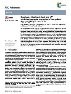
INTRODUCTION Saccharin (1,2-benzisothiazole-3(2H)-one 1,1 -dioxide) (Fig. 1) is about 500 times sweeter than sugar but is practically insoluble in the water at room temperature. Therefore its water-soluble sodium salt is the most frequently used artificial sweetener for diabetics as well as a dietetic food additive. Due to the presence of three different functional groups (imino, carbonyl and sulfonyl) in the structure of saccharin, its deprotonated form serves as a versatile polyfunctional ligand forming complexes with various metals [1]. The versatile ligation properties of saccharin [e.g. 2–16] and the physiological activity [e.g. 17] of saccharin and its metal complexes have been widely studied mainly due to its proven cancerous nature in rats [e.g. 18]. Consequently, in 1977 saccharin joined the list of human potential cancer-causing substances in USA, the products containing saccharin carrying a special warning label [1, 19]. Since in the meantime numerous epidemiological studies did not provide a clear evidence that saccharin indeed causes cancer in humans, in 2001 the use of the warning label on saccharin-containing products was discontinued [20, 21].
Fig. 1. Structural formula of saccharin with the atom labeling
Continuing our systematic study of the coordination properties of saccharin in various metal saccharinates, in addition to the numerous structural and spectroscopic studies of other saccharinate compounds, we have undertaken a study of the structural and/or spectroscopic characteristics of saccharin compounds with metals of group 2 (Mg, Ca, Sr and Ba). Aside of this study, the reported structural and vibrational spectroscopic data on Mg and earth metal (Ca, Sr and Ba) saccharinates are rather scarce. Thus, the crystal structure of magnesium disacchari-
nate heptahydrate has been reported [4] and the infrared spectra of the saccharinates of Ca, Sr and Ba in the regions of the OH, OD, CO and SO2 stretching modes (together with their X-ray powder diagrams) have been studied [22]. Here we report the results of vibrational investigations and complete assignment of the infrared and Raman spectra of magnesium disaccharinate heptahydrate, Mg(sac)2⋅7H2O, as well as the correlation of the spectroscopic data with the previously published (by us) structural data for this compound. Special attention is paid to the discussion concerning the H2O, HOD, CO and SO2 stretching vibrations. EXPERIMENTAL The studied compound was synthesized from warm aqueous solution of saccharin and MgCO3. Partially deuterated analogues were obtained by recrystallization of the protiated sample from D2O. The room temperature (RT) and liquid nitrogen temperature (LNT) infrared spectra in the 4000– 380 cm–1 region were recorded on a Perkin-Elmer 580 infrared spectrophotometer using KBr pellets. A variable temperature cell (RIIC VLT-2) was used for the LNT measurements. The roomtemperature Raman spectra in the 4000–100 cm–1 region were recorded on a micro-Raman multichannel spectrometer – Horiba JobinYvon LabRam Infinity (f × 100) operating at 532 nm YAG laser line. The GRAMS/32 software package was applied for the spectral manipulations. STRUCTURAL DATA The results of the crystal structure determination of magnesium disaccharinate heptahydrate, Mg(sac)2⋅7H2O, have already been published [4]. Here, only some important general characteristics of the structure as well as details concerning the H2O, CO and SO2 groups are mentioned. Maced., J. Chem. Chem. Eng., 27 (1), 1–8 (2008)
Vibrational study and spectra-structure correlations in magnesium disaccharinate heptahydrate, Mg(sac)2⋅7H2O
The crystals of the title compound belong to the triclinic system, space group P 1 , Z = 2. The structure consists of two crystallographically different saccharinate anions, one type of magnesium cations and seven independent types of water molecules. Each magnesium ion is octahedrally coordinated by six oxygen atoms, one of them belonging to the carbonyl group of the saccharinate anion [labeled O(13) in [4]] and the remaining five to the water molecules. The remaining two water molecules are non-coordinated to the metal atom. The Mg–O distances range from 203.0 to 211.3 pm. Only one of the two independent saccharinate ions is coordinated to the magnesium cation, the second one being at a distance from Mg larger than 340 pm. The carbonyl oxygen of the non-coordinated saccharinate anion, {labeled O(23) in [4]}, is a proton acceptor involved in hydrogen bonding with the neighboring water molecules. The two C–O distances of the non-coordinated and the coordinated carbonyl group are very close to each other (124.2 and 124.0 pm, respectively). Both O–S–O angles in the two crystallographically different sulfonyl groups are practically identical (114.4 and 114.7 o), whereas the S–O distances are 144.5 and 144.9 pm in the first and 142.2 and 144.5 pm in the second SO2 group. All seven independent water molecules (five coordinated and two non-coordinated) are involved in the hydrogen bonding with the neighboring water molecules and/or with the electron-donor atoms from the saccharinate anions (N, OCO, OSO2), except the coordinated carbonyl oxygen [O(13)] (see Table 1).
Table 1 Scheme of the hydrogen bonding in the Mg(sac)2⋅7H2O Hydrogen bond Ow(1)⋅⋅⋅N(2) Ow(1)⋅⋅⋅Ow(5)ii Ow(2)⋅⋅⋅O(22) Ow(2)⋅⋅⋅O(21)i Ow(3)⋅⋅⋅Ow(6) Ow(3)⋅⋅⋅Ow(7) Ow(4)⋅⋅⋅Ow(6)i Ow(4)⋅⋅⋅N(1)iii Ow(5)⋅⋅⋅O(23)ii Ow(5)⋅⋅⋅N(2)i Ow(6)⋅⋅⋅O(23)iv Ow(6)⋅⋅⋅Ow(7)v Ow(7)⋅⋅⋅O(12)iii Ow(7)⋅⋅⋅O(11)v i
Distance/pm 292.6(3) 304.6(3) 277.2(3) 281.8(2) 271.4(3) 273.2(3) 285.2(3) 293.9(3) 275.4(2) 289.8(3) 275.7(4) 276.3(3) 277.0(3) 283.2(3)
= x, y, z – 1; ii = 1 – x, – y, – z; iii = 1 – x, 1 – y, – z; = 1 – x, – y, 1 – z; v = 1 – x, 1 – y, 1– z
iv
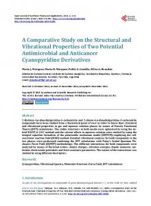
Fig. 2. The RT (a) and LNT (b) infrared spectra of Mg(sac)2⋅7H2O
RESULTS AND DISCUSSION The RT and LNT infrared spectra of the protiated and the partially deuterated forms of the studied compound are presented in Figs 2 and 3 respectively, whereas the RT Raman spectrum of the protiated compound is given in Fig. 4. The O–D stretching region in the RT and LNT infrared spectrum of the partially deuterated sample is shown in Fig. 5, and Fig. 6 gives the infrared spectral appearance in the region of the SO2 stretching vibrations. The assignment of the experimental frequencies in the infrared and Raman spectra of magnesium disaccharinate heptahydrate in the 4000–100 cm–1 region is presented in Table 2.
Maced., J. Chem. Chem. Eng., 27 (1), 1–8 (2008)
3
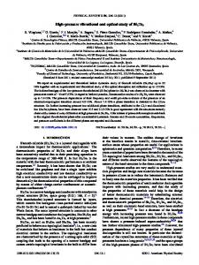
Fig. 3. The RT (a) and LNT (b) infrared spectra of highly deuterated (ca. 60 % deuterium content) Mg(sac)2⋅7H2O
4
G. Jovanovski, P. Makreski, B. Šoptrajanov
Table 2 Assignment* of the experimental frequencies in the infrared and Raman spectra of Mg(sac)2⋅7H2Oa Experimental
Ab initio [8]
Raman
Approximate assignmentc
b
No.
Infrared
1.
3570 sh
2.
3450 vs
3459 w
ν(H2O)
3.
3380 sh
3389 vw
ν(H2O)
4.
3240 sh
A
ν(H2O)
ν(H2O) 3099 m
ν(CH)
6.
3075 s
ν(CH)
7.
3061 m
ν(CH)
8.
3040 w
ν(CH)
5.
1648 w
1700
495.6
ν(CO) + ν(CN)
9.
1660 sh
10.
1645 s
11.
1627 vs
1620 vw
12.
1587 s
1589 m
1596
17.7
ν(CC)+ δ(CCC)
13.
1462 m
1460 w
1468
4.5
δ(HCC)+ ν(CC)
14.
1458 m
1454 sh
1459
13.2
δ(HCC)+ ν(CC)
15.
1414 vw
1415 vw
16.
1360 sh
1359 m
δ(H2O)d ν(CO)+ν(CN)
17.
1350 m
1352 sh
18.
1338 sh
1338 sh
19.
1300 sh
1303 sh
20.
1293 m
1293 m
1285
9.5
21.
1265 vs
1268 w
1289
297.6
22.
1258 s
1251 w
1246
46.2
23.
1217 sh
1216 vw
24.
1168 sh
1171 m
25.
1160 sh
26.
1155 vs
27.
1142 s
1146 vs
δ(HCC)+δ(SCC)+ν(CC) νas(SO2) δ(HCC)+ ν(CC)
1201
450.3
ν(CN)+ν(PhC)+δ(CSN)
1160
349.4
νs(SO2)+ δ(CSN)+ν(CC)
1135
2.4
ν(CC)+δ(HCC)
1127
0.6
ν(CC)+δ(SCC)+δ(HCC)
1117
40.9
28.
1130 m
29.
1121 m
1119 vw
30.
1054 m
1055 vw
ν(CS)
31.
1015 vw
1017 m
δ(CCC)
32
1000 vw
33.
958 s
961 vw
νas(CNS)
34.
890 vw
888 vw
35.
795 sh
801 vw 776 vw
δ(CO)
707 vs
δ(CCC)
36.
775 m
37.
753 s
38.
708 m
δ(CCC)+δ(HCC)
δ(CCC)
39.
678 m
40.
647 w
41.
610 m
609 w
42.
560 vw
553 sh
δ(SO2)
Maced., J. Chem. Chem. Eng., 27 (1), 1–8 (2008)
Vibrational study and spectra-structure correlations in magnesium disaccharinate heptahydrate, Mg(sac)2⋅7H2O
Experimental No.
Infrared
Raman
43.
543 w
539 m
Ab initio [8] A
5
Approximate assignmentc
b
δ(CNS) δ(CCC)
44.
532 vw
45.
440 w
446 vw
46.
393 w
396 m
47.
355 w
355 w
48.
300 vw
299 vw
49.
257 w
50.
243 w
ρ(SO2) τ(SO2)
51.
156 sh
Lattice vibrations
52.
142 sh
Lattice vibrations
53.
131 vs
Lattice vibrations
54.
117 s
Lattice vibrations
* Based on the literature theoretical infrared data for the deprotonated saccharin (saccharin anion) and the literature data for the earlier empirical assignments of the vibrational spectra of various metal saccharinates. a Except for column denoted by A, the data are given in cm–1; vw – very weak; w – weak; m – medium; s – strong; vs – very strong; sh – shoulder. b IR intensities (in km mol–1) for saccharin anion. c Vibrational modes: ν, stretching; δ, bending (all kinds of); ρ, rocking; subscripts: as, antisymmetric; s, symmetric. d Assignment based on the band intensity decreasing in the IR spectrum of the deuterated sample (see Fig. 6).
General considerations
The O–H and O–D stretchings
The assignment of the bands in the RT and LNT IR and RT Raman spectra of the studied compound was based on the earlier theoretical HF/3-21G(d) results for the free deprotonated saccharin species and the experimental values for its DMSO solution [8], as well as the literature data for the earlier empirical assignments of vibrational spectra of various saccharinates [e.g. 1, 11, 14, 23– 27]. Here, the characteristic OH and OD stretching vibrations of the water molecules as well as the CO and SO2 stretching modes are discussed and correlated with the crystallographic values of the corresponding interatomic distances.
As it was already mentioned, all seven crystallographically non-equivalent water molecules in the structure of Mg(sac)2⋅7H2O are involved in hydrogen bonding with carbonyl and sulfonyl oxygens or with nitrogen atoms from the saccharinate anions [4]. The Ow⋅⋅⋅O and Ow⋅⋅⋅N distances range from 271.4 to 304.6 pm and from 289.8 to 293.9 pm, respectively (Table 1). Some of the distances are very close (practically equal) to each other [e.g. 275.4(2) and 275.7(2) pm, or 277.0(3) and 277.2(3) pm].
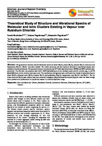
Fig. 4. The RT Raman spectrum of Mg(sac)2⋅7H2O Maced., J. Chem. Chem. Eng., 27 (1), 1–8 (2008)
The appearance of only eight bands in the region of the OD stretching vibrations of the isotopically isolated HDO molecules (Fig. 5), instead of the fourteen expected based on the structural characteristics (Table 1) obviously results from the overlap of the bands due to the existence of some very close Ow⋅⋅⋅O distances in the structure. The non-equal intensity of the bands in the O–D stretching region (Fig. 5) is in line with such a conclusion. All together, it is evident that the existence of a considerable number of expected bands in a relatively narrow spectral region makes the spectra–structure correlation rather difficult.
6
G. Jovanovski, P. Makreski, B. Šoptrajanov
Fig. 5. The O–D stretching region in the RT (b) and LNT (c) infrared spectrum of partially deuterated (ca. 3 % deuterium content) Mg(sac)2⋅7H2O. Curve (a) corresponds to the LNT spectrum of the protiated form of the studied compound
The C=O stretchings The stretching vibrations of the carbonyl group are considered as characteristic vibrational modes and are manifested by very strong bands in the vibrational Raman and, especially, infrared spectra of the corresponding compounds including the metal saccharinates [e.g. 1, 8, 26]. Therefore they are very often used to correlate the predicted and the experimentally observed spectroscopic evidence for the carbonyl stretches with the corresponding data obtained by structure determination. The assignment of the carbonyl stretching bands in magnesium disaccharinate heptahydrate is not straightforward since the bands due to vibrations are localized mainly in the six-membered aromatic ring [ν(CC)+ δ(CCC)] and the bands originating from the bending vibrations of the water molecules [δ(H2O)] are found in the same spectral region [1, 23, 26, 28]. The former bands, however, are usually sharper and appear at somewhat lower frequencies (Figs 2 and 3, Table 2), whereas the latter bands can be eliminated almost completely by recording the spectra of highly deuterated compounds (Fig. 3), or can be recognized by decreasing of their intensity in the spectra of the partially deuterated samples. Two well separated bands (1645 and 1627 –1 cm ) and one shoulder (1660 cm–1) are registered in the RT infrared spectrum of Mg(sac)2⋅7H2O in the region where the stretching C=O modes are
expected to appear (Fig. 2a, Table 2). The number of the observed bands in the corresponding region of the RT Raman spectrum is also two (1648 and 1620 cm–1) (Fig. 4, Table 2). By lowering the temperature, the number of IR bands in this region increases to five (1718, 1668, 1653, 1641 and 1630 cm–1) (Fig. 2b). Finally, only two bands (at 1660 and 1627 cm–1) are observed in the RT and LNT IR spectrum of the highly deuterated analogue (ca. 60 % deuterium content) of the studied compound (Fig. 3a and 3b, respectively). Having in mind that the bands which disappear (1718, 1653 and 1641 cm–1) belong to the δ(HOH) modes, whereas the lower-frequency band at 1586 cm–1 (IR) and at 1589 cm–1 (Raman) is due to the [ν(CC)+ δ(CCC)] mode, obviously the two remaining bands at 1660 and 1627 cm–1 are due to the carbonyl stretching modes. This observation shows that, despite the fact that both carbonyl distances [C–O(13) and C–O(23)] in the structure of Mg(sac)2⋅7H2O are practically the same (124.2 and 124.0 pm, respectively) [4], two well resolved ν(C=O) bands appear in its IR (and Raman) spectrum. Here, it should be mentioned that few new bands were observed in the 1480–1380 cm–1 region where the δ(HOD) vibrations are expected to take place in the spectrum of the highly deuterated analogue (Fig. 2 and Fig. 3). The pronounced frequency lowering of the two IR carbonyl stretching bands (from 1725 cm–1 in the spectrum of saccharin [27] down to 1660 cm–1; Δν = 65 cm–1 and to 1627 cm–1; Δν = 95 cm–1) in the IR spectrum of Mg(sac)2⋅7H2O agrees well with the previously determined ionic character of the magnesium-to-deprotonated saccharin bonding in the studied compound [4]. Similar spectral behavior is observed in the case of the ionic saccharinates of sodium, potassium and lead [1, 11, 28]. This is in line with the earlier conclusions [1] that the extent of the frequency lowering of the ν(C=O) mode depends on the character of the metal-to-deprotonated saccharin bonding, and it is more pronounced in the ionic than in the covalent saccharinates. It is worth mentioning that significant frequency difference is also observed in the case of the Raman active ν(C=O) modes observed in the spectrum of saccharin [27] as compared with the corresponding modes in the Raman spectrum of the presently studied compound which appear at 1648 cm–1 (Δν = 49 cm–1) and 1620 cm–1 (Δν = 77 cm–1) (Fig. 4, Table 2). Maced., J. Chem. Chem. Eng., 27 (1), 1–8 (2008)
Vibrational study and spectra-structure correlations in magnesium disaccharinate heptahydrate, Mg(sac)2⋅7H2O
7
The SO2 vibrations The theoretical and empirical analysis of the SO2 stretching vibrations in various compounds containing SO2 groups has shown that, similarly to CO group, the SO2 group can also be considered as a characteristic group vibration [1, 8, 25, 29, 30]. The situation is complicated by the fact that four bands originating from the saccharinate ring vibrations are expected in the region (1335–1130 cm–1) where the antisymmetric, νas(SO2), and symmetric, νs(SO2) sulfonyl stretches are predicted to appear [8] or are experimentally observed [1, 25, Table 2]. The saccharinate ring bands are found to be sharper and less intense than those arising from the sulfonyl stretching vibrations. Two strong bands in the RT infrared (at 1265 and 1258 cm–1) and also two weaker bands in the RT Raman spectrum (at 1268 and 1251 cm–1) of the studied compound are registered in the νas(SO2) region (Figs. 4 and 6, Table 2). Taking into account the results of the theoretical analysis [8] as well as the empirical results for the series of metal saccharinates [25], the bands at 1265 cm–1 in RT infrared spectrum and at 1268 cm–1 in the Raman spectrum are attributed to the antisymmetric SO2 stretching vibrations. The assignment of the band arising from the symmetric SO2 stretches is much easier. Namely, only one strong band (at 1155 cm–1) accompanied by a shoulder at the high-frequency side (at 1160 cm–1) is present in the RT infrared spectrum of Mg(sac)2⋅7H2O in the region where this type of vibrations are expected to appear (Fig. 6). Similarly, one strong band (at 1146 cm–1) is observed in the RT Raman spectrum in the same spectral region (Fig. 4). Undoubtedly they originate from the symmetric SO2 stretching mode (Table 2). The presence of a single pair of ν(SO2) bands in the vibrational (infrared and Raman) spectra of the studied compound is consistent with the crystallographic data for the studied Mg(sac)2⋅7H2O [4] as well as with the detailed analysis of the SO2 stretches in metal saccharinates [25] which has shown that the ν(SO2) frequencies are dependent on the O–S–O angle values rather than on the S–O distances or on the type of the metal-to-deprotonated saccharin type of bonding. Namely, as it was previously mentioned, the O–S–O angles (114.4 and 114.7o) in both crystallographically different sulfonyl groups are practically the same.
Maced., J. Chem. Chem. Eng., 27 (1), 1–8 (2008)
Fig. 6. The region of the SO2 stretching vibrations in the infrared spectrum of Mg(sac)2⋅7H2O
In the compounds containing SO2 groups, in addition to the SO2 stretches, the bands due to the δ(SO2) as well as to ρ(SO2), ω(SO2) and τ(SO2) modes are expected in the region below 610 cm–1. Following Quinzani et al. [31], the bands at 610 cm–1 (infrared) and at 609 cm–1 (Raman) are attributed to the δ(SO2) mode, those at 393 cm–1 (infrared) and at 396 cm–1 (Raman) to the ρ(SO2) librational mode, whereas the bands at 300 cm–1 (infrared) and at 299 cm–1 (Raman) are assigned to the τ(SO2) librational modes (Figs. 2, 3 and 4) . Acknowledgment: The financial support from the Ministry of Education and Science of the Republic of Macedonia is gratefully acknowledged.
REFERENCES [1] G. Jovanovski, Metal Saccharinates and Their Complexes with N-donor Ligands, Croat. Chem. Acta, 73, 843–868 (2000). [2] B. Kamenar, G. Jovanovski, Manganese(II) Saccharinate Hexahydrate, Mn(C7H4NO3S)2·6H2O, and Isomorphism with the Analogous Fe, Co, Ni, Zn and Cd Complexes, Cryst. Struct. Comm. 11, 257–261 (1982). [3] B. Kamenar, G. Jovanovski, D. Grdenić, Mercury(II) Saccharinate, Hg(C7H4NO3S)2, Cryst. Struct. Comm. 11, 263– 268 (1982). [4] G. Jovanovski, B. Kamenar, Two Ionic Saccharinates: (1a) Sodium Saccharinate 2/3 Hydrate, C7H4NO3SNa⋅2/3H2O, (1b) Magnesium Disaccharinate Heptahydrate, (C7H4NO3S)2Mg⋅7H2O, Cryst. Struct. Commun., 11, 247– 255 (1982). [5] F. A. Cotton, G, E. Lewis, C. A. Murillo, W. Schwotzer, G. Valle, Comparative Study of Structures, Including Jahn–Teller Effects, in the Saccharinate Complexes, [M(C7H4NO3S)2(H2O)4]⋅2H2O, of Chromium and Zinc, as
8
G. Jovanovski, P. Makreski, B. Šoptrajanov
well as Other Divalent Metal Ions, Inorg. Chem., 23, 4038–4041 (1984). [6] S. Z. Haider, K. M. A. Malik, S. Das, M. B. Hursthouse, Structural Studies of Tetraaquabis(saccharinatoN)zinc(II) Dihydrate, [Zn(C7H4NO3S)2(H2O)4]⋅2H2O, and Tetraaquabis(saccharinato-N)cadmium(II) Dihydrate, [Cd(C7H4NO3S)2(H2O)4]⋅2H2O, Acta Crystallogr., C40, 1147–1150 (1984). [7] G. Jovanovski, A. Hergold-Brundić, B. Kamenar, Structure of Lead(II) Disaccharinate Monohydrate, Acta Crystallogr., C44, 63–66 (1988). [8] I. G. Binev, B. A. Stamboliyska, E. A. Velcheva, The Infrared Spectra and Structure of o-sulfobenzimide (Saccharin) and of Its Nitranion: An ab initio Force Field Treatment, Spectrochim. Acta, A52, 1135–1143 (1996). [9] P. Naumov, G. Jovanovski, On the Coordination in Metal Saccharinates, J. Coord. Chem. 54, 63–79 (2001). [10] P. Naumov, G. Jovanovski, An Update to the Combined Vibrational-Diffraction Experimental and Theoretical Studies of Small Biologically Important Cyclic Imides: Reference to Saccharin, Curr. Org. Chem. 5, 1059–1077 (2001). [11] G. Jovanovski, B. Kaitner, O. Grupče, P. Naumov, Crystal Structure, Infrared and Raman Spectra of Tripotassium Trisaccharinate Dihydrate, K3(C7H4NO3S)3⋅2H2O, Cent. Eur. J. Chem. 2, 254–275 (2004). [12] P. Naumov, G. Jovanovski, O. Grupče, B.Kaitner, D. A. Rae, S. W. Ng, Solid-State Structure and Temperature/ Evacuation-induced Dehydration of Sodium Saccharinate 1.875 hydrate, Angew. Chem. 44, 1251–1254 (2005). [13] E. J. Baran, The Saccharinate Anion: A Versatile and Fascinating Ligand in Coordination Chemistry, Quim. Nova, 28, 326–328 (2005). [14] P. Naumov, G. Jovanovski, S. Tančeva, S. W. Ng, Crystal Structure and Spectroscopic Characterization of Lithium Saccharinate 11/6 Hydrate, Hygroscopic and Potentially Physiologically Active Compound, Z. Anorg. Allg. Chem. 632, 454–460 (2006). [15] E. J. Baran, V. T. Yilmaz, Metal Complexes of Saccharin, Coord. Chem. Rev., 250, 1980–1999 (2006). [16] P. M. Bhatt, G. R. Desiraju, Crystal Structure of Na4Li4(saccharinate)8⋅14H2O and its Comparison with Other Alkali Metal Saccharinates, J. Mol. Struct., 871, 73–79 (2007). [17] W. C. Groutas, J. B. Epp, R. Venkataraman, R. Kuang, T. M. Truong, J. J. McClenahan, O. Prahash, Design, Syn-
thesis, and In Vitro Inhibitory Activity Toward Human Leukocyte Elastase, Cathepsin G, and Proteinase 3 of Saccharin-Derived Sulfones and Congeners, Bioorg. Med. Chem., 4, 1393–1400 (1996). [18] J. M. Price, C. G. Biava, B. L. Oser, E. E. Vogin, J. Steinfeld, H. L. Ley, Bladder Tumors in Rats Fed Cyclohexylamine or High Doses of a Mixture of Cyclamate and Saccharin, Science, 167, 1131–1132 (1970). [19] http://www.fda.gov/fdac/features/1999/699_sugar.html [20] http://www.en.wikipedia.org/wiki/Sugar_substitute [21] http://www.saccharin.org/facts_policy.html [22] G. Jovanovski, D. Spasov, S. Tančeva, B. Šoptrajanov, Structural Characteristics of the Hydrates of the Saccharinates of Calcium, Strontium and Barium, Acta Chim. Slov. 43, 41–50 (1996). [23] G. Jovanovski, B. Šoptrajanov, Bonding of the Carbonyl Group in Metal Saccharinates: Correlation with the Infrared Spectra, J. Mol. Struct., 174, 467–472 (1988). [24] A. J. Jubert, R. P. Diez, S. B. Etcheverry, E. J. Baran, Raman, Pre-Resonance Raman and Electronic Spectra of Iron(II) Saccharinate, J. Raman Spectrosc., 23, 15–20 (1991). [25] G. Jovanovski, S. Tančeva, B. Šoptrajanov, The SO2 Stretching Vibrations in Some Metal Saccharinates: Spectra-structure Correlations, Spectrosc. Lett., 28, 1095–1109 (1995). [26] P. Naumov, G. Jovanovski, Spectra-structure Correlations in Solid Metal Saccharinates. I. The Carbonyl Stretchings, J. Mol. Struct., 563–564, 335–339 (2001). [27] P. Naumov, G, Jovanovski, Vibrational Study and Spectra-structure Correlations in Ammonium Saccharinate: Comparison with the Alkali Saccharinates, Spectrochim. Acta, A56, 1305–1318 (2000). [28] G. Jovanovski, B. Šoptrajanov, B. Kamenar, Spectrastructure Correlations in Some Metal Saccharinates, Bull. Chem. Technol. Macedonia, 8, 47–66 (1990). [29] W. R. Feairheller, J. E. Katon, The Vibrational Spectra and Molecular Configuration of Sulfolane, Spectrochim. Acta, 20, 1099–1108 (1964). [30] T. Uno, K. Machida, K. Hanai, Vibrational Spectra of Dimethyl Sulpholane and Dimethyl Sulphone-d6, Spectrochim. Acta, A27, 107–118 (1971). [31] O. V. Quinzani, S. Tarulli, O. E. Piro, Ej. J. Baran, E. E. Castellano, Crystal Structure, Vibrational Spectra and Thermal Analysis of Bis(saccharinato)bis(pyridine)zinc(II), Z. Naturforsh., B52, 183–187 (1997).
Maced., J. Chem. Chem. Eng., 27 (1), 1–8 (2008)