doi:10.1510/mmcts.2004.000448
Video robotic lobectomy Franca M.A. Melfi*, Marcello C. Ambrogi, Marco Lucchi, Alfredo Mussi Division of Thoracic Surgery - Cardiac and Thoracic Department of Surgery, University of Pisa, Via Paradisa 2, 56124 Pisa, Italy Video-assisted thoracoscopic surgery (VATS) is beneficial to the patient but challenging for the surgeon. Recently, robots have been introduced into surgical procedures in an attempt to facilitate surgical performance. The da Vinci姠 Robotic System (Intuitive Surgical, Inc, CA, USA) is one of these robots. It consists of a console and a surgical cart supporting three articulated robotic arms. The surgeon sits at the console where he manipulates the joystick handles while observing the operating field through binoculars that provide a three-dimensional image. Improved ergonomic conditions and instrument mobility at the level of distal articulation seem beneficial in thoracic procedures. After a period of technical development and training we used the robotic systems to treat patients with various thoracic diseases. We focused our efforts on the development of this technique in thoracic surgery particularly to perform video robotic lobectomy (VRL).
Keywords: Robotic surgery; Thoracoscopy; Lobectomy; VATS Introduction Video-assisted thoracic surgery (VATS) in the last decade has allowed surgeons to perform an increasing number of operations with minimal tissue trauma, standardization of many procedures and a progressive broadening of the indications. This technique has become widespread and is currently used in a wide range of surgical procedures, but many major difficulties remain. Sensory information is restricted to a two-dimensional image, and effectors instruments have limited manoeuvrability due to the rigid shaft axis fixed to the thorax or abdominal wall by the entry trocar. Advanced engineering technology makes it possible to overcome these difficulties. Robotic surgery is the most recent and advanced stage of this process thanks to ‘micro-mechatronic’ instruments introduced through traditional trocars. * Corresponding author: Tel.: q39-050-995211; fax: q39-0509957239. E-mail:
[email protected] 䉷 2005 European Association for Cardio-thoracic Surgery
Historical notes The early robotic systems employed in surgery were relatively simple – programmed to handle the scope only or to maintain an endoscopic instrument in a fixed position during surgery w1x. These elementary systems have given way to extremely complex and sophisticated robots. Human robotic surgery was introduced by Cadiere’s team in March 1997 w2x. A thoracic procedure was performed using a voice-controlled robot (Zeus姠, MMCTSLink 30) w 3,4x and, in the same period, a different robotic device was used by other surgical teams w5–7x. At the present time, different types of robotic devices are used in clinical practice w8–10x (Table 1). The da Vinci姠 Robotic System (MMCTSLink 17) represents a complete device currently applied in the field of cardiac and general surgery. Although there is a real difference between thoracic surgery and other disciplines, an application in thoracic surgery seemed realistic. Therefore in the last years an increasing number of thoracic surgeons have used the robot device to perform thoracic procedures some of which included 1
F.M.A. Melfi et al. / Multimedia Manual of Cardiothoracic Surgery / doi:10.1510/mmcts.2004.000448 Table 1. Surgical robots systems System
Discipline
Aesop EndoAssist Zeus Da Vinci CyberKnife Novac 7 Robodoc Neuromate
Endoscopy Endoscopy Cardiac/thoracic surgery Cardiac/thoracic surgery Radiosurgery Radiosurgery Orthopedics Neurosurgery
Video 1. The surgeon sits at the master console located at a distance from the patient with eyes focused downwards toward the operative field which appears as an open surgical technique and a robotic unit provides a ‘tele-presence’ within the chest for the manipulation of micro-instruments.
major lung resections (video robotic lobectomy (VRL) in NSCLC-stage I patients) w11–15x.
Method Compared to conventional surgery and VATS, robot surgery demands a new set of manual skills and eye– hand coordination. The transition from traditional surgery to advanced totally robotic surgery is not immediate. Just as it was necessary to follow certain precise organizational and didactic routes in passing from open surgery to minimally-invasive technique, here too, the same process is necessary. It is essential to be familiar with the device and that all members of the surgical team (surgeons, scrub nurses, and technicians) have undergone specific training. The learning curve is relatively short since the surgeons have a solid background in conventional and thoracoscopic surgery. Robot-system in the operating room The system employed to perform video robot lobectomy (VRL) is the da Vinci姠 Robotic System (MMCTSLink 17). It is a complete robot device which comprehends a master remote console, a computer controller and a three-arms surgical manipulator with fixed remote centre kinematics connected via electrical cables and optic fibres (Video 1). 2
Photo 1. Motion scaling. (Reprinted with permission from Intuitive Surgical Inc, CA, USA.) In terms of motion, the mechanical wrists of the instruments have 6 degrees of freedom. Tip articulations mime the up/down (‘pitch’) and the side-to-side (‘yaw’) flexibility of the human wrist.
The master console is connected to a surgical manipulator with two instrument-arms and a central arm to guide the endoscope. Two master handles at the surgeon’s console are manipulated by the user. The position and the orientation of the surgeon’s hands on the handles trigger highly-sensitive motion sensors which transfer the surgeon’s movements to the tip of the instrument at a remote location. The surgical arm cart provides three degrees of freedom (pitch, yaw, insertion). Attached to the robot arm is the surgical instrument, the tip of which is provided by a mechanical cable-driven wrist (EndoWrist䊛, MMCTSLink 31). This adds four more degrees of freedom (internal pitch, internal yaw, rotation and grip). To increase precision, the system uses downscaling from the motion of the handles to that of the surgical arms. In addition, unintended movements caused by human tremor are filtered by a 6-Hz motion filter (Photo 1).
F.M.A. Melfi et al. / Multimedia Manual of Cardiothoracic Surgery / doi:10.1510/mmcts.2004.000448
Video 2. The robot’s arms are draped in special disposable nylon covers (for the sterile operating field) which contain the microchips to connect the arms to the robotic instruments. The insertion of electronic microcircuit plates establishes a connection with other robotic instruments.
require few accurate manoeuvres – dissection/coagulation – in a restricted and well-defined field: enucleation of condroma, excisions of mediastinal masses, thymectomies. On the contrary, during major resections such as lobectomies, some manoeuvres must necessarily be performed by the assistant surgeon, given the need of a fourth arm. In fact, maintaining a correct position with appropriate tension of the lung parenchyma is a top priority for identifying and dissecting the hilum structures (vessels and bronchus), as is suction, passing the sutures in the chest cavity, and appropriate positioning of the stapler. In order to perform these manoeuvres the role of the assistant surgeon is mandatory, and he must always be at hand at the operating table. Like VATS, few absolute contraindications are applicable to this surgical procedure (Table 2).
Video 3. In order to avoid collisions between the mechanical arms, the correct placement of the robot arm cart and of the trocars is essential. The trocars must be positioned at a greater distance from each other than they normally would be in standard thoracoscopic procedures. Physical orientation and optimal working angles between the instruments are important issues which must be considered. Table 2. Selection criteria Size Stage Anatomical features
-4 cm (max diameter) Clinical stage I NSCLC Absence of chest wall involvement Absence of pleural symphysis Complete or near complete interlobar fissures
In order to perform robotic surgery in a safe and straightforward manner, it is necessary to standardize procedures and establish operative schemes. This robotic device requires meticulous preparation in terms of set-up of the system and its placement at the operating table (Video 2). The main body of the machine (robot cart) and the robotic arms are placed in relation to the side of the lesion. When the robotic cart has been positioned and the patient placed in the chosen position, the robotic arms are brought into the operative field (Video 3).
Operative technique Many of the robotic procedures can be carried out by a single operator. This is true for procedures which
During the video robotic lobectomy, a single-lung anaesthesia is achieved via a double lumen endotracheal tube. Patients are prepared and draped for a posterior lateral thoracotomy so that the procedure can be converted in the event of intraoperative complications or in case a video robot lobectomy is not possible (Video 4). Instruments Few robotic instruments are used during robotic lobectomy. To handle the lung parenchyma safely a Cadiere forcep (MMCTSLink 32) is advisable because it is an a-traumatic instrument. In contrast, other robotic grasps are too small to handle the lung. Dissection of structures is performed with a combination of electrocautery and Debakey forceps (MMCTSLink 32) mounted on the robotic arms. Accessory endoscopic instruments handled by an assistant surgeon at the operative site are inserted through the minithoracotomy (‘service entrance’) or through an additional small incision (5 mm), when necessary (Video 5). Incisions The location of the incisions is critical for the successful identification and dissection of the interlobar artery – technically the most difficult aspect of video robot lobectomy. The best positioning of the system and of the robotic arms are established in relation to the side of the lesion in order to have an excellent, unobstructed view of the chest cavity without arm impingement and interference. However, the exact position of the operating ports is best assessed during the operation when suitable points of entry in rela3
F.M.A. Melfi et al. / Multimedia Manual of Cardiothoracic Surgery / doi:10.1510/mmcts.2004.000448 Table 3. Clinical features of patients undergoing robotic lobectomy Age (yr)/sex
Preoperative conditions
Procedure
Pathological findings
Postoperative course
Dicharge p.o
64/F
Diabetes
RL lobectomy
Uneventful
5
41/M 66/M
Unremarkable Unremarkable
66/M
Prostatism/ hypertension Cough
Uneventful Sputum retention Uneventful
4 6
70/M
LL lobectomy LL lobectomy (converted) LL lobectomy
Adenocarcinoma T1N0 Typical carcinoid Typical carcinoid
Unremarkable
Sputum retention Uneventful
6
64/F
RL lobectomy (converted) LL lobectomy
4
60/M
Hypertension
LL lobectomy
Uneventful
4
61/F
Unremarkable
RL lobectomy
Air leaks
10
61/M
Unremarkable
LL lobectomy
Uneventful
5
69/M
Hypertention
LL lobectomy
65/M
Unremarkable
LL lobectomy
66/F
Diabetes
M lobectomy
58/F
Unremarkable
LR lobectomy
67/M
Cough
LR lobectomy
70/M
M lobectomy
69/F
Sigmoid colectomy 2 years before Unremarkable
UR lobectomy
69/M
Unremarkable
LR lobectomy
74/M
Cough/hemoptysis
LL lobectomy
65/F
Unremarkable
LR lobectomy
59/M
Unremarkable
LR lobectomy
67/F
Cough
M lobectomy
71/M
Unremarkable
LL lobectomy
60/M
Hypertension
LL lobectomy
Sq. carcinoma T1N0 Sq. carcinoma T1N0 Adenocarcinoma T1N0 Adenocarcinoma T1N1 Adenocarcinoma T1N0 Sq. carcinoma T1N0 Adenocarcinoma T1N0 Sq. carcinoma T1N0 Sq. carcinoma T1N1 Adenocarcinoma T1N0 Atypical carcinoid T1N0 Sq. carcinoma T1N0 Sq. carcinoma T1N0 Adenocarcinoma T1N0 Sq. carcinoma T1N0 Adenocarcinoma T1N0 Adenocarcinoma T1N0 Sq. carcinoma T1N0 Sq. carcinoma T1N0 Adenocarcinoma T1N0
Video 4. The patient is placed in the lateral position with general anaesthesia and single-lung ventilation
4
Acute kidney failure IV p.o Uneventful
5
Remarks
Retained in unit (angina)
Dead XII p.o. 4
Atrial fibrillation Uneventful
5
Sputum retention Sputum retention Uneventful
5 6
Air leaks
6
Uneventful
4
Uneventful
4
Uneventful
5
Sputum retention Uneventful
6
Uneventful
5
4
5
4
tion to the shape of each patient’s chest cavity are made. The standard layout is the following: the first port is placed at the 7th or 8th space in the mid-axillary line (for the 08 3-D scope), the other at the 6th or 7th intercostal space in the post-axillary line (for the left robotic arm), a ‘service entrance’ is made at the 4th or 5th intercostal space in the anterior axillary line (where the right robot arm is placed). An additional small incision is made (between the ‘service entrance’ and the 3-D scope) for the assistant surgeon to insert conventional
F.M.A. Melfi et al. / Multimedia Manual of Cardiothoracic Surgery / doi:10.1510/mmcts.2004.000448
Video 5. The robotic instruments currently used during the VRL are Cadiere and Debakey forceps, electrocautery and micro scissors (EndoWrist䊛, MMCTSLink 31). In addition, thoracoscopic instruments and a full thoracotomy instrument set is opened and kept on hand, in case of preoperative complications.
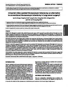
Photo 3. CT scan. A computed tomogram of the chest demonstrating a speculated mass in the lower lobe of the left side.
Video 6. The first incision is made at the 7th space in the mid-axillary line to verify the feasibility of the robot procedure using a standard endoscopic optic.
(in the anterior axillary line). It provides an easy direct access to the hilum; to insert standard endoscopic instruments when the robotic instruments are not suitable; moreover, it is wider than the posterior space and facilitates later retrieval of the specimen (Photo 2).
Surgical steps Video-robotic left lower lobectomy Video robotic lobectomy follows the standard surgical steps of open thoracic surgery and implies the isolation and resection of the vascular and bronchial hilar elements. Usually the artery is dealt with before the vein and eventually the bronchus is resected. However, priorities are not strictly set. Frequently, as is the case of open thoracic surgery, due to surgical strategies the ligature of the vein precedes that of the artery. In some cases it is preferable to resect the bronchus before resecting the artery branch.
Photo 2. Chest incisions. Incision layout during a robot left lower lobectomy.
endoscopic instruments only when strictly necessary (Video 6). The routine VATS exploration prior to the operation could yield important information that would markedly alter the treatment strategy. If there is no contraindication to proceed, the minithoracotomy ‘service entrance’ (approximately 4 cm in length) is placed over the 4th or 5th intercostal space
Here below is described a robot-left lower lobectomy in a 64-year-old female. She was a non-smoker with small lung mass without mediastinal lymphadenopathy on CT scan, with normal bronchoscopic appearances, judged to have clinical stage I (positive cytology for adenocarcinoma at needle aspiration/CTguide) (Photo 3). Arterial phase Dissecting around the pulmonary vessel is basically the same as in conventional open surgery. The Cadiere forceps and robot electrocautery (MMCTSLink 32), connected to the robot arms, are 5
F.M.A. Melfi et al. / Multimedia Manual of Cardiothoracic Surgery / doi:10.1510/mmcts.2004.000448
Video 7. The dissection of the fissure with robotic instruments to expose the interlobar artery.
Video 11. A double tie is advisable in all cases, even in small calibre vessels to ensure a safe ligation. A resection is made between the two ligations by micro scissors (EndoWrist䊛, MMCTSLink 31).
priate traction of the lung parenchyma and helps the surgeon to position the lobes so that the hilum and the vessels can be easily accessed.
Video 8. After dissection of the fissure, the pulmonary artery branches are carefully identified and isolated.
Video 9. A sling is passed to lift the vessels separately to obtain a safer ligation.
If the interlobar fissure is complete or nearly complete, the incision of the visceral pleura with robotic electrocautery or blunt dissection with a pledget mounted on the Cadiere forceps (EndoWrist䊛, MMCTSLink 31), allows the pulmonary artery to be easily identified. In this case, the electrocautery at a low setting is useful and safe (Videos 7 and 8). When the fissure is incomplete the upper lobe is retracted upward and forward, the artery appears from the posterior aspect of the lung root. The artery is then identified within the fissure by careful dissection and a sling is passed between the two points of the arterial access to elevate the fused posterior fissure, which is divided by stapling. The same procedure is carried out for the resection of the anterior fissure. The mechanical stapler (Endopath ATB45, MMCTSLink 34) is used by the assistant surgeon through one of the other two ports (depending on the alignment). Currently the apical and main-stem lower vessels are taken separately and tied with Linen 2.5 (Videos 9, 10 and 11).
Video 10. The vessels are taken separately and tied with Linen 2.5. Due to their rough texture, linen as well as silk are ideal because the ligation does not come undone.
introduced through the minithoracotomy and posterior incision for the dissection of the hilum and the fissure. A standard endoscopic holding forceps (Babcock 5BB, MMCTSLink 33) can be introduced by the assistant surgeon through the service entrance or (rarely) through an additional incision. This provides appro6
Instead of a double tie with Linen, conventional endoscopic clips (MMCTSLink 35) can be used on the distal part of the artery. However, this technique is not very safe because these endoscopic clips must be applied at the operating site by the assistant surgeon, who has a bi-dimensional vision and does not have sufficient coordination. On the other hand, the available robotic clips are too small to be used for pulmonary vessels. In this regard, a possible complication is that the clips can lacerate the vessel during the surgery or that they can slip off (Video 12). Vein phase Usually the surgical time sequence implies treating the vein as a second step. The pulmonary ligament is
F.M.A. Melfi et al. / Multimedia Manual of Cardiothoracic Surgery / doi:10.1510/mmcts.2004.000448
Video 12. This is an example of the laceration of a pulmonary artery due to the imprecise positioning of the clips solved by further isolating the artery peripherally and placing additional clips.
Video 13. The vein is cleared and separated from the lymph node, which is removed, by using electrocautery and the Cadiere forceps (MMCTSLink 32). A blunt dissection can be useful.
Video 16. The resected lobe is removed in sterile plastic bags through the ‘service entrance’.
through a 4th incision, when it is made, and stitch it by using the robot Debakey forceps and a large needle holder (EndoWrist䊛, MMCTSLink 31) (Polypropylene Monofilament 4/0). This is more difficult and not safe, considering that both the stapler and the clamp have to be placed by the assistant surgeon (at hand at the operating table), whose hand–eye orientation (bi-dimensional vision) is less precise compared to the surgeon who, at the console, has a different depth perception and optical resolution. Consequently poor coordination between the surgeon and the assistant can jeopardize the success of the operation. Bronchus phase The last step consists of isolation and resection of the lobar bronchus by using the stapler (Endopath ATB45, MMCTSLink 34), necessarily performed by the assistant surgeon. This is the only possible way to resect and suture the bronchus with this approach.
Video 14. After isolation, a sling is passed and the vein is double tied with Linen 2.5 and resected between the ligations.
Although the robot wrists are able to simulate even fine physiological movements, the surgeon cannot make a running stitch when dealing with the bronchus as the robotic instruments in current use are too small to handle such a thick structure (Video 15). Blunt dissection is particularly useful when dissecting and for sweeping tissue along the lobar bronchus peripheral to the line of intended bronchial division so that all the lobar bronchial nodes can be included in the operative specimen.
Video 15. The bronchus is isolated and cleaned by dissection manoeuvres. Here too, a sling is used to better isolate the bronchus and to position the stapler correctly.
The lobectomy specimen is placed in a sterile plastic bag and removed through the minithoracotomy. The bronchial stump is then tested under water for air leaks with 20 cm of positive airway pressure (Video 16).
incised and the lower vein cleared from the surrounding tissues and divided (Videos 13 and 14). When it is particularly thick it is advisable to use a mechanical stapler, although this increases the cost. Another way of handling the vein is to place a vascular clamp
At the end of the operation all the accessible nodal stations are systematically sampled to ensure proper staging of the lung cancer. Currently the lymph-node samplings are made at stations that are more likely involved for tumours originating from a particular lobe 7
F.M.A. Melfi et al. / Multimedia Manual of Cardiothoracic Surgery / doi:10.1510/mmcts.2004.000448 the lower left (ns11), the lower right (ns9), the middle lobe w3x, the upper right (ns1).
Table 4. Mean data ("SD) of robotic lobectomy Operative time (h) Operative blood loss (ml) Post-operative morphine consumption (mg/h) Postoperative (VAS) pain score first 24 h (mm:range 0–100) Chest tube (days) Hospital stay (days)
3.2 ("0.6) 103 ("28.1) 0.47 ("0.1) 1.3 ("0.7) 2 ("1.4) 5 ("1.3)
w16x: the right upper lobe (prevascular and retro tracheal N3 and lower paratracheal N4R), the middle lobe (N3 and subcarinal N7), the right lower lobe (N7), the left upper lobe (sub-aortic N5 and para-aortic N6), the left lower lobe (N7). If a complete lymph node dissection is considered necessary it should be done through a thoracotomy. Unlike VATS approach, there are no limitations regarding accurate lymph-node sampling, given that this is carried out at the end of the operation when a part of the lung has been removed. In fact, there is no need to move the entire pulmonary parenchyma in order to access all lymphnode stations. Two 24 French chest drains (through the previous camera and instrument ports) and closure of the minithoracotomy wound complete the operation.
Personal experience Since February 2001 we have used a robotic system to operate 85 patients with a range of thoracic diseases. There were 54 men and 31 females aged 19– 71 years (mean age 61). We applied the da Vinci姠 Robotic System (MMCTSLink 17) to perform various thoracic operations, ranging from the simplest procedures, such as benign tumour enucleations/ excisions, to very complex ones, such as major pulmonary resections. Video robotic lobectomies (VRL) were performed in 23 good-risk patients selected by pre-operative investigations. There were 15 men and 8 females aged 41 to 78 years (mean age 64.4). The patients were referred to our Department with a pulmonary opacity on chest radiographs and normal bronchoscopic appearances. In accordance with our then normal practice for small lesions without mediastinal lymphadenopathy on the CT scan, mediastinoscopy was not undertaken. These patients were judged to have clinical stage I (NSCLC). Arterial blood gases where within normal limits, and pulmonary function demonstrated adequate pulmonary reserve to undergo a planned lobectomy (forced expiratory volume in 1 s (FEV1) )1.5 l). Specific consent was obtained for attempted robotic resection. The lobes resected were 8
Results Details of patients who underwent robotic lobectomy are summarized in Table 4. In this highly selected group of good-risk patients no technical operative mishaps related to manoeuvres of the instrumentarms occurred. None of the patients had problems related to operative bleeding. All patients tolerated the procedure well and the post-operative course was satisfactory, requiring less analgesic compared to conventional surgery. In two patients the procedure was converted to a minimal thoracotomy. In these cases we began the lobectomies by isolating and stitching the transected lower vein with the robot. However, we had to complete the operations using the ‘service entrance’ (enlarged by about 2 cm) due to hilar calcified lymph nodes, since these rendered the dissecting of the pulmonary artery unsafe. In another two patients we had air leaks. In both cases mechanical staplers were used to complete the fissure. There was one death on the 12th p.o. day (not related to the surgical technique) due to pulmonary embolus. After an initial excellent postoperative recovery the patient had acute kidney failure on the 4th p.o. day which led to a worsening of the clinical conditions. Chest tubes were removed in mean 2 p.o. days and the patients were discharged in mean 5 postoperative days. Operative time varied between 2.5 to 5 h, of which 1 h was used to do the self-test of the machine and instrument set-up. This time was considerably longer than that for standard open surgery or VAT procedure, but it decreased with experience so that the last cases averaged 3 h. All patients were discharged in good condition and returned to preoperative levels of physical activity within 10 days of the operation.
Comments and controversies As far as is known, few video-assisted robotic lobectomies are performed, consequently few surgeons have experience in this field w11–16x. Currently, many of the limitations of robotics surgery are related to the imperfections of the system. At present the greatest difficulties of the VRL are associated with the available arm instruments, which are designed for cardiac or general surgery and are not adequate for thoracic surgery. Consequently some procedures become more
F.M.A. Melfi et al. / Multimedia Manual of Cardiothoracic Surgery / doi:10.1510/mmcts.2004.000448 complicated especially for the lung major resection. This accounts for the extreme difficulty in performing upper lobectomies, which can only be carried out with the aid of the assistant surgeon, who is in a different ergonomic position. In our experience we performed only one upper right lobectomy and no upper left lobectomy. This is because a fourth arm would have to be available to allow the surgeon to easily access the hilum of the upper lobe and to handle the pulmonary parenchyma. Therefore, lower-lobe lobectomies, especially left lower lobectomy – also in conventional thoracic surgery – are technically the most straight-forward resection to carry out. Other limitations are system-related. At present, blocking of the robotic arms or working against strong resistance is experienced at the console. The surgeon does not receive information on the amount of force applied to tissue or sutures, and therefore is dependent on visual feedback. This is sufficient in the majority of the manoeuvres, but not when it comes to suturing delicate structures and tying knots, or when the surgeon wants to distinguish tissue characteristics. This poor tactility impairs the surgeon’s ability to judge the amount of tension applied during the manoeuvres of suture/ligation tensioning w10x. Considering all these limitations, robotic procedures may be technically feasible only in highly selected cases and in the hands of an experienced thoracic surgeon. Like the VAT, this new surgical technique should only be undertaken by surgeons trained in thoracic surgery w17x. Besides a perfect knowledge of topographical anatomy and broad experience in conventional surgery, training in specific thoracoscopic skills is required. Nonetheless, we believe that many of the current limitations can be overcome in the near future, and that, as the da Vinci System is improved and its instruments better adapted to thoracic surgery, their application will be extended to a wider range of operations.
w4x
w5x
w6x
w7x
w8x
w9x
w10x
w11x
w12x
w13x
References w1x Osmote K, Feussner H, Ungeheurer A, Arbter K, Wey GQ, Siewert JR. Self-guided robotic camera control. Am J Surg 1999;177:321–324. w2x Cadiere GB, Himpens J, Vertruyen M, Favretti F. The world’s first obesity surgery performed by a surgeon at a distance. Obes Surg 1999;9:206– 209. w3x Vassiliades TA Jr, Nielsen JL. Alternative approaches in off-pump redo coronary artery
w14x
w15x
bypass grafting. Heart Surg Forum 2000;3: 203–206. Stephenson ER, Sankholkar S, Ducko CT, Damiano RJ. Robotically assisted microsurgery for endoscopic coronary artery bypass grafting. Ann Thorac Surg 1998;66:1064–1067. Loulmet D, Carpantier A, d’Attelis N, Berrebi A, Cardon C, Ponzio O. Endoscopic coronary artery bypass grafting with the aid of robotic assisted instruments. J Thoracic Cardiovasc Surg 1999; 118:4–10. Falk V, Diegeler A, Walther T, Bannusch J, Autschbach R, Mohr FW. Total endoscopic coronary artery bypass grafting. Eur J Cardiothoracic Surg 2000;17:38–45. Schurr MO, Arezzo A, Buess GF. Robotics and systems technology for advanced endoscopic procedures: experiences in general surgery. Eur J Cardiothorac Surg 1999;16 Suppl 2:S97–105. LaPietra A, Grossi EA, Derivaux CC, Applebaum RM, Hanjis CD, Ribakove GH, Galloway AC, Buttenheim PM, Steinberg BM, Culliford AT, Colvin SB. Robotic-assisted instruments enhance minimally invasive mitral valve. Ann Thorac Surg 2000;70:835–838. Rininsland HH. Basics of robotics and manipulators in endoscopic surgery. Endosc Surg Allied Technol 1993;1:154–159. Reichenspurner H, Damiano RJ, Mack M, Boehm DH, Gulbins H, Detter C, Meiser B, Ellgass R, Reichart B. Use of the voice-controlled and computer-assisted surgical system ZEUS for endoscopic coronary artery bypass grafting. J Thorac Cardiovasc Surg 1999;118:11–16. Okada S, Tanaba Y, Yamauchi H, Sato S. Singlesurgeon thoracoscopic surgery with a voicecontrolled robot. Lancet 1998;351:1249. Melfi FM, Menconi GF, Mariani AM, Angeletti CA. Early experience with robotic technology for thoracoscopic surgery. Eur J Cardiothorac Surg 2002;21:864–868. Okada S, Tanaba Y, Sugawara H, Yamauchi H, Ishimori S, Satoh S. Thoracoscopic major lung resection for primary lung cancer by a single surgeon with a voice-controlled robot and an instrument retraction system. J Thorac Cardiovasc Surg 2000;120:414–415. Morgan JA, Ginsburg ME, Sonett JR, Morales DLS, Kohmoto T, Gorenstein LA, Smith CR, Argenziano M. Advanced thoracoscopic procedures are facilited by computer-aided robotic technology. Eur J Cardiothorac Surg 2003;23:883–887. Bodner J, Wykypiel H, Wetscher G, Schmid T. 9
F.M.A. Melfi et al. / Multimedia Manual of Cardiothoracic Surgery / doi:10.1510/mmcts.2004.000448 First experiences with the da VinciTM operating robot in thoracic surgery. Eur J Cardiothorac Surg 2004;25:844–851. w16x Naruke T, Tsuchiya R, Kondo H, Nakayama H, Asamura H. Lymph node sampling in lung cancer: how should it be done? Eur J Cardiothorac Surg
10
1999;16:S17–24. w17x McKneally MF, Lewis RJ, Anderson RJ, Fosburg RG, Gay WA Jr, Jones RH, Orringer MB. Statement of the AATS/STS Joint Committee on Thoracoscopy and Video Assisted Thoracic Surgery. J Thorac Cardiovasc Surg 1992;104:1.