relative preservative content into the lumen surface, in the S2 cell wall layer and in the middle lamella. Keywords: Bamboo cells, chemical preservatives, ...
Current Research Journal of Biological Sciences 7(1): 6-10, 2015 ISSN: 2041-076X, e-ISSN: 2041-0778 © Maxwell Scientific Organization, 2015 Submitted: March 19, 2014 Accepted: April 15, 2014
Published: January 20, 2015
Comparative Distribution of Chemicals in Treated Gigantochloa Scortechinii Through Soaking, Vacuum Pressure and High-Pressure Sap-Displacement 1
Razak Wahab, 2Mahmud Sudin, 1,3Izyan Khalid, 1Mohammed Abdus Salam, 1Shafiqur Rahman and 4 Hashim W. Samsi 1 Universiti Malaysia Kelantan (UMK), Jeli Campus, 17600 Jeli, Kelantan, 2 Universiti Malaysia Sabah, 88999 Kota Kinabalu, 3 Universiti Sains Malaysia, 11800 Penang, 4 Forest Research Institute Malaysia (FRIM), Kepong 52109, Kuala Lumpur, Malaysia
Abstract: The chemicals distribution of preservatives treated bamboo Gigantochloa scortechinii through soaking, vacuum pressure and high-pressure sap displacement were studied. The distribution of chemicals in the bamboo influences their durability against the attacks of the insects and fungi. This largely depends on the chemicals penetration, location and retention at the tissue and cell wall levels in the bamboo. At optimum retention levels, the preservative performance should be comparable unless the preservative's distribution, substrate susceptibility or fixation product had altered. In evaluating the performance of various treatment methods employed, the distribution of preservatives within the cell walls of treated bamboo must be considered. The results from the energy dispersive x-ray analysis on the bamboo treated with copper chrome arsenic and ammoniacal copper quaternary are analyzed. The G. scortechinii samples were treated by soaking, vacuum pressure impregnating and high-pressure sapdisplacement. Observations were carried out using the transmission electron microscope linked system to an energy dispersive x-ray analyzer. The system enables to detect preservative distribution at the cellular level and measured relative preservative content into the lumen surface, in the S 2 cell wall layer and in the middle lamella. Keywords: Bamboo cells, chemical preservatives, chemicals distribution, energy dispersive x-ray analyzer, transmission electron microscope, treated bamboo results from the energy dispersive x-ray analysis on Gigantochloa scortechinii culms treated with Copper Chrome Arsenic (CCA) and Ammoniacal Copper Quaternary (ACQ) at 4% of concentration. This study was designed to determine the presence of different chemicals in the preservatives after each treatment at the tissue and cell walls levels. Transmission Electron Microscope (TEM) linked to an energy dispersive x-ray analyzer was used to detect the chemicals distribution at the cellular level. It was also used in measuring the relative chemicals content in the lumen surface, in the S 2 cell wall layer and in the middle lamella.
INTRODUCTION Bamboo behaves differently from timber when treated with preservatives especially in their distribution of chemicals. The distribution of chemicals in the treated bamboo indisputably influences the preservatives performance against insects and fungi (Daniel and Nilsson, 1989; Razak et al., 2004). The actual protections, however, depends largely on the chemical penetration, location and retention at the tissue and cell wall levels in the bamboo (Jayanetti and Follett, 1998). At optimum retention levels, the preservative performance should be comparable unless the preservative distribution, substrate susceptibility or fixation product had altered (Liese, 1985; Liese and Satish, 2003; Razak, 2004; Razak et al., 2013). In evaluating the performance of bamboo through various treatment methods employed, consideration must be given to the distribution of chemicals within the cell walls of the treated bamboo. Three methods of preservatives treatment were employed in treating the bamboo in this study. These are soaking, vacuum pressure impregnating and highpressure sap-displacement. This paper present the
MATERIALS AND METHODS This study was conducted during the year 2012 to 2013 at the University Malaysia Kelantan (UMK) and Forest Research Institute Malaysia (FRIM). The TEM linked system to an energy dispersive x-ray analyzer was used in detecting the presence of various chemicals and their distribution at the cellular level and measured relative preservative content into the lumen surface, in the S 2 cell wall layer and in the middle lamella.
Corresponding Author: Razak Wahab, Faculty of Earth Science, Universiti Malaysia Kelantan, Kelantan, Malaysia
6
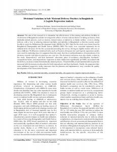
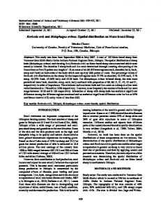
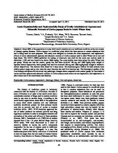
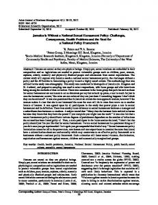
Curr. Res. J. Biol. Sci., 7(1): 6-10, 2015 prepared and analyzed. The match sticks selected at the middle portion of the treated culm samples were prepared and selected using the procedure adopted by Newman (1994) with some modification (Table 1). All samples were analyzed on a PHILLIPS 400T with a Link QX 200 analyzers. The specimen holder was fitted with a Beryllium (Be) low background windowed detection. Operating and analysis conditions was standardized as follows: beam current 17 pA, accelerating voltage 80 kV, live time 200 sec and magnification 17,000 times, spot size 200 nm, specimen angle 20°. The types of cells chosen for the x-ray analysis were a vessel, 3 fibres and parenchyma cells. The vessel was chosen because this is where the major uptake of preservative was expected to take place. From here the preservative was distributed to the surrounding cells and it was considered that cells near the vessel might absorb more preservative than cells further away. The cells analyzed were all located in one vascular bundle (Fig. 1). The first fibre was located in the first layer of fibres around the vessel. The second fibre was in the third layer around the vessel and the third fibre was located in the second layer in the free fibre strand closest to the vessel. The parenchyma was located in the third layer of the parenchyma ground tissue closest to the vessel. These cells were all about in one line to the chosen vessel. The location and types of cells selected for this investigation are shown as in Fig. 2.
Fig. 1: Vascular bundles of bamboo found in G. scortechinii taken at the center of the duct cross-section with two large metaxylem and phloem cells surrounding the fibers and parenchyma
Prior to that, bamboo culms of G. scortechinii were harvested from the FRIM Natural Bamboo Plot Areas in Jeli, Kelantan, Malaysia. These bamboo culms were then taken to the UMK and FRIM for preservative treatments with CCA and ACQ at 4% concentration using soaking process. The samples were then air-dried for three weeks. Procedure developed by Razak et al. (2005) was used with some modification in this study. Match sticks of size 0.5 mm×0.5 mm×10.0 mm were cut from the treated G. scortechinii culm sample blocks of selected blocks treated with CCA and ACQ by soaking, vacuum pressure impregnating and highpressure sap-displacement. Thirty six match sticks blocks consisting one age group, three treatments methods, 2 type of preservatives and 6 replicates were
(a): Bamboo culm wall
(b): A bamboo vascular bundles
Fig. 2: Location of cells within each vascular bundle for the TEM-EDXA study
7
Curr. Res. J. Biol. Sci., 7(1): 6-10, 2015 Table 1: Sample fixation, embedding and cutting The match sticks were: ----------------------------------------------------------------------------------------------------------------------------------Fixed in: 3% glutaraldehyde in 0.1M phosphate buffer (pH 7.2) Dehydrated in: 0.1M phosphate buffer (pH 7.2) 3×20 min 50% / 60% / 70% / 80% / 90% ethanol 1×10 min each 100% ethanol 2×15 min Propylene oxide 2×10 min Embedded in : Propylene oxide : Spurr (2:1) 1h Propylene oxide : Spurr (1:1) 1h Propylene oxide : Spurr (1:2) 48 h Spurr (1) 30 h Spurr (2) 30 h Cast in : Spurr (3) Spurr: ERL 4206 (10 g) DER 736 (4 g) NSA (26 g) DMAE (0.4 g) Cutting :
Diatome 45° diamond knife 6° clearance angle 1 mm per second cutting speed Sections relaxed under chloroform
RESULTS
Table 2: Mean elemental peak: Background ratios in 2 year-old G. scortechinii treated with ACQ soaking treatment Peak: Background ------------------------------------------------Type of cell Location Copper Vessel Lumen 4.38 S2 3.84 Lamella 3.77 Fibre 1 Lumen 2.96 S2 2.45 Lamella 3.04 Fibre 2 Lumen 2.34 S2 2.36 Lamella 2.78 Parenchyma Lumen 1.81 S2 1.19 Lamella 2.75 Fibre 3 Lumen 2.25 S2 2.06 Lamella 2.67
Considerable variation was recognised between analyses carried out on different cells and in order to overcome this, where, possible analyses were conducted on cell walls that were nearer to the vessels. The estimated concentrations of the preservative of the preservative elements present were determined using a peak-to-background ratio P/B, where P and B are, respectively the areas under the peak and of the spectral background, within the same range of the energies. For each peak, a number of selected channels of interest were used in the area determination Eq. (1): 40 for Cu Kα, 44 for Cr Kα and 24 for As Lα (1) The background area was determined from a background line by linear interpolation of the value of the background spectrum in the extremes of each peak. In this condition, for each characteristic line of each active element, the P/B ratio is related to the concentration of the element in the block. Thus, for each active element, it is possible to make a semiquantitative comparison between the several fields of analysis. The mean values in the P/B ratio obtained for copper, chrome and arsenic elements are presented in Tables 2 to 7. As already referred, this energy dispersive x-ray technique used gives only a semiquantitative information on the concentration of the active elements in the treated G. scortechinii. The results of the TEM-EDXA analyses on blocks treated with ACQ by soaking, vacuum pressure and high pressure sap-displacement processes are presented in Tables 2 to 4. Only copper could be analyzed since it is the only metallic element found in the ACQ formulation. The results obtained shows that the Peak to Background counts decreased from the vessel to the fibre 1, fibre 2, fibre 3 and parenchyma. Vacuum
Table 3: Mean elemental peak: Background ratios in 2 year-old G.scortechinii treated with ACQ by vacuum pressure treatment Peak: Background ------------------------------------------------Type of cell Location Copper Vessel Lumen 6.27 S2 6.08 Lamella 4.71 Fibre 1 Lumen 3.76 S2 3.16 Lamella 4.30 Fibre 2 Lumen 3.31 S2 2.96 Lamella 3.36 Parenchyma Lumen 2.68 S2 1.98 Lamella 2.76 Fibre 3 Lumen 2.92 S2 2.79 Lamella 3.48 Mean of 5 replicates
pressure process recorded highest count and soaking process followed closely behind (Table 3). High pressure sap-displacement recorded the lowest P/B counts (Table 4). 8
Curr. Res. J. Biol. Sci., 7(1): 6-10, 2015 Table 4: Mean elemental peak: Background ratios in 2 year-old G. scortechinii treated with ACQ through high pressure sapdisplacement treatment Peak: Background -----------------------------------------------Type of cell Location Copper Vessel Lumen 3.53 S2 3.25 Lamella 3.14 Fibre 1 Lumen 2.42 S2 2.19 Lamella 2.92 Fibre 2 Lumen 2.15 S2 1.91 Lamella 2.25 Parenchyma Lumen 1.42 S2 1.18 Lamella 1.97 Fibre 3 Lumen 1.62 S2 1.30 Lamella 1.66
Table 7: Mean elemental peak : Background ratios in G. scortechinii treated with CCA by high pressure sap-displacement treatment. Peak: Background ---------------------------------------------------------------Cell Location Copper Chrome Arsenic Vessel Lumen 3.45 1.73 2.51 S2 3.12 1.72 1.49 Lamella 2.66 1.35 1.30 Fibre 1 Lumen 2.44 1.54 1.28 S2 1.65 0.92 0.98 Lamella 3.12 1.68 1.72 Fibre 2 Lumen 1.98 1.27 1.10 S2 1.86 0.60 1.40 Lamella 2.42 1.39 1.66 Parenchyma Lumen 1.95 0.84 0.85 S2 1.86 0.39 0.67 Lamella 2.76 1.09 1.64 Fibre 3 Lumen 2.35 0.94 1.40 S2 2.14 0.72 0.87 Lamella 2.48 1.06 1.34 Mean of 5 replicates
Table 5: Mean elemental Peak: Background ratios in G. scortechinii treated with CCA by soaking treatment Peak: Background ---------------------------------------------------------------Type of cell Location Copper Chrome Arsenic Vessel Lumen 4.49 1.73 2.30 S2 3.83 0.99 1.44 Lamella 3.33 0.82 0.95 Fibre 1 Lumen 2.58 0.96 1.14 S2 1.68 0.63 0.91 Lamella 3.83 1.21 2.06 Fibre 2 Lumen 2.25 0.95 1.19 S2 1.76 0.37 0.61 Lamella 2.61 1.15 1.44 Parenchyma Lumen 1.14 0.69 0.75 S2 1.77 0.35 0.36 Lamella 3.14 0.99 1.07 Fibre 3 Lumen 2.60 0.90 0.94 S2 2.43 0.66 0.84 Lamella 2.77 0.96 1.02 Mean of 5 replicates
Similar observations were made in the P/B counts in the CCA treated bamboo culms where counts decreased from the vessel to the fibre 1, fibre 2, fibre 3 and parenchyma. Vacuum pressure process recorded highest count and soaking process followed closely behind (Table 6). High pressure sap-displacement recorded the lowest P/B counts (Table 7). DISCUSSION Complimentary TEM-EDXA studies conducted on the match transverse faces for the distribution of ACQ and CCA elements gave results showing that the copper, chrome and arsenic reading was found in the more lignified regions. The highest count were generally recorded for the middle lamella followed by the lumen and the lastly the S 2 regions of the fibres and parenchyma cells in G. scortechinii blocks. Similar findings also observed by Newman (1994), Daniel and Nilsson (1989), Othman et al. (2006) and Razak et al. (2013) in their separate studies on CCA treated, Betula papyriferra, B. verrucosa, B. papyrifera and Pinus nigra var. maritima respectively. For the vessels, the highest count was recorded in the lumen areas where, the liquid absorption took place followed by the middle lamella and the S 2 layers. The results gathered during the TEM-EDXA experiment shows that the preservatives are not evenly distributed in the cross-section of the bamboo culm walls. The P/B ratios of the metallic elements tended to decrease as the cells were located further away from the vessels. The vessels played an important role in preservative's treatment (Razak et al., 2013). The penetration of liquids into the bamboo culms takes place through the vessels in the axial direction, from the end to end (Razak et al., 2005). From the vessels, the liquids are distributed to the surrounding fibres and parenchyma cells.
Table 6: Mean elemental peak : Background ratios in G. scortechinii treated with CCA by vacuum pressure treatment Peak: Background ---------------------------------------------------------------Type of cell Location Copper Chrome Arsenic Vessel Lumen 6.39 1.84 2.37 S2 5.84 1.73 1.90 Lamella 4.46 1.66 1.70 Fibre 1 Lumen 3.56 1.54 1.94 S2 3.05 1.49 1.91 Lamella 4.17 1.95 2.34 Fibre 2 Lumen 3.13 0.81 0.92 S2 2.67 0.58 0.70 Lamella 4.12 1.12 2.12 Parenchyma Lumen 2.69 0.39 0.50 S2 2.17 0.31 0.42 Lamella 3.66 0.99 1.85 Fibre 3 Lumen 2.73 0.69 0.71 S2 2.35 0.51 0.62 Lamella 3.47 0.49 0.85
The results of the TEM-EDXA analyses on blocks treated with CCA by soaking, vacuum pressure and high pressure sap-displacement processes are presented in Tables 5 to 7. 9
Curr. Res. J. Biol. Sci., 7(1): 6-10, 2015 The parenchyma cell, which was located second last away from the vessel, were observed to have the lowest count for the preservative elements. The third fibre located further away from the vessel recorded a higher count number that the parenchyma. This indicated that the cell wall thickness is an important factor in influencing, absorbing and retaining the preservative solutions. Comparison between different treatment processes indicated that vacuum pressure gives the higher number of count for the metallic elements followed by soaking treatment and high pressure sap-displacement, respectively. CCA treated blocks recorded a higher count number for the preservative elements than the ACQ treated blocks. The distribution of copper in ACQ treated blocks however was slightly more uniform than between the middle lamella, S 2 and lumen in the CCA treated blocks. This was clearly shown in Tables 2 to 4 where there were slight differences in copper count between lumen, S2 layers and the middle lamella of the fibres and parencyma cells' wall as well as the vessel.
REFERENCES Daniel, G.F. and T. Nilsson, 1989. Interactions between soft rot fungi and CCA preservatives in Betula verrucosa. J. I. Wood Sci., 11: 162-171. Jayanetti, D.J. and P.R. Follett, 1998. Bamboo in construction an introduction. Proceeding of the Trada Technology and International Network for Bamboo and Rattan (INBAR), pp: 120. Liese, W. and K. Satish, 2003. Bamboo Preservation Compendium. Published by Centre for Indian Bamboo Resource and Technology, American Bamboo Society and International Network for Bamboo and Rattan (INBAR), pp: 231. Liese, W., 1985. Bamboos: Biology, Silvics, Properties, Utilization. Deutsche Gesellschaft für Technische Zusammenarbeit, Eschborn. Newman, P.R., 1994. Effect of application method on the performance of a CCA wood preservative. Ph.D. Thesis. Imperial College of Science, Technology and Medicine. Othman, S., Rokiah, H., W. Razak, W.S. Hashim and M. Azmy, 2006. Evaluation of shear strength of oil treated laminated bamboo. J. Bioresour. Technol., 97(18): 2466-2469. Razak, W., W.S. Hashim, S. Mahmud and M. Janshah, 2004. Strength and durability of bamboo treated through an oil-curing process. J. Biol. Sci., 4(5): 658-663. Razak, W., S. Mahmud, M. Janshah and M.Y. Awang, 2005. Penetration class and net dry salt retention of ammoniacal copper quarternary, borax boric acid and copper chrome arsenic in 2 and 4 year-old bamboo Gigantochloa scortechinii. J. Biol. Sci., 5(4): 511-518. Razak, W., S. Othman, M. Mohd Tamizi, F. Norashikin Mohd and K. Izyan, 2013. Properties and Utilization. Universiti Malaysia Kelantan, Bamboo, pp: 204.
CONCLUSION The middle lamella recorded the highest count of the metallic elements followed by the lumen and S 2 layers for the fibres and parenchyma cells. The vessel lumen areas recorded the highest reading counts and the vessel are considered the primary site of preservative absorption and uptake. The lowest counts were recorded for the parenchyma S 2 cell wall layers where the cell wall thickness was thinner compared to that of fibre. Vacuum pressure treated blocks recorded the highest counts. This was followed by soaking and highpressure sap-displacement respectively. The CCA treated blocks recorded the higher mean counts than the ACQ treated blocks. The ACQ treated blocks recorded a slightly more uniform count for the presence of copper between the lumen, S 2 and middle lamella. The P/B ratio values tend to decrease with the radial distance from the vessel though occasionally the behavior was not consistent.
10