Joseph Curran and Daniel Kolakofsky. Department of Microbiology, University of Geneva School of. Medicine, C.M.U., 9 ave de Champel, 1211 Geneva 4, ...
The EMBO Journal vol.8 no.2 pp.521 - 526, 1989
Scanning independent ribosomal initiation of the Sendai virus Y proteins in vitro and in vivo
Joseph Curran and Daniel Kolakofsky Department of Microbiology, University of Geneva School of Medicine, C.M.U., 9 ave de Champel, 1211 Geneva 4, Switzerland
Communicated by D.Kolakofsky
The Sendai virus P/C mRNA contains five ribosomal initiation sites between positions 81 and 201 from the 5' end. One of these sites initiates in the P open reading frame (ORF) (ATG/104), whereas four initiate in the C ORF (ACG/81 and ATGs/114, 183, 201), to give a nested set of C proteins (C', C, Yl, Y2). Introduction of new ATGs or physically breaking the mRNA upstream of these natural sites was used in vitro to prevent ribosomal scanning downstream. The results suggest that a minority of the ribsomes which initiate C (ATG/114) and all of those which initiate YV and Y2 (ATGs/183 and 201) do so by a scanning independent mechanism. When the leaky ACG/81 site is changed to a non-leaky ATG site in in vivo experiments, ribosomal initiation at Y is again not diminished, whereas that at C as well as at P becomes undetectable. Ribosomal iniitiation at Y appears to be scanning independent in vitro and in vivo. That at C is partly independent in vitro, but completely dependent in vivo. These results are discussed in terms of a model of internal initiation at Y. Key words: internal initiation/ribosomes/Sendai virus
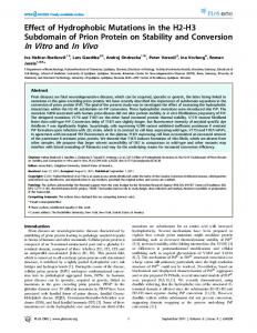
Introduction Sendai virus, a paramyxovirus, contains a non-segmented minus strand RNA genome of 15.3 kb, from which six mRNAs are transcribed (see Kolakofsky and Roux, 1987, for a recent review). Five of the mRNAs are monocistronic and code for only a single primary translation product. The P/C mRNA, however, is polycistronic and contains two overlapping ORFs which start near the 5' end of the chain (Giorgi et al., 1983). The longer P ORF begins with the 5' most proximal ATG at position 104 (ATG/104) and continues for 568 amino acids, yielding the P protein (Figure 1). The last 95 amino acids of this ORF are also expressed as a separate protein (X), which is independently initiated by ribosomes > 1500 nt from the 5' end (Curran and Kolakofsky, 1988b). The shorter C ORF is also responsible for a nested set of proteins, called C', C, Y1 and Y2, which are carboxy co-terminal, and which we refer to collectively as the C proteins. Site directed mutagenesis has shown that C' is initiated on a non-ATG codon, ACG/81, whereas C, Y1 and Y2 start on ATGs at positions 104, 183, and 201 respectively. When each of these codons is changed to noninitiators in a SP6 vector carrying the gene, only the expression of its cognate protein is ablated in in vitro translations (Curran and Kolakofsky, 1988a; Gupta and )IRL Press
Patwardhan, 1988; Patwardhan and Gupta, 1988). Including P (ATG/104), five proteins (C', P, C, Y1 and Y2) are therefore initiated near the 5' end and one (X) near the 3' end of a single mRNA. Only the P protein has so far been detected in mature virions; the others are considered to be non-structural proteins. Eukaryotic ribosomal initiation sites are generally the 5' most proximal ATGs, whose surrounding nt or context conforms to a weak but recognizable consensus, of which the most important elements in the model pre-pro-insulin mRNA, are a purine at position -3, followed by a G at +4 (Kozak, 1983, 1986). The initiation codons are thought to be chosen by a scanning mechanism, in which the 43S ribosomal pre-initiation complex, or initiation factors of the eIF4 series, or both, traverse the mRNA from its capped 5' end, and initiate translation when they reach the first such site. To account for unusual situations such as viral mRNAs with overlapping genes, Kozak (1986) proposed a modified or 'leaky' scanning model. Ribosomes could scan through the first ATG some of the time if it were in an unfavourable or poor context for initiation, and initiate on the next ATG, if it was in a good context. These terms refer to how well the key positions (-3 and +4) conform to the consensus. An expanded version of this model could then explain the first three sites of the P/C mRNA; C' starts on a non-ATG codon, P on an ATG in a poor context (a Py at position -3), and C on an ATG in a good context (an A at -3). Moreover, as the C/ATG is not in the context considered to be the most favourable (it contains a C rather than a G at +4), it is not impossible that Y1 and Y2 are also initiated by leaky scanning. Initiation of the Y proteins, on the other hand, could be due to a scanning independent mechanism, which takes place during X protein synthesis, at least in vitro (Curran and Kolakofsky, 1988b). Whether a particular initiation codon is in a favourable context or not, however, cannot always be predicted from the immediate context. Examples of overlapping genes are known in which the simple context of the initiation codons does not conform to the leaky scanning model (Bos et al., PROTEIN CODING REGIONS ON THE P/C
1028
104 p
5'
C' 81
Xba
I
1523 X
1808 .
."
C 114
,RNA
Y 1/Y2
183/281
3'
726
Fig. 1. Schematic representation of the P/C mRNA. The mRNA is shown as a horizontal line. Open boxes above the line refer to proteins coded by the P ORF; those below the line to those coded by the C ORF. The initiation sites are indicated by letters. Numbers refer to the position of the first base of the initiation or termination codons, or the unique XbaI site, relative to the 5' end of the natural mRNA.
521
J.Curran and D.Kolakofsky
1981; Shaw et al., 1983), and in some viral mRNAs the context can be changed in either direction without affecting relative translational efficiency (Munemitsu and Samuel, 1988; Williams and Lamb, 1989). In these cases, other sequences or structures of the mRNA must play a dominant role. This paper reports a study of how the Sendai virus C', P, C, and Y proteins are initiated, both in vitro and in vivo, in which the context of the natural initiation codons has not been altered.
Pulse
'x~~~~~~~~~~~~~~~~~~~~~~ ,.1,
(h.p,i)