logic provides a mathematical framework to capture the uncertainties associated with ..... mathematical operation in a non-fuzzy neuron may be defined in terms of ..... 105 (1999) 143-202. [24] D. Hugh, Physiology of eye, Churchill Livingstone,.
Visual Representation Model for Motion Direction Selectivity Xiaoxing Wang, Jesse S. Jin School of Information Technologies The University of Sydney, NSW 2006, Australia {wxx, jesse}@it.usyd.edu.au
Abstract In this paper we proposed a visual representation model for motion direction selectivity in the Superior Colliculus (SC) with a fuzzy neural networks system. The SC is a midbrain nucleus that contains a number of horizontally oriented cellular layers receiving and integrating information from the visual, auditory and somatosensory systems. The discharge of cells in the superficial layer of the SC is tightly linked to the direction of visual motion. The availability of a biologically plausible computational model of the SC may open the door for the modeling of cognitive interactions with visual sensory systems. We have simulated the model and the outputs are in line with those observed physiologically. Keywords: Visual representation, Fuzzy neural networks; Visual motion directional selectivity
1
Introduction
Over the last decade or so, significant advances have been made in two distinct technological areas: fuzzy logic and computational neutral networks. The theory of fuzzy logic provides a mathematical framework to capture the uncertainties associated with human cognitive processes, such as thinking and reasoning. On the other hand, the computational neural network paradigms have evolved in the process of understanding the incredible learning and adaptive features of neuronal mechanisms inherent in certain biological species. The integration of these two fields, fuzzy logic and neural networks; has given birth to an emerging technological field, the fuzzy neural networks [1]. To enable a visual system to deal with motion directional selectivity in a manner more like human brain, we incorporate the fuzzy theory into a biological neural network, the motion direction selectivity model in the Superior Colliculus.
Copyright © 2003, Australian Computer Society, Inc. This paper appeared at the Pan-Sydney Area Workshop on Visual Information Processing (VIP2002), Sydney, Australia. Conferences in Research and Practice in Information Technology, Vol. 22. J. S. Jin, P. Eades, D. D. Feng, H. Yan, Eds. Reproduction for academic, not-for profit purposes permitted provided this text is included.
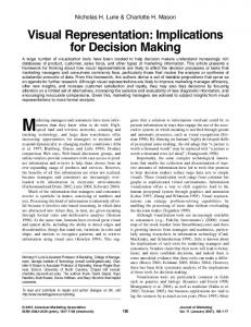
The Superior Colliculus (SC) is a midbrain nucleus, which contains a number of horizontally oriented cellular layers receiving and integrating information from the visual, auditory and somatosensory systems. A new model for SC has been proposed by Wang, Jin and Jabri [2] recently. The discharge of the cells in the superficial layer of the SC is tightly linked to the direction of the visual motion. The availability of a fuzzy biologically plausible computational model of the SC will open a door for the modeling of cognitive interactions with visual sensory systems. The computational process envisioned for neuro-fuzzy systems is as follows. It starts with the development of a fuzzy neuron based on the understanding of biological neuronal morphologies, followed by learning mechanisms. This leads to the following three steps in a fuzzy-neural computational process: (a) development of fuzzy neural models motivated by biological neurons, (b) models of synaptic connections which incorporates fuzziness into neural network, and (c) development of learning algorithms. We propose in this paper a visual representation model for orientation and direction selective motion detection in the SC. The overall model consists of three layers, shown in Figure 1, with the first representing retina ganglion cells receiving inputs from light receptors and the two subsequent layers representing cells in the SC. The first layer is subdivided into ON-cell and OFF-cell sub-layers. The models of the ON-cells include an off-response that plays an important role in the direction selectivity of our model. The second layer represents superficial layer cells of the SC. The weights of the synapses between ganglion cells and the second layer embed the characteristics of the preferred direction for visual motion detection. We have simulated this model and the outputs are in line with those observed physiologically.
Receptors
ON-cells
OFF-cells Ganglion cell layer
Fuzzy synaptic weights Superficial layer cells of SC
Directionally selective cells of SC
Figure 1. The structure of the directionally selectivity model. The first layer represents retina ganglion Figure 1. The structure of the direction selectivity model. cells receiving inputs from light receptors and is subdivided into ON-cell and OFF-cell sub-layers. The second layer represents the superficial-layer-cells of the SC. The weights of the synapses between ganglion cells and the second layer embed the characteristics of the preferred direction for visual motion detection. The third layer represents the directionally selective cell of the SC.
2 The biological model for motion directional selectivity in SC Since the discovery of direction selective ganglion cells in the retina of rabbits [3], many models have been proposed to explain the mechanisms of directional selectivity [4]. Barlow and Levick [5] originally proposed a scheme for directionally selective motion detection in terms of three interconnected general functional elements, and the details of the functions were later elaborated upon (e.g. Wyatt and Daw [6]). A variety of circuits have been proposed to account for these functions. Barlow and Levick [5] proposed two types of models for explaining the mechanism of direction selectivity. One is constructed with cells that have inhibitory connections to the neighbouring cells in the non-preferred direction. The other is constructed with cells that have excitatory connections to the neighbouring cells in the preferred direction. Our fuzzy neural network model for motion direction detection is different from previous models. There are three mainly mechanisms our model used. Firstly, a space-time mapping is established in the ganglion cell layer based on the overlapping properties of population of ganglion cells in the model. Secondly, we use offresponse properties of On-center cells to judge the direction of a moving light bar. Thirdly, the matched weight-vectors between ganglion cells and the direction selective cells embed the characteristics of the preferred direction for visual motion detection. When the activity vectors of the ganglion cells match with the weight
vectors, the direction selective cell fries. The overall proposed fuzzy network model is shown in Figure 1 and consists of three layers. The first layer represents retina ganglion cells receiving inputs from light receptors and is subdivided into ON-cell and OFFcell sub-layers. The models of the ON-cells include an off-response that plays an important role in the direction selectivity of our model. The second layer represents the superficial-layer-cells of the SC. The fuzzy weights of the synapses between ganglion cells and the second layer embed the characteristics of the preferred direction for visual motion detection. To explain our model we start with the response of a superior colliculus superficial direction sensitive cell: N
S = w! f =
wn ! f n
(1)
n=1
where S is the output activity of a cell; w is a fuzzy weight vector of the synapses between ganglion cells and the second layer; and f is the activity vector of the ganglion cells in response to a light stimulus. According to the property of the inner product, the value S reaches the maximum S * when f is directly proportional to w , that is, w = " ! f , where " is a constant. We define a threshold # = c ! S * , where c is a constant, and define that a direction sensitive cell fires when its output S is greater than # .
From an information processing point of view an individual neuron consists of the following three parts The receptive field of the ON-cell is coved by light bar
Motion direction
activity off-response
The receptive field of the OFF-cell is coved by light bar
t Figure 3. Off-response of an ON-cell. The off-response of an ON-cell is the activity of the ON-cell due to the light moving out of the surround area of its receptive field.
Figure 2 The activity of ON-cell (top) and OFF-cell (bottom). When light shines on the centre of the receptive field, the ON-cell responds vigorously (second graph on top), the OFF-cell is depressed (second graph on bottom). When the light shines on the surround, the opposite effect is achieved. Under diffuse illumination of both the centre and the surround, both cells respond weakly (the first graph on both top and bottom).
each associated with a particular mathematical function. (a) the synapses are a storage area of the past experience (the preferred direction) and receive information from the ganglion neurons; (b) the cell body, called soma, receives synaptic information and performs further processing of information; and (c) the neuron transmits information to other neurons through a single fiber called axon. The junction point of an axon with a dendrite is called synapse. Synapses provide long term memory to the past accumulated experience and this is storage for the knowledge base.
2.1 Ganglion cells layer The ganglion cells layer is subdivided into an ON-cells (ganglion cells with ON-center) and OFF-cells sublayers. In the model each ganglion cell is located on a mesh grid point with a two-dimensional index. ON-cells and OFF-cells with the same mesh grid index have their receptive field completely overlapping. When light shines on the center of the receptive field, the ON-cell responds vigorously, the OFF-cell is inactive. When the light shines on the surround, the opposite effect is achieved. Under diffuse illumination of both the center and the surround, both cells respond weakly [7]. Note that the ON-cell fires after the light moves out of its surround area.
The activity of a ganglion cell depends on the area of its receptive field covered by light (see Figure 2). Rodieck and Stone [8] proposed that a pair of Gaussian sensitivity surfaces could model the receptive fields of retinal ganglion cells. The case of the rear-edge of light bar moving across the receptive fields will be discussed in our model since the small parts of an edge of a complex light stimulus can be regarded as a light bar. The behavior of a center/surround ganglion cell when a light bar moves across its receptive fields can be described approximately by an integral of algebraic sum of both Gaussian sensitivity surfaces: u
F (u ) =
f1 $ f 2 d% = D
rs2 $ x 2
$
kc e
x2 + y 2 rc2
$
$ ks e
x2 +y2 rs2
dxdy
$ rs $ r 2 $ x 2 s
u & ( $ rs , rs )
(2)
Surface f1 represents the receptive field center and has a radius, rc, and sensitivity, kc. Surface f2 with opposite sign, represents the surround and has a radius, rs, and sensitivity, ks. The two surfaces are assumed to be circular, concentric and 180° out of phase with each other: Although more complex models have been developed, the original model continues to be useful in characterizing the responses of retinal cells [9].
2.2 Off response of ON-cell The off-response of an ON-cell is the activity of the ONcell due to the light moving out of the surround area of its receptive field [10] (see Figure 3). The off-response appears for a moving light stimulus. For example, if a light bar moves from left to right the off-response appears on the right side as it leaves the surround receptive field of the ON-cell, as shown in Figure 3. If the light bar moves in the opposite direction the off-response appears on the left side of the main activity of the ON-cell. Hence, the off-response can be utilized to determine the direction of the moving light bar. The biological basis of the off-response of an ON-cell is that the off-surround reacts to the disappearance of light in its area, which in turns excites the off-surround inputs
leading to the ganglion cell activation. That is, when a light bar covers the surround area, light hyperpolarizes the receptors, turning them off [7]. Once the light bar moves out of the surround area, the disappearance of the light breaks the antagonism balance and the ON-cell fires again in the dark. As this activity is a transient response of the ON-cell, the activity of the ON-cell decreases then ceases in a short time. The off-response activity can be described as follows: f ( x) =
ax b + cx 3
(3)
where a, b, and c are constants; x presents the position of the left edge of the moving bar.
2.3 Representation of the output of the ganglion cells h
Motion direction
t
' b
c
i
c d1 d2
Figure 4. ON-cell and the moving light bar. The centre of the ganglion cell is located at point (i , j).
There are three areas where an ON-cell is active when a light bar moves over its receptive field. The first area is located around the front edge of the light bar. The second area is located around the rear edge of the light bar. The third area is near the second area, in which the activity of the cell is evoked by its off-response. To determine the direction of the moving light bar we only use the second and third areas. The activity of the cells in the two areas is described by fon and foff-response respectively. Following from equations 2 and 3 we have j $ c $ h ! csc' $ (a + r)2 ) 2s when c + h ! csc' < j < c + h ! csc' + d1
fon (i, j) = exp($
f off $ response(i, j ) =
a1 ( j $ c $ d 2 $ h ! csc' $ 1) b1 + c1 ( j $ c $ d 2 $ h ! csc' )3
j $ c $ h ! csc ' $ (a + r ) 2 ) 2s when c + h ! csc ' + d 1 < j < c + h ! csc ' + d 2
f off (i , j ) = exp( $
(6)
3 Fuzziness of the biological neural network The concept of graded membership in fuzzy sets was introduced by Zadeh [11] in 1973. This notion of graded membership was introduced in order to provide a mathematical precision to information arising from our cognitive process. Fuzzy set definition. Let X be a space of points (or objects) with a generic element of X denoted by x. X is often referred to as the universe of discourse. A fuzzy set (class) A in X is characterized by a membership (characteristic) function µA(x) which associates with each point in X a real number in the interval [0, 1], with the value of µA(x) representing the grade of membership’ of x in A. Thus, the nearer the value of µA(x) to unity, the higher the grade of membership of x in A. A fuzzy set A is a subset of the universe of discourse X that admits partial membership. The fuzzy set A is defined as an ordered pair A={x, µA(x) }
j
b
Similarly, for OFF-cells we have
(7)
where x & X and 0 ) µA(x) ) 1, The membership function µA(x) describes the degree to which the object x belongs to the set A. µA(x) is also referred to as the characteristic function graded membership of x in A. If µA(x) = 0 then it is certain that x is not in A, and µA(x) = 1 then it is certain that x is in A. For x over 0< µA(x) b 1 if a ) b b / a if a > b
a' 2 b =
for T3=,.
The synapses of the cell in the superficial layer of the SC are a storage area of the synapse strength and receive information from the ganglion neurons. The activity S(i) of a cell in the superficial layer of the SC is:
won (i, j) ' f ont (i, j) +
j =1
+ woff (i, j) ' f offt (i, j) +
(12)
+ woff $response(i, j) ' f offt $response(i, j) where f t is the transpose of f. ' is the '-operator '3. In addition, we also have D (i ) =
1
if
0
otherwise
S (i ) $ # > 0
D (i ) = I
(13)
where # is a threshold. Here we assume the cells are arranged in an array. The output of the model, the direction selective cells (DS) is connected to a population
(14)
i =1
0
else
Note, for simplicity, we assume that only when all of the superficial-layer-cells are active that the directional selective cell is active. Actually, within a certain error range, the model could be set so that some predefined level of activity at the superficial-layer-cells would make the DS cells active. Using the activity of a DS cell we can determine the error between the direction of a moving light bar and the preferred direction according to: OD (' ) =
1
if
D (i ) = N (' ) < I
(15)
i =1
0
(11)
From the mathematical point of view, it may be concluded that the processing of information within a neuron involves two distinct mathematical operations namely (a) the synaptic operation: The strength (weight) of the synapse is a representation of the stored knowledge. The synaptic operation provides a weight to the neural inputs. Thus, the synaptic operation assigns a relative weight (significance) to each incoming signal according to the past experience (knowledge or memory) stored in the synapse. (b) the somatic operation: This provides aggregation, threshold and nonlinear activation to the synaptic inputs. If the weighted aggregation of the neural inputs exceeds a certain threshold, soma will produce an output signal.
J
if
I
where T3=,. indicates,. a b = max(0,a+b-1), + a,b&[0,1], and '3 corresponds to Lukasiewicz infinite valued logic.
S (i) =
1
for T2 = •
(10) a '3 b = min ( 1, 1 - a + b )
DS =
else
where OD(') is another directional selective cell tuned to angle ' with respect to the preferred direction. N(') is the activity of the preferred DS cell when a light bar moves along a direction that has an angle ' with its preferred direction.
4
Development of learning algorithms
The mechanism of the model is based on the specific property of the direction selective superior colliculus cells. It is known that the preferred directions of superior colliculus cells are not present in all species. For example, in the iguana [13], the hamster [14], and the mouse [13] the optimal direction is upward, or upward and nasal. In contrast, the optic direction in goldfish [15] and pigeon [16] responds best to stimuli moved from temporal to nasal along the horizontal meridian, and in the cat the opposite direction, nasal to temporal, is most frequently preferred [17]. Some researchers have suggested that the hunting and feeding habits of a species might determine the directions of movement that are most important to detect [18]. This interesting property of the directional selective collicular cells suggests that some part in the brain could encode the preferred direction. In our model, we propose that the synaptic weights between ganglion cells and the collicular cells encode the preferred direction. A population of direction sensitive cells converges on direction selective cells that are active when the light stimulus is moving in their preferred direction and orientation. The weighting and spatio-temporal aggregation operations performed by the synapses and soma, respectively, provide a similarity measure between the input vector A (new neural information) and the synaptic weight vector B (accumulated knowledge base). When a new input pattern, A’, that is significantly different from the previously learned patterns, is presented to the neural network, the similarity between this input and the existing knowledge base is small. As the neural network learns this new pattern A’, by changing the strength of the synaptic weight B’, the distance between the new information and accumulated knowledge decreases. In other words, to adapt the synapse weights (connection
strengths) leads the problem, if A implies B, and A’ is different from A, what B’ should be implied by A’? This question involves the approximate reasoning methods. Among the various approximate reasoning methods, the fuzzy reasoning with linguistic truth value (RLV) [19] has its interesting feature. It converts the fuzzy sets under reasoning into the so-called linguistic truth value fuzzy set, and then carries out the reasoning using fuzzy set. Unlike many other fuzzy sets, this RLV fuzzy set is independent of the antecedent and the conclusion sets. In most cases, Lukasiewicz infinite valued logic has been adopted [20]. It would be interesting to extend the method to the '-operator that is more generals, and includes the Lukasiewicz infinite valued logic. We now give an equivalent definition of the '-operator in (8). Let T is a continuous t-norm. + a, b & [0, 1 ], the following operator is equivalent to the '- operator given in (8). a'b =
1 sup{ x aTx = b }
if a ) b
(18)
where R is the fuzzy relation (A/ B) that depends on both the antecedent A and conclusion B. However, this expression is inconvenient for applications as compared with the crisp logic. In the crisp logic, the reasoning function is defined by the truth table consisting of 0 or 1 and is independent of the antecedent and the conclusion. To overcome this shortcoming, the method of fuzzy reasoning with linguistic truth value has been developed by J.F. Baldwin [21], M. Mizumoto [22], Y. Tsukamoto[20] and X. Wang [23]. Let us consider the fuzzy general modus ponens A’, A/ B B’, where A, A’ & F(U); B, B’ & F(V) and A(u), A’(u), B(v) and B’(v) are membership functions of A, A’, B and B’, respectively. The process of RLV is decomposed into the following three steps [21]: Step 4.1 Find A, given A and A’, +x & [ 0,1 ], A( x ) = sup A' ( u ) A( u )= x
(19)
A/ B = R = x ' y, + x, y & [ 0, 1 ]
+ v & V,
B’(v) = B ( B (v) )
(21)
To apply the approximate reasoning methods to adapt the synapse weights, an equivalent RLV method has been developed by which the conclusion set B’ can be obtained from A, A’ and B directly [23], that is +v & V ,
B' ( v ) = sup A' ( u )
(22)
u& B( v )
where B(v) = { u | A’(u) T A(u) = B(u)} is a nonfuzzy subset of U. The reason is as follow .
+v & V ,
sup
A' (u )
u&B ( v ) ={u A' ( u )TA ( u ) = B ( v )}
sup
A' (u )
x = A ( u ),u&B ( v ) ={u A '( u ) Tx = B ( v )}
(16) where { x | a T x = b } implies that x is a solution of equation a T x = b. The following properties are obvious. Let x’ = sup{ x | a T x = b }. If a - b, then x’ ) a ' b . a T x’ ) b, x’ > a ' b . a T x’ > b. (17) and + y & [ 0, 1 ], x ' y is non-increasing. The basic approximate reasoning using composition rule of inference can be expressed by the following equation:
Step 4.2 Determine B using A and A/ B, let
(20)
Step 4.3 Find B’ using B and B,
=
if a > b
B’=A’oR
+y&[0,1], B(y)=sup[A(x)|(A(x)Tx)=y}
sup
=
A' (u )
x = A ( u ),u&{u sup A '( u )Tx = B ( v )}
={
sup A' (u ) ( sup A' (u )Tx ) = B(v)} x = A( u )
x = A(u )
= { A( x) A( x)Tx = B(v)} = sup { A( x) , [ x'B(v)]} x&[ 0 ,1]
= B ( B(v)) = B' (v) (23) The Equation 23 looks rather complex. However, it can be given a simple explanation. B’ is a function of B. The relationship B between B and B’ can be described by the difference of A and A’. That is, if A then B, now the A changes into A’, so B changes into B’. B’ is effected by B and alter between A and A’.
5
Simulations
Figure 6. The top plot shows the weights between the DS cells and the ON cells that encode preferred direction 0°. The other 4 plots show the activities of 60 DS cells when a light bar moves along 0°, 0.88°, 10°, and 35° respectively.
angle is from 0 to 45 degree
10 5 0 0 0
50
10
20
30
40
50
100 degree
6 ang le= 40 4 deg ree 2
0
0
10
20
30 row
40
50
60
Figure 7. The relationship between ' and the response of direction selective cells.
We have simulated the complete model, the ON-cells and OFF-cells, the off-response of the ON-cell and the activity of superficial-layer-cells and DS cells using MATLAB. The viewing angle of the diameter of the center of the receptive field of a ganglion cell was about 2° (30 pixels), and viewing angle of the diameter of the surround was about 3.5° (48 pixels) [24]. The size, row by column, of the visual field (receptors) of the ganglion cells was 80x200 pixels. The ON-cell and OFF-cell sublayers have 60x120 cells each. The distance between two neighboring receptive fields of the ganglion cells was set to one pixel. For a given preferred direction a DS cell connects to 60 superficial-layer-cells, and every superficial-layer-cell connects to population of ON-cells and OFF-cells that align on one row on ON-cell sublayer and OFF-cell sublayers respectively. Therefore, every superficial-layer-cells detects the activities of 120 ONcells and 120 OFF-cells whose receptive fields align on one row. The top plot of Figure 6 shows the weights between the superficial-layer-cells and the ON-cells that encode preferred direction 0°. The curve on the left in that plot corresponds to the activity of the on center, and that on the right corresponds to the activity due to the offresponse of ON-cells. The other 4 plots of Figure 6 show the activities of 60 DS cells when a light bar moves along 0°, 0.88°, 10°, and 35° respectively. According to Equation 14, the DS cell is active only when the angle of the moving light bar is within ±0.88° of its preferred direction. The top plot of Figure 6 shows the relationship between ' and the response of a DS cell: the DS cell has maximum response to the moving light bar along its preferred direction, and its activity decays when the motion direction is further away from its preferred direction. Figure 7(b) is a cross section of the surface in figure 7(a).
6
Conclusions
operation provides a measure of similarity between the neural inputs and accumulated stored experience in synaptic weights, and the activation function provides a graded output to similarity measure. The model has three special features. Firstly, the off-response of ON-cells is used as a basic motion direction detection feature. Secondly, the synapses weights between ganglion cell and the direction selective cells encode the preferred direction. Thirdly, the learning algorithm of the neural network is based on a fuzzy reasoning with linguistic truth value. We simulate the behavior of the model using a simulator MATLAB. The results showed that the DS cell is active only when the angle of the moving light bar is within about 1° of its preferred direction. The DS cell does not show a response for opposite direction with preferred direction. The model we proposed is not unique to the superior colliculus but can be applied as a general model of direction/orientation selectivity based on the generic properties of ganglion cells.
7
References
[1] M. M. Gupta and D. H. Rao, On the principles of fuzzy neural networks, Fuzzy Set and Systems, 61 (1994) 1-18. [2] X. Wang, J Jin, and M. Jabri, (2002) Neural Network Models for the Gaze Shift System in the Superior Colliculus and Cerebellum, Neural Networks, Vol. 15, No.7, pp. 811-832. [3] H. B. Barlow and R. M. Hill, Selective sensitivity to direction of motion in ganglion cells of the rabbit's retina, Science Wash. DC, 139 (1964) 412-414. [4] N. M. Grzywacz And F.R. Amthor, Facilitation in ON-OFF directionally selective ganglion cells of the rabbit retina, Journal of Neurophysiology 69(6) (1993) 2188-2199. [5] H. B. Barlow and W. R. Levick, The mechanism of directionally selective units in rabbit's retina, J. Physiol. Lond. 178 (1965) 477-504. [6] H. J. Wyatt and N. W. Daw, Specific effects of neurotransmitter antagonists on ganglion cells in rabbit retina, Science, 191 (1976) 204-205. [7] D. H. Hubel, Eye, brain, and vision, New York : Scientific American Library, 1995. [8] R. W. Rodieck and J. Stone, Analysis of receptive fields of cat retinal ganglion cells in moving visual patterns, J. Neurophysiol. 20 (1965) 819-832. [9] G. E. Irvin, T. T. Norton, and V. A. Casagrande, Receptive field properties derived from spatial contrast sensitivity measurements of primate LGN cells, Invest. Ophthalmol. Vis. 27 (1986) 16-26. [10] B. Dreher and K. J. Sanderson, Receptive field analysis: Responses to moving visual contours by single lateral geniculate neurons in the cat, J. Physiol. 234 (1973) 95-118.
We have proposed in this paper a visual representation model for direction selectivity in the superior colliculus. It [11] L. A. Zadeh, Outline of a new approach to the is postulated that the fuzzy neural networks can learn by analysis of complex systems and decision processes, experience if their synaptic connections between the IEEE Trans. Systems Man Cybernet 3(1) (1973) 28external inputs and the dendritic inputs. The confluence 44.
[12] W. M. Wu, Fuzzy reasoning and fuzzy realational equations, Fuzzy Sets and Systems 20 (1987) 67-78. [13] U. C. Drager and D. H. Hubel, Responses to visual stimulation and relationship between visual, auditory and somatosensory inputs in mouse superior colliculus, J. Neurophysiology 38 (1975) 690-713. [14] B. E. Stein and J. P. Dixon, Properties of superior colliculus neurons in golden hamster, J. Comp. Neurol. 183 (1978) 269-284. [15] J. R. Cronly-Dillon, Units sensitive to direction of movement in goldfish optic tectum, Nature, 203 (1964) 214-215. [16] B. J. Frost and D. E. DiFranco, Motion characteristic of single units in the pigeon optic tectum. Vision Res. 1 (1976) 1220-1234. [17] P. Sterling and B. G. Wickelgren, Visual receptive fields in the superior colliculus of the cat, J. Neurophysiol. 32 (1969) 1-15. [18] D. J. Ingle, Evolutionary perspectives on the function of the optic rectum. Brain Behav. Evol. 8 (1973) 211-237. [19] D. Dubois and H. Prade, Fuzzy sets in approximate reasoning, Part 1: inference with possibility distributions, Fuzzy Sets and Systems 40 (1991) 143202. [20] Y.Tsukamoto, An approach to fuzzy reasoning method, Advances in Fuzzy Set Theory and Applications, North-Holland Publishiog Compang (1989) 137-149. [21] J. F. Baldwin, Fuzzy logic and fuzzy reasoning, Fuzzy Reasoning and its Applications , Academic Press (1981). [22] M. Mizumoto, Some Methods of Fuzzy Reasoning, Advances in Fuzzy Set Theory and Applications, North-Holland Publishing Company (1989) 117-186. [23] X. Wang and X. Hu, Approximate reasoning based on linguistic truth value with '-operator, Fuzzy Sets and Systems 105 (1999) 143-202. [24] D. Hugh, Physiology of eye, Churchill Livingstone, 1980.