Downloaded by US DEPT AGRCLT NATL AGRCLTL LBRY on September 25, 2015 | http://pubs.acs.org Publication Date (Web): June 18, 2015 | doi: 10.1021/bk-2015-1192.ch007
Chapter 7
Water-Insoluble Glucans from Sucrose via Glucansucrases. Factors Influencing Structures and Yields Gregory L. Côté* and Christopher D. Skory Renewable Product Technology Research Unit, National Center for Agricultural Utilization Research, Agricultural Research Service, United States Department of Agriculture, 1815 North University Street, Peoria, Illinois 61604 *E-mail:
[email protected].
Dextrans and related glucans produced from sucrose by lactic acid bacteria have been studied for many years and are used in numerous commercial applications and products. Most of these glucans are water-soluble, except for a few notable exceptions from cariogenic Streptococcus spp. and a very small number of Leuconostoc mesenteroides strains. The ability to produce water-insoluble glucans in situ may be of value in enhanced oil recovery, encapsulation technology and in the production of biocompatible films and fibers. There are several different ways in which these enzymes can be exploited to produce water-insoluble glucans with varying properties. In previous work, we found that modifying a single amino acid from a L. mesenteroides glucansucrase significantly altered the proportions of linkage types, with correlating changes in the physical properties of the polysaccharide. Here we present results of our studies on factors affecting the structures and yields of these insoluble glucans. For example, addition of soluble dextran or an enzyme that produces a soluble dextran can increase the yield of insoluble glucan. However, the insoluble glucan product contains a lower proportion of α(1→3) linkages. We also show that the yield is drastically affected
Not subject to U.S. Copyright. Published 2015 by American Chemical Society In Green Polymer Chemistry: Biobased Materials and Biocatalysis; Smith, et al.; ACS Symposium Series; American Chemical Society: Washington, DC, 2015.
by both substrate and enzyme concentrations. Studies using cloned enzymes indicate no significant differences between the insoluble-glucan producing enzymes from L. mesenteroides strains NRRL B-523 and B-1118.
Downloaded by US DEPT AGRCLT NATL AGRCLTL LBRY on September 25, 2015 | http://pubs.acs.org Publication Date (Web): June 18, 2015 | doi: 10.1021/bk-2015-1192.ch007
Introduction The type strain of Leuconostoc mesenteroides (NRRL B-1118; also known as ATCC 8293) has been known for many years to make an insoluble dextran (1, 2). Recently, this strain and the closely related strain, NRRL B-523, have received attention for their potential in microbially enhanced oil recovery (MEOR), in which the insoluble glucans are exploited for bioplugging (3–6). In particular, the ability to generate polymers in situ renders these systems particularly interesting (4, 7). We initially decided to examine the structures and biosynthesis of the extracellular glucans, since they are the components of interest in MEOR. However, applications in many other fields have also been considered. A number of glucansucrases involved in synthesizing various glucan linkages have been characterized from both Streptococcus and Leuconostoc (8, 9). Because of their importance in the etiology of dental caries, streptococcal glucansucrases have been studied in great detail (10, 11). Broadly speaking, there are two types of streptococcal glucansucrases: GTF-S (glucosyltransferase, soluble), which synthesize water-soluble glucans, and GTF-I (glucosyltransferase, insoluble), which synthesize water-insoluble glucans. The former glucans are predominantly α(1→6)-linked, with various amounts of branching through 1,3,6-substituted D-glucopyranosyl units, and are called dextrans. The latter glucans are water-insoluble due to the presence of long sequences of α(1→3)-linked D-glucopyranosyl units. Because of the structural and functional differences, the insoluble streptococcal glucans are often referred to as “mutan” (12). Many of the GTF enzymes from various species of Streptococcus have been sequenced, cloned, and characterized. Similar glucansucrases have also been identified in L. mesenteroides, a lactic-acid bacterium involved in fermentation of plant matter, including kimchi and sauerkraut, and a frequent contaminant of sugar mills and processing plants. L. mesenteroides is the source of glucansucrases for the commercial production of dextran, which is used for blood plasma extenders and related pharmaceutical applications, and cross-linked dextran gels for chromatographic supports (13). A few strains of L. mesenteroides are known to produce insoluble glucans similar to those produced by dental plaque streptococci (1, 2), but aside from one strain, namely NRRL B-523 (5), very little research has been carried out on the glucansucrases responsible for their synthesis. Three putative dextransucrases (Dsr) (GenBank accession YP_819207, YP_818338, and YP_819212) were previously identified (14) from the genome sequence of the L. mesenteroides type strain NRRL B-1118 (15) and found to have significant sequence identity to DsrS from strain B-512F (16), DsrE from strain B-1299 (17), and DsrP from L. mesenteroides IBT-PQ (18), respectively. DsrS from B-512F is known to catalyze the formation of predominantly 102 In Green Polymer Chemistry: Biobased Materials and Biocatalysis; Smith, et al.; ACS Symposium Series; American Chemical Society: Washington, DC, 2015.
α(1→6)-linked glucans and DsrE from B-1299 synthesizes a mixture of α(1→6) and α(1→2)-linkages. It has been reported that YP_818338 codes for a 2-domain glucansucrase, which is closely related to DsrE from L. mesenteroides B-1299 (19).
Downloaded by US DEPT AGRCLT NATL AGRCLTL LBRY on September 25, 2015 | http://pubs.acs.org Publication Date (Web): June 18, 2015 | doi: 10.1021/bk-2015-1192.ch007
Pure versus Mixed Enzyme Systems We previously cloned and characterized the glucansucrase YP_819212 (DsrI) and demonstracted that it produced a water-insoluble glucan (20). However, the glucan differed somewhat in its structure from that previously described for the native strain as it occurred in nature (2). In particular, methylation analysis indicated small but significant differences in the percentage of α(1→3)-linked and α(1→6)-linked D-glucopyranosyl linkages. The glucan from our cloned enzyme contained a higher percentage of α(1→3)-linkages compared to that produced by the parent strain (2). We hypothesized that the difference may be due to the fact that the glucan produced in the native system is the product of a mixture of enzymes. Here we present evidence to support this idea, using cloned DsrI and DsrS enzymes from L. mesenteroides NRRL B-1118 to synthesize similar co-polymers. Cloning and Expression of L. mesenteroides dsrS Gene The dsrS gene was PCR amplified from L. mesenteroides NRRL B-1118 DNA using primers DsrS P1 5′- CGC GAA CAG ATT GGA GGT gac agt agt gta cct gat gtg agt g -3′ and DsrS P2 5′- CGA ATT CGG ATC CTC TAG cag aaa gct tat gct gac aca g -3′, which were designed to amplify the entire coding region for protein YP819207 except for a 42 amino acid signal peptide sequence predicted by the SignalP 3.0 Server (21). The 4,502-bp amplicon was then cloned into an E. coli expression vector pE-SUMOpro Kan (LifeSensors, Malvern, PA), a plasmid that utilizes an N-terminal small ubiquitin-like modifier (SUMO) to enhance solubility and contains an N-terminal polyhistidine (His6) tag for purification. Sequence ligation independent cloning (22), SLIC, was utilized to assemble the dsrS amplicon with vector pE-SUMOpro Kan digested with BsaI. The sequence of resulting plasmid, pDsrS-SUMO, was confirmed before proceeding to protein isolation. Expression of the DsrI (20) and DsrS were performed using E. coli BL21(DE3) grown at 37°C in LB broth to an OD600=0.5. Cells were induced with isopropyl β-D-1-thiogalactopyranoside (IPTG) to a final concentration of 1 mM and then shifted to 15°C with continued shaking at 200 rpm for 18 hrs. Cells were harvested and suspended in a minimal volume of 20 mM NaH2PO4 (pH 8.0). Benzonase nuclease and phenylmethanesulfonylfluoride (PMSF), a serine protease inhibitor, were then added to a final concentration of 25 units/ml and 1.25 mM, respectively, before lysing cells with a French press at an internal pressure of 16,000 psi (110,000 kPa). After centrifugation, the cell-free supernate was adjusted to 500 mM NaH2PO4 (pH 8.0) and 300 mM NaCl. Protein was bound to PrepEase nickel resin (Affymetrix, Santa Clara, CA) by gently mixing 1 103 In Green Polymer Chemistry: Biobased Materials and Biocatalysis; Smith, et al.; ACS Symposium Series; American Chemical Society: Washington, DC, 2015.
Downloaded by US DEPT AGRCLT NATL AGRCLTL LBRY on September 25, 2015 | http://pubs.acs.org Publication Date (Web): June 18, 2015 | doi: 10.1021/bk-2015-1192.ch007
g resin per gram of original wet cell pellet for 15 min. The suspension was poured onto a column with a 50µm filter frit, washed twice with four volumes of buffered saline (50 mM NaH2PO4 pH8.0, 300 mM NaCl) and eluted with three volumes of elution buffer (50 mM NaH2PO4 pH8.0, 300 mM NaCl, 250 mM imidazole). Fractions containing glucansucrase protein were combined and diluted with buffered saline to reduce imidazole concentration to below 150 mM. The SUMO fusion was cleaved according to manufacturer’s recommendations using SUMO Protease 1, which recognizes the tertiary structure of SUMO and does not cleave within the protein of interest. Protease cleavage and removal of the SUMO fusion from the purified protein should yield DsrI and DsrS protein identical to that of the mature enzymes secreted by L. mesenteroides NRRL B-1118. Uncleaved glucansucrase protein and released N-terminal SUMO modifier, both of which contain the His6, were removed by passing the digested protein back over the PrepEase resin. Enzymes thus prepared were electrophoretically pure. The buffer for the recovered protein was finally exchanged with 20 mM sodium acetate (pH 5.5) containing 2mM CaCl2 with 0.01% sodium azide as a preservative. Analytical Enzyme assays were based on the incorporation of 14C-labelled glucose from 14C-(U)-labeled sucrose (PerkinElmer-NEN, Waltham, MA) into alcohol-insoluble polysaccharide, according to a modification of the method described by Germaine et al. (23). In a typical example, 30µL of 0.3M 14C-sucrose in acetate buffer was incubated with 60µL of enzyme solution at 30°C. At timed intervals, 15 µL aliquots were withdrawn and absorbed onto 1.5cm squares of Whatman 3MM chromatography/filter paper. The squares were immediately dropped into a beaker containing approximately 150-200 mL of stirred methanol. A metal screen was used to protect the paper squares from maceration by the stir bar. After washing with three changes of methanol for ten minutes per wash, they were dried under a heat lamp and counted for 14C content in Ecolume cocktails (MPBio, Solon, OH) using a Beckman-Coulter (Brea, CA) LSC-6500 liquid scintillation counter. An enzyme unit is defined here as the amount of enzyme activity that incorporates one µmole of glucose into glucan in one minute. For water-soluble glucan analysis, 1 mL of enzyme solution (0.8 U/mL) was mixed with 15 mL of 1M sucrose in acetate buffer, and diluted to 50 mL. After standing overnight at room temperature (22°C), the dextran was collected by mixing with an equal volume of ethanol, followed by centrifugation. The dextran was redissolved and reprecipitated twice, then dissolved in water and freeze-dried. Structural analyses of the glucans were carried out by methylation, hydrolysis, and gas-liquid chromatography of the peracetylated aldononitrile derivatives as described by Seymour et al. (24). How Mixed Enzyme Systems Affect Product Structure The recombinant enzyme DsrS produced water-soluble glucan from sucrose. Methylation analysis results from the soluble glucan product from DsrS are shown in Table 1. The peracetylated aldononitrile (PAAN) derivatives of 2,3,4-tri-O104 In Green Polymer Chemistry: Biobased Materials and Biocatalysis; Smith, et al.; ACS Symposium Series; American Chemical Society: Washington, DC, 2015.
Downloaded by US DEPT AGRCLT NATL AGRCLTL LBRY on September 25, 2015 | http://pubs.acs.org Publication Date (Web): June 18, 2015 | doi: 10.1021/bk-2015-1192.ch007
methyl glucose constitute 73% of the dextran, indicating a predominantly α(1→6)linked glucan. Branching occurs through 3,6-disubstituted residues as indicated by the presence of 11 mole percent of 2,4-di-O-methyl PAAN derivative. It is likely that the insoluble glucan occurring in cultures of L. mesenteroides NRRL B-1118 grown on sucrose is the product of two or more glucansucrases acting together to produce a hybrid product. Such a product could arise from the action of DsrI acting via acceptor reactions on soluble dextran formed by DsrS, to produce a glucan with sequences of 3-linked α-D-glucopyranosyl units attached to a backbone of 6-linked α-D-glucopyranosyl units. The data shown in Table 1 support such a hybrid type of structure. A similar mechanism has been proposed for insoluble glucans formed by mixed-enzyme systems of GTF-I and GTF-S from Streptococcus spp. (25–28) and subsequently demonstrated by NMR spectroscopy of 13C-labelled products (29). Although the hybrid polymer has a structure that yields analytical results that fall between those of the soluble dextran and the insoluble glucan, it is not a precise 50/50 average of the two individual glucans, as seen in Table 1. It is likely that the proportions of linear 3-linked α-D-glucopyranosyl and 6-linked α-D-glucopyranosyl units may vary considerably, as well as the extent of branching, depending on concentrations of the two enzymes and their individual products, among other factors. Indeed, the two analyses reported in the literature for native insoluble glucan differ significantly (2, 30). It known that the degree of branching in highly branched dextrans from both Leuconostoc and Streptococcus can vary considerably, depending on conditions of biosynthesis (31, 32). This work demonstrates that the products of multi-enzyme glucansucrase systems are hybrid copolymers, and suggests approaches for customized glucan synthesis using multi-enzyme systems or polysaccharide acceptors.
Table 1. Methylation Analysis Results of NRRL B-1118 Glucans Produced by Various Enzyme Preparations Methylated PAAN product
Mole percent DsrS
DsrIa
Mixtureb
Nativec
Natived
2,3,4,6-tetra-O-Me Glc
16
15
13
4
11
2,3,4-tri-O-Me Glc
73
29
71
69
66
2,4,6-tri-O-Me Glc
ND
44
10
22
13
2,4-di-O-Me Glc
11
9
6
5
10
2,6-di-O-Me Glc
ND
4
ND
ND
ND
From Côté and Skory (20). b Insoluble glucan synthesized by a mixture of equal units of DsrI and DsrS. c From Pearce et al. (2). d From Slodki et al. (30). ND = none detected. a
105 In Green Polymer Chemistry: Biobased Materials and Biocatalysis; Smith, et al.; ACS Symposium Series; American Chemical Society: Washington, DC, 2015.
Downloaded by US DEPT AGRCLT NATL AGRCLTL LBRY on September 25, 2015 | http://pubs.acs.org Publication Date (Web): June 18, 2015 | doi: 10.1021/bk-2015-1192.ch007
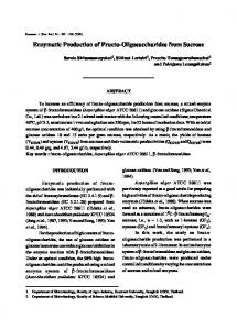
Influence of Substrate and Enzyme Concentrations Variable yields of insoluble glucan with DsrI in some of our early experiments led us to investigate the roles of enzyme and sucrose concentrations on the structure and yield of water-insoluble glucan. To measure the effect of enzyme and sucrose concentration, a series of reactions were carried out containing 7.5 mL of buffer with concentrations of sucrose ranging from 67mM to 933 mM and enzyme ranging from 0.017 U/mL to 1.667 U/mL. Reactions were monitored by TLC, and when all of the sucrose had been consumed, the insoluble glucan product was isolated, dried, and weighed.
Figure 1. Absolute yields of water-insoluble glucan as a function of sucrose and enzyme (DsrI) concentration.
Figure 1 shows the effect of sucrose concentration and enzyme activity on the amount of glucan produced, expressed as weight of glucan. From these data, it is evident that the optimum sucrose concentration for maximum yield depends partially on the amount of enzyme used. At higher levels of enzyme activity, the optimum sucrose concentration is approximately 0.3 – 0.4 M. At lower enzyme levels, the optimum occurs at lower sucrose concentrations. Regardless of sucrose concentration, the yield is directly proportional to the amount of enzyme present. If 106 In Green Polymer Chemistry: Biobased Materials and Biocatalysis; Smith, et al.; ACS Symposium Series; American Chemical Society: Washington, DC, 2015.
Downloaded by US DEPT AGRCLT NATL AGRCLTL LBRY on September 25, 2015 | http://pubs.acs.org Publication Date (Web): June 18, 2015 | doi: 10.1021/bk-2015-1192.ch007
one calculates the percent of theoretical yield from sucrose rather than the absolute amount of glucan formed, Figure 2 shows that the optimum yield occurs at high enzyme levels and low sucrose concentrations. Furthermore, the maximum yields do not exceed 25% of the theoretical maximum yield from sucrose, despite the fact that all of the sucrose was consumed in the reactions.
Figure 2. Relative yield of water-insoluble glucan as a function of sucrose and enzyme (DsrI) concentration.
These results can be interpreted in light of the fact that the insoluble glucan is not the sole product of the enzyme. We have examined the reaction products by TLC, and find that two phenomena occur that diminish the yield of glucan. Firstly, fructose released during the glucosyl transfer reaction participates in what are known as acceptor reactions (33). The major product of glucosyl transfer to fructose is leucrose (34), with isomaltulose being a minor product (35). Furthermore, as the concentration of glucan containing a high proportion of α(1→3) linkages increases, it eventually reaches a saturation point, whereupon it precipitates from the aqueous solution phase. The precipitated glucan may bring associated enzyme out of solution with it, resulting in what is essentially an immobilized enzyme surrounded by gel-like glucan. At that point, the acceptor 107 In Green Polymer Chemistry: Biobased Materials and Biocatalysis; Smith, et al.; ACS Symposium Series; American Chemical Society: Washington, DC, 2015.
Downloaded by US DEPT AGRCLT NATL AGRCLTL LBRY on September 25, 2015 | http://pubs.acs.org Publication Date (Web): June 18, 2015 | doi: 10.1021/bk-2015-1192.ch007
reactions would become more favorable, since the small oligosaccharide products can freely diffuse out of the gel matrix. Thus, the yield of water-insoluble glucan is limited by the fact that it is insoluble and closely associated with the enzyme. We have been able to overcome this drawback to some extent by performing the reactions in dialysis tubing. An enzyme solution is placed in a length of dialysis tubing, which is immersed in a vessel of buffered sucrose solution. This prevents the fructose concentration from building up as the reaction proceeds, thus diminishing the relative extent of acceptor reactions. A similar approach has been described by O’Brien and Payne (36). Another approach to increasing yields is to add a small amount of water-soluble dextran. Figure 3 shows results of an experiment in which 4 units of DsrI was incubated with 684 mg of sucrose in 6 mL of acetate buffer until all sucrose was consumed. Various amounts of water-soluble commercial dextran (Mw=2,000,000) were added, and the yields of water-insoluble glucan were determined as above. The structures of the products were analyzed by measuring their susceptibility to endodextranase hydrolysis, as previously described (20).
Figure 3. Increase in yield of water-insoluble glucan produced by DsrI upon addition of water-soluble dextran. Filled circles: Yield of insoluble glucan. Open circles: Percent remaining insoluble after dextranase digestion. 108 In Green Polymer Chemistry: Biobased Materials and Biocatalysis; Smith, et al.; ACS Symposium Series; American Chemical Society: Washington, DC, 2015.
Downloaded by US DEPT AGRCLT NATL AGRCLTL LBRY on September 25, 2015 | http://pubs.acs.org Publication Date (Web): June 18, 2015 | doi: 10.1021/bk-2015-1192.ch007
The yield of water-insoluble glucan was increased from 82 mg to 99 mg (17 mg, for a 20% increase) by addition of as little as 5 mg of soluble dextran. Such increases in yields have been exploited for streptococcal glucansucrases, and have been interpreted as a “priming” mechanism for the enzyme (37). Although the actual mechanism of so-called “primer reactions” is not well understood, it is likely that the actual mechanism is a combination of allosteric activation and graft copolymer formation, since the so-called “primers” are not essential for glucan formation (38–40). Evidence for this comes from the observation that the glucan formed in the presence of dextran is more susceptible to hydrolysis and solubilization by endodextranase, indicating a higher percentage of α1→6-linkages (Figure 3). This could arise from formation of a graft, or hybrid, polymer structure, as discussed in the preceding section describing dextrans formed by enzyme mixtures. Thus, we can conclude that the presence of water-soluble dextran, either added exogenously or synthesized endogenously by dextransucrase, can influence both the yields and structures of insoluble glucans formed by DsrI. The addition of dextran may increase the yield of water-insoluble glucan, but the resulting glucan contains a lower proportion of α(1→3)-linkages.
Influence of Enzyme Source Jeanes and her colleagues (1) showed that the bacterial source strain has a marked influence on the structure of the dextran produced. Pearce et al. (2) demonstrated that the same holds true for water-insoluble glucans. To determine if this was the case when pure, cloned enzymes were used, we compared the water-insoluble glucans produced by DsrI from strains NRRL B-523 and NRRL B-1118. These two enzymes have a 94.8% amino acid sequence homology, and are essentially identical in the region surrounding the active site. The insoluble-glucan producing glucansucrase DsrI from L. mesenteroides NRRL B-523 was cloned and expressed in E. coli as previously described for strain NRRL B-1118 (20). The isolated SUMO-His6-tagged enzymes were either used without further treatment, or were proteolyzed to remove the SUMO-His6 tag. The tagged and the proteolyzed enzymes from both strains were used to synthesize water-insoluble glucan from sucrose under identical conditions. One unit of enzyme was incubated with 684 mg of sucrose in 6 mL of buffer, and the reactions were mixed by gentle rotation until TLC indicated all sucrose had been consumed. The insoluble glucan was isolated by centrifugation, washed and dried, then weighed. Yields were not significantly different between the strains, nor were they different between the enzymes with or without the SUMO-His6 tag. The glucans were also analyzed by dextranase digestion, and by methylation and NMR as previously described (20). All three methods indicated no statistically significant differences among the four different enzyme preparations in the relative proportions of α(1→3) and α(1→6)-linked glucosyl units present in the insoluble glucan products. 109 In Green Polymer Chemistry: Biobased Materials and Biocatalysis; Smith, et al.; ACS Symposium Series; American Chemical Society: Washington, DC, 2015.
Downloaded by US DEPT AGRCLT NATL AGRCLTL LBRY on September 25, 2015 | http://pubs.acs.org Publication Date (Web): June 18, 2015 | doi: 10.1021/bk-2015-1192.ch007
In their 1990 paper, Pearce et al. (2) presented data showing small differences between the insoluble glucans produced by strains NRRL B-523 and B-1118. No estimates of error were given, so it is not known if the differences were significant. However, Slodki et al. (30) in an earlier paper presented data showing a much larger difference in the percentage of α(1→3) linkages between the same two strains. We can only provide conjecture as to the reasons for this discrepancy, but it may be due to batchwise variation in the glucans resulting from variations in the relative activities of DsrI and DsrS enzymes. It should be noted that the products they examined were from crude cultures of the wild-type bacteria, which contain multiple glucansucrases. Based on the similarity of the product glucans, and on the high degree of sequence homology, we can conclude that the purified or cloned insoluble-glucan producing enzymes from L. mesenteroides NRRL B-523 and B-1118 are essentially interchangeable.
Summary and Outlook This work represents a first step toward a better understanding of the factors influencing the enzymatic synthesis of water-insoluble glucans from sucrose. The cloning of glucansucrases and expression as secreted enzymes will contribute significantly to the eventual commercialization of these glucans, but much work needs to be done to increase the yields from sucrose, either by reaction optimization or by enzyme engineering. Co-production of prebiotic or functional oligosaccharides in addition to the insoluble glucans could represent one possible approach. We anticipate that a wide variety of products could be developed using the materials described herein, including functional food and feed ingredients, wound dressings, absorbent polymers, biocomposites, bioplastics, biodegradable films and fibers, and adhesives, to mention just a few.
Acknowledgments We thank Suzanne Unser and Kristina Glenzinski for their many hours of technical assistance, and Dr. Karl Vermillion for his expert advice and assistance pertaining to NMR spectroscopy. We also thank Drs. Joseph Rich and Ryan Cormier for valuable advice and support. This work was supported and carried out by the U.S. Department of Agriculture. Mention of trade names or commercial products in this publication is solely for the purpose of providing specific information and does not imply recommendation or endorsement by the U.S. Department of Agriculture. USDA is an equal opportunity provider and employer.
References 1.
Jeanes, A.; Haynes, W. C.; Wilham, C. A.; Rankin, J. C.; Melvin, E. H.; Austin, M. J.; Cluskey, J. E.; Fisher, B. E.; Tsuchiya, H. M.; Rist, C. E. J. Am. Chem. Soc. 1954, 76, 5041–5052. 110 In Green Polymer Chemistry: Biobased Materials and Biocatalysis; Smith, et al.; ACS Symposium Series; American Chemical Society: Washington, DC, 2015.
2. 3. 4.
Downloaded by US DEPT AGRCLT NATL AGRCLTL LBRY on September 25, 2015 | http://pubs.acs.org Publication Date (Web): June 18, 2015 | doi: 10.1021/bk-2015-1192.ch007
5. 6. 7. 8. 9. 10. 11. 12. 13.
14. 15.
16. 17.
18. 19.
20. 21.
Pearce, B. J.; Walker, G. J.; Slodki, M. E.; Schuerch, C. Carbohydr. Res. 1990, 203, 229–246. Wu, Y.; Surasani, V. K.; Li, L.; Hubbard, S. S. Geophysics 2014, 79, E61–E73. Surasani, V. K.; Li, L.; Ajo-Franklin, J. B.; Hubbard, C.; Hubbard, S. S.; Wu, Y. Energy Fuels 2013, 27, 6538–6551. Padmanabhan, P. A.; Kim, D.-S. Carbohydr. Res. 2002, 337, 1529–1533. Padmanabhan, P. A.; Kim, D.-S.; Pak, D.; Sim, S. J. Carbohydr. Polym. 2003, 53, 459–468. Vilcaez, J.; Li, L.; Wu, D.; Hubbard, S. S. Geomicrobiol. J. 2013, 30, 813–828. Monchois, V.; Willemot, R.-M.; Monsan, P. FEMS Microbiol. Rev. 1999, 23, 131–151. van Hijum, S. A.; Kralj, S.; Ozimek, L. K.; Dijkhuizen, L.; van Geel-Schutten, I. G. Microbiol. Mol. Biol. Rev. 2006, 70, 157–76. Montville, T. J.; Cooney, C. L.; Sinskey, A. J. Adv. Appl. Microbiol. 1978, 24, 55–84. Hamada, S.; Slade, H. D. Microbiol. Rev. 1980, 44, 331–384. Guggenheim, B.; Newbrun, E. Helv. Odontol. Acta 1969, 13, 84–97. Leathers, T. D. In Polysaccharides I. Polysaccharides from Prokaryotes; Vandamme, E. J., DeBaets, S., Steinbüchel, A., Eds.; Biopolymers, Wiley-VCH: Weiheim, Germany, 2002; Vol. 5, pp 299−321. Olvera, C.; Centeno-Leija, S.; Lopez-Munguía, A. Antonie van Leeuwenhoek 2007, 92, 11–20. Makarova, K.; Slesarev, A.; Wolf, Y.; Sorokin, A.; Mirkin, B.; Koonin, E.; Pavlov, A.; Pavlova, N.; Karamychev, V.; Polouchine, N.; Shakhova, V.; Grigoriev, I.; Lou, Y.; Rohksar, D.; Lucas, S.; Huang, K.; Goodstein, D. M.; Hawkins, T.; Plengvidhya, V.; Welker, D.; Hughes, J.; Goh, Y.; Benson, A.; Baldwin, K.; Lee, J. H.; Diaz-Muniz, I.; Dosti, B.; Smeianov, V.; Wechter, W.; Barabote, R.; Lorca, G.; Altermann, E.; Barrangou, R.; Ganesan, B.; Xie, Y.; Rawsthorne, H.; Tamir, D.; Parker, C.; Breidt, F.; Broadbent, J.; Hutkins, R.; O’Sullivan, D.; Steele, J.; Unlu, G.; Saier, M.; Klaenhammer, T.; Richardson, P.; Kozyavkin, S.; Weimer, B.; Mills, D. Proc. Natl. Acad. Sci. U.S.A. 2006, 103, 15611–15616. Monchois, V.; Remaud-Simeon, M.; Russell, R. R. B.; Monsan, P.; Willemot, R.-M. Appl. Microbiol. Biotechnol. 1997, 48, 465–472. Bozonnet, S.; Dols-Laffargue, M.; Fabre, E.; Pizzut, S.; RemaudSimeon, M.; Monsan, P.; Willemot, R. M. J. Bacteriol. 2002, 184, 5753–5761. Olvera, C.; Fernandez-Vazquez, J. L.; Ledezma-Candanoza, L.; LopezMunguía, A. Microbiology 2007, 153, 3994–4002. Kralj, S.; Grijpstra, P.; Van Leeuwen, S. S.; Kamerling, J. P.; Dijkhuizen, L. Proceedings of the Eighth Carbohydrate Bioengineering Meeting, Naples, Italy. May 10-13, 2009; Abstract number P49. Côté, G. L.; Skory, C. D. Appl. Microbiol. Biotechnol. 2012, 93, 2387–2394. Bendtsen, J. D.; Nielsen, H.; von Heijne, G.; Brunak, S. J. Mol. Biol. 2004, 340, 783–95. 111 In Green Polymer Chemistry: Biobased Materials and Biocatalysis; Smith, et al.; ACS Symposium Series; American Chemical Society: Washington, DC, 2015.
Downloaded by US DEPT AGRCLT NATL AGRCLTL LBRY on September 25, 2015 | http://pubs.acs.org Publication Date (Web): June 18, 2015 | doi: 10.1021/bk-2015-1192.ch007
22. Li, M. Z.; Elledge, S. J. Nat. Methods 2007, 4, 251–256. 23. Germaine, G. R.; Schachtele, C. F.; Chludzinski, A. M. J. Dent. Res. 1974, 53, 1355–1360. 24. Seymour, F. R.; Slodki, M. E.; Plattner, R. D.; Jeanes, A. Carbohydr. Res. 1977, 53, 153–166. 25. Fukui, K.; Moriyama, T.; Miyake, Y.; Mizutani, K.; Tanaka, O. Infect. Immun. 1982, 37, 1–9. 26. Hanada, N.; Takehara, T.; Itoh, M.; Saeki, E. FEMS Microbiol. Lett. 1986, 36, 173–175. 27. Walker, G. J.; Schuerch, C. Carbohydr. Res. 1986, 14, 259–270. 28. Takehara, T.; Ansai, T.; Yamashita, Y.; Itoh-Andoh, M.; Kunimori, A. Oral Microbiol. Immunol. 1992, 7, 155–158. 29. Mukasa, H.; Tsumori, H.; Shimamura, A. Carbohydr. Res. 2001, 333, 19–26. 30. Slodki, M. E.; England, R. E.; Plattner, R. D.; Dick, W. E. Carbohydr. Res. 1986, 156, 199–206. 31. Côté, G. L.; Robyt, J. F. Carbohydr. Res. 1983, 119, 141–156. 32. Côté, G. L.; Robyt, J. F. Carbohydr. Res. 1984, 127, 95–107. 33. Koepsell, H. J.; Tsuchiya, H. M.; Hellman, N. N.; Kazenko, A.; Hoffman, C. A.; Sharpe, E. S.; Jackson, R. W. J. Biol. Chem. 1953, 200, 793–801. 34. Stodola, F. H.; Koepsell, H. J.; Sharpe, E. S. J. Am. Chem. Soc. 1952, 74, 3202–3203. 35. Sharpe, E. S.; Stodola, F. H.; Koepsell, H. J. J. Org. Chem. 1960, 25, 1062–1063. 36. O’Brien, J. P.; Payne, M. S. U.S. Patent Appl. 2013/0244288A1, 2013. 37. Payne, M. S.; Brun, Y.; He, H.; Scholz, T. U.S. Patent Appl. 2014/ 0087431A1, 2014. 38. Robyt, J. F.; Corrigan, A. J. Arch. Biochem. Biophys. 1977, 183, 726–731. 39. Robyt, J. F.; Martin, P. J. Carbohydr. Res. 1983, 113, 301–315. 40. Robyt, J. F.; Yoon, S.-H.; Mukerjea, R. Carbohydr. Res. 2008, 343, 3039–3048.
112 In Green Polymer Chemistry: Biobased Materials and Biocatalysis; Smith, et al.; ACS Symposium Series; American Chemical Society: Washington, DC, 2015.