Scientific Correspondence
When Simpler Is Better. Unicellular Green Algae for Discovering New Genes and Functions in Carbohydrate Metabolism Glenn R. Hicks*, Catherine M. Hironaka, David Dauvillee, Roel P. Funke, Christophe D’Hulst, Sabine Waffenschmidt, and Steven G. Ball Plant Genetics, Exelixis, Inc., 170 Harbor Way, P.O. Box 511, South San Francisco, California 94083–0511 (G.R.H., C.M.H., R.F.); Laboratoire de Chimie Biologique, Unite Mixte de Recherche du Centre National de la Recherche Scientifique Number 111, Universite des Sciences et Technologies de Lille Flandres-Artois, 59655 Villeneuve d’Ascq cedex, France (D.D., C.D., S.G.B.); and Institut fur Biochemie, Universitat zu Koln, Greinstr. 4, 50939 Koln, Germany D–50931 (S.W.) Of all algae, those in the division chlorophyta (green algae) display the closest relationship to the vascular plants. Chlorophytes harbor a chloroplast that is considered to have originated from a single endosymbiotic event and contain the same types of photosynthetic pigments as land plants. At variance with other divisions of algae, starch is produced within chlorophyte plastids and displays a structure very similar to that described for vascular plants. Considerable variation is found in the organization and composition of chlorophyte cell walls. Some are characterized by the presence of a simple glycoprotein wall, whereas others synthesize elaborate walls with a polysaccharide composition like that of land plants. Algae often contain pulsatile vacuoles that in some cases allow growth in the absence of a normal cell wall structure, a useful feature for studying wall biology. Because of the presence of plastids and plant-like cell walls, unicellular chlorophytes may be considered true plant-like eukaryotic microorganisms. Their microbial nature permits the use of extremely powerful genetic techniques akin to those in yeast (Saccharomyces cerevisiae) for the dissection of plant pathways. In this scientific correspondence, we will emphasize the potential of using unicellular chlorophytes to understand plant pathways, including polysaccharide and cell wall metabolism. We hope to encourage plant biologists to consider these species because of the potential for rapid progress in understanding many basic plant pathways. Among unicellular green algae, Chlamydomonas reinhardtii is by far the most studied system. Many recent reviews describing the speed and ease of C. reinhardtii genetics and molecular biology have appeared. In fact, within this issue of Plant Physiology, several detailed research articles depict the use of C. reinhardtii to study chloroplast biogenesis and cell motility. This alga grows rapidly in defined medium * Corresponding author; e-mail
[email protected]; fax 650 – 837– 8122. www.plantphysiol.org/cgi/doi/10.1104/pp.010821. 1334
both in liquid and on agar, and its sexual cycle can be as easily controlled as that of yeast. Single colonies grow within 5 d and crosses can be analyzed in less than a month. In addition, the nuclear genome can be efficiently transformed and gene replacement via homologous recombination in the chloroplast genome is a routine method. Unlike land plants, C. reinhardtii has the remarkable ability to dispense with photosynthesis and to use acetate as a carbon source; this has allowed the isolation of mutations in both nuclear and plastid genes that affect all possible aspects of chloroplast biogenesis. Similarly, the ease with which cell motility mutants can be isolated has allowed a thorough description of the structure, assembly, and function of flagella. These major achievements have overshadowed many aspects of plant metabolism for which C. reinhardtii could be an extremely useful model system such as commercially valuable pathways leading to carbohydrates, carotenoids, lipids, and secondary products, as well as other essential pathways. UNRAVELING STARCH METABOLISM
An excellent example of the power of unicellular algae is the use of C. reinhardtii to understand starch metabolism, which is resulting in the discovery of new functions even within enzymes that are well characterized. Such knowledge can guide rational efforts to manipulate starch composition for practical purposes (Slattery et al., 2000). Starch is an extremely valuable polymer both nutritionally and as an industrial raw material. It is stored in photosynthetically active leaf chloroplasts as transient starch and in seeds or tubers of economically important crops such as maize (Zea mays), rice (Oryza sativa), and potatoes (Solanum tuberosum) as storage starch. Starch is an insoluble crystalline granule composed of two polysaccharide fractions. Amylopectin is the predominant high-Mr polymer in storage granules and contains an abundance of branched glucans having ␣-1,6 linkages. Amylose is a lesser component of the gran-
Plant Physiology, December 2001, Vol. 127, pp. 1334–1338, www.plantphysiol.org © 2001 American Society of Plant Biologists
Scientific Correspondence
ule having relatively few branch points (Ball et al., 1998; Buleon et al., 1998). Storage starch has been studied extensively in higher plants particularly in maize, rice, pea (Pisum sativum), and potato, whereas mutants in leaf starch have been identified and studied in Arabidopsis (for example, see Casper, 1994; Zeeman et al., 1998; Yu et al., 2001). Such efforts have resulted in a relatively good description of the major biosynthetic enzymes in the pathway. It is unfortunate that this has also led to the impression among many scientists that little remains to be understood. Quite to the contrary, however, many aspects of starch metabolism remain poorly understood, including granule nucleation and assembly, regulation of synthesis and turnover, mechanisms of starch modification, and the contributions of the many enzyme isoforms to starch composition and crystalline structure. C. reinhardtii produces starch granules that are similar to those in other plants morphologically as well as in composition and fine structure (Buleon et al., 1997). An extremely valuable feature of the alga is the ability to induce granule formation easily within several days by simple nutrient limitation as opposed to flowering plants where storage granule development requires seed set. Under nitrogen-limited conditions, algal colonies can be scored directly for starch composition by staining or biochemical methods. As mentioned, a key feature of C. reinhardtii is its microbial nature, which should permit large-scale screens that can be automated by the adoption of existing colony picking and screening robots. The ability to rapidly screen tens of thousands of colonies for mutants make algae an excellent complement to research in crop species where such efforts require significantly more time and labor. To date, 12 loci have been identified genetically in C. reinhardtii that are involved in starch biosynthesis using as mutagens UV, x-ray, and, in some cases, insertional disruption (Buleon et al., 1998). These loci define most of the components of the pathway known in land plants and orthologs can be readily identified by sequence comparisons, indicating the extremely high degree of conservation within the plant kingdom. Mutations at these loci were identified by a sensitive and straightforward iodine vapor screen for altered starch structure (Delrue et al., 1992). The method results in a variety of colors that are indicative of the length of the glucan chains and starch structure, and mutants are easily identified compared with wild-type cells that stain violet (Fig. 1A). In the examples presented, mutations in AGPase, granule-bound starch synthase I (GBSSI), and soluble starch synthase result in essentially no starch, low amylose, and high amylose, respectively. As an illustration to the general reader of the utility of C. reinhardtii in dissecting starch metabolism, we performed a modest screen of 5,600 methyl methanesulfonic acid mutants. We initiated the screen to estabPlant Physiol. Vol. 127, 2001
lish methods that could be adapted for highthroughput screening of a large mutant collection in C. reinhardtii and to search for novel mutations using a chemical mutagen, which had not been tried previously in the alga to dissect the starch pathway. Cells were mutagenized and grown in the light, then inoculated into microtiter plates (Fig. 1B). The cells were arrayed onto plates containing standard medium (stock plate) or onto medium without nitrogen to induce starch formation then stained by iodine vapor. Starch prepared from induced cultures of the mutants was examined for structure and relative amylose and amylopectin (Libessart et al., 1995). The spectral properties of the mutants, another indication of altered structure, were also measured. Several mutants displayed an apparent increase in amylose content; whereas other mutants could not be associated with known defects based upon starch analysis and other approaches such as isozyme assays, indicating the possibility of novel mutations. More informative in the short term are mutations within characterized genes, which provide comparative results. Therefore, we focused attention upon mutants defective in the well-characterized enzyme GBSSI, which is essential for the synthesis of amylose. Waxy starch from maize, a commercially valuable starch, is the result of natural mutations in GBSSI leading to reduced function and a high relative content of amylopectin due to a decrease in amylose biosynthesis. Three mutants were defective in GBSSI as indicated by the characteristic loss of amylose, altered spectral properties of the amylopectin fraction (Delrue et al., 1992), and isozyme analysis. It is interesting that one of the mutants retains granule-associated GBSSI protein, and subsequent mapping suggests the presence of at least one mutation within the protein structure that was not associated previously with loss of function. Detailed characterization is in progress, but here is an excellent example where an algal system may lead to the discovery of new regions necessary for function.
NEW TOOLS
Algae such as C. reinhardtii offer many advantages, yet have lacked the coordinated development of resources aimed at sequencing the genome or producing genetic tools that are useful to the scientific community, such as large numbers of single nucleotide polymorphisms and insertion and expression tagged lines as are available in Arabidospsis. This is now changing with the development of genome-wide single nucleotide polymorphisms (Vysotskaia et al., 2001). To speed the discovery of novel components and functions in the starch pathway, we have constructed a collection of 50,000 insertion lines in C. reinhardtii using a vector that contains a gene essential for Arg biosynthesis as the selectable marker for insertion. The collection is in a genetic background containing the sta2-1 mutation in the GBSSI gene 1335
Scientific Correspondence
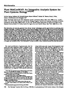
Figure 1. C. reinhardtii can be screened efficiently for mutations in starch. A, Distinct mutants define the starch pathway. Previously characterized mutants (Buleon at el, 1998) illustrate the color-based detection of mutants for altered starch (sta1, no color; sta2, red; sta3, olive; and cc1928 wild type, violet). Left, The starch biosynthetic pathway is depicted with intermediates indicated and addition of ␣-linked Glc (n) to the lengthening glucan chain (n ⫹ 1). Mutations resulting in loss of enzyme function are indicated (X). Right, The affected enzymes are shown along with their respective mutant loci in C. reinhardtii. From top to bottom the loci are: sta1-1, sta2-1, sta2-2, sta3-1, sta3-2, and sta3-3, respectively. The sta2-1 mutation results in less than 5% (w/w) total starch. B, Mutagenized cells can be arrayed for efficient screening. Mutagenized cells are grown on standard medium for 7 d then inoculated into microtiter plates containing liquid medium and grown for an additional 5 d. Once in microtiter plates, cells are easily arrayed on solid medium to induce starch for screening. All steps indicated can be automated by the use of robots to increase the throughput.
(and Arg auxotrophy). Because the sta2-1 allele causes the cells to stain red by iodine vapor, it will be possible to detect a broader range of mutations, such as those selectively defective for amylopectin synthesis, that cannot be detected easily in a wild-type background. Because disrupted genes will usually be linked to the selection marker, rapid cloning of affected genes should be possible. The results may aid in the discovery of useful genes that can be engineered into crops directly or provide valuable information about metabolism that can help focus our efforts in crop species. Although the collection was designed with starch metabolism in mind, many unrelated screens are possible for lipids or other valuable metabolites. UNDERSTANDING THE CELL WALL
The plant cell wall is a complex and dynamic mosaic of three coextensive and interactive networks of cellulose/xyloglucan, pectin, and proteins (Showalter, 1993). Although significant inroads have been made recently in understanding the molecular details of cell wall synthesis (for example, see Faik et al., 2000), much of this research has focused on proteins 1336
in the Hyp-rich glycoprotein (HRGP) superfamily. In the last decade, numerous HRGPs have been characterized at both the protein and DNA level. As a consequence, a lot is known about the structure and regulation of these proteins, but the precise function of any particular HRGP remains unclear. An alternative approach is to develop a model system for studying wall assembly using C. reinhardtii, whose walls are composed almost exclusively of HRGPs. In the walls that surround vegetative cells and gametes (Goodenough et al., 1986; Snell and Adair, 1990), the HRGPs are arranged in two major layers: an inner layer (W2) whose constituent HRGPs are insoluble and a contiguous outer crystalline layer (W6) of HRGPs that are soluble but salt extractable. The assembly of the vegetative wall can be induced by treating cells with a cell wall lytic enzyme produced by gametes (Claes, 1971; Kinoshita et al., 1992). Within several hours, the protoplasts secrete a new and insoluble wall. Analysis of soluble enzyme activity combined with studies in which inhibitors that interfere with crosslinking are applied reveal that a peroxidase and a transglutaminase are involved in wall regeneration (Waffenschmidt et al., 1993, 1999). Because C. reinPlant Physiol. Vol. 127, 2001
Scientific Correspondence
hardtii is haploid, mutations will produce a phenotype directly. Thus, mutagenesis and screening for defects in wall regeneration provide information about essential structural proteins or cross-linking enzymes as well as perception or signaling in response to stresses such as wall rupture. In contrast to vascular plants, most C. reinhardtii mutants defective in wall regeneration will remain viable due to the presence of pulsatile vacuoles, which obviates the need for tedious selection of conditional phenotypes. In fact, cell wall-defective colonies of C. reinhardtii can be distinguished easily by their mucoid morphology. Although C. reinhardtii is useful for understanding HRGPs, its wall lacks several components of vascular plants, including cellulose, xyloglucan, and pectin. However, other chlorophyte cell walls do possess elaborate vascular plant-like organization. The desmidiales are unicellular organisms with highly ornate walls organized in two symmetrical semicells. They synthesize cellulose through hexagonal arrays of rosettes and their walls contain pectins and arabinogalactan proteins. Several heterothallic species of desmidiales are available and conjugation has been mastered in the laboratory. Thus, desmidiales are a potentially useful system to study plant cell wall morphogenesis, although much work remains to develop them as genetic and molecular models equivalent in utility to C. reinhardtii.
NEW MODELS
An important and perhaps overlooked aspect of unicellular green algae is the potential for reduced functional gene redundancy, which is displayed by higher plant genomes. Whereas C. reinhardtii displays a genome complexity approaching that of Arabidopsis, some recently characterized microalgae may have genomes simpler than that even of yeast. Ostreococcus tauri, a unicellular chlorophyte belonging to the prasinophyceae, defines the smallest eucaryote known to date (0.8 m in diameter) (Courties et al., 1994; Chre´ tiennot-Dinet et al., 1995). It also has one of the smallest genomes (10.2 Mbp; Courties et al., 1998). Yet O. tauri, recently identified as a picophytoplanktonic organism, displays all major features of chlorophytes and other plant cells. Picophytoplanktonic organisms were discovered only 2 decades ago, when it was realized that cell counts based on chlorophyll measurements from the surface of the open seas did not agree with the cell counts performed by classical techniques. It was then discovered that the seas contain tiny planktonic cells in abundance. Among these picophytoplanktonic organisms (between 0.3–3 m in diameter), phycologists found a great diversity of picoeukaryotes. Although axenic cultures of O. tauri are not yet available, other picochlorophytes appear to grow well both on defined solid media and in liquid cultures. Small size seems to offer a selective advantage Plant Physiol. Vol. 127, 2001
to oceanic planktonic species. To achieve such a small size, other picochlorophytes will likely have simplified genomes with reduced nDNA content. It is unlikely that O. tauri and its picoeukaryotic cousins, with roughly one-tenth the nDNA content of Arabidopsis, could maintain a level of “functional” redundancy equivalent to that of C. reinhardtii or Arabidopsis. This is particularly true given the need to maintain chloroplast, mitochondrion, and other major aspects of the eukaryotic way of life. It is more likely that these organisms will have streamlined pathways and will have reduced functional gene redundancy to core functions. Thus, a green Escherichia coli might very well be lurking out in the open seas, an organism that could turn out to greatly simplify functional studies of vascular plant pathways.
A PROMISING FUTURE
We are just beginning to see the true value of algal species for the elucidation of important pathways in plants. Classical discoveries in chloroplast and flagella function in C. reinhardtii were only the beginning. Recent results in understanding carbohydrate metabolism indicate that chlorophytes like C. reinhardtii provide the efficiency and speed necessary to permit us to saturate mutationally basic pathways such as starch or cell wall synthesis more fully. This will lead to a new understanding of these and other important pathways. It will be essential ultimately to understand such pathways in vascular plants if we are to reap the practical benefits from our research. However, algae can serve as powerful microbial models for the rapid identification of novel genes and functional domains much as yeast has elucidated many core functions that are essential in animals. In fact, examples of the use of microbial genes for crop improvement are well known. To move forward requires a concerted effort to fully develop algae such as C. reinhardtii in terms of genome sequence and tools to further increase the speed of large genetic screens. We are developing tools such as insertion collections, and as we approach the completion of genome sequencing for a number of higher plant species, the opportunity to expand our list to include unicelluar plants will present itself. As we have discussed, the identification of the picoeukaryotic algae such as O. tauri highlights the potential to develop models that avoid the functional gene redundancy found in vascular plants. Given the indications that their genomes will be quite small, sequencing should be relatively straightforward compared to the efforts applied in Arabidopsis and rice. The comparison of simple unicellular plant genomes to those in multicellular plants will in itself be quite illuminating. 1337
Scientific Correspondence
ACKNOWLEDGMENTS The authors are very grateful to Drs. Claude Courties and Herve´ Moreau (Observatoire Oceanologique de Banyuls, Laboratoire Arago, Banyuls-sur-mer, France) for their generous sharing of O. tauri data prior to publication. We also wish to thank Jacqueline McLaughlin (Exelixis, South San Francisco, CA) for excellent graphic arts. Received September 7, 2001; returned for revision September 14, 2001; accepted September 20, 2001. LITERATURE CITED Ball G, van de Wal MHBJ, Visser RGF (1998) Trends Plant Sci 3: 462–467 Buleon A, Colonna P, Planchot V, Ball SG (1998) Int J Biol Macromol 23: 85–112 Buleon A, Gallant D-J, Bouchet B, Mouille G, D’Hulst C, Kossman J, Ball SG (1997) Plant Physiol 115: 949–957 Casper T (1994) In EM Meyerowitz, C Somerville, eds, Arabidopsis. Cold Spring Harbor Laboratory Press, Cold Spring Harbor, NY, pp 913–936 Chre´ tiennot-Dinet MJ, Courties C, Vaquer A, Neveu J, Claustre H, Lautier J, Machado C (1995) Phycologia 4: 285–292 Claes H (1971) Arch Microbiol 78: 180–188 Courties C, Perasso R, Chre´ tiennot-Dinet MJ, Gouy M, Guillou L, Trousselier M (1998) J Phycol 34: 844–849 Courties C, Vaquer A, Trousselier M, Lautier J, Chre´ tiennot-Dinet MJ, Neveu J, Machado C, Claustre H (1994) Nature 370: 255
1338
Delrue B, Fontaine T, Routier F, Decq A, Wieruszeski J-M, Van den Koornhuyse N, Maddelein M-L, Fournet B, Ball S (1992) J Bacteriol 174: 3612–3620 Faik A, Bar-Peled M, DeRocher AE, Zeng W, Perrin RM, Wilkerson C, Raikhel NV, Keegstra K (2000) J Biol Chem 275: 15082–15089 Goodenough UW, Gebhart B, Mecham RP, Heuser JE (1986) J Cell Biol 103: 405–417 Kinoshita T, Fukuzawa H, Shimada T, Saito T, Matsuda Y (1992) Proc Natl Acad Sci USA 89: 4693–4697 Libessart N, Maddelein M-L, Van den Koornhuyse N, Decq A, Delrue B, Mouille G, D’Hulst C, Ball S (1995) Plant Cell 7: 1117–1127 Showalter AM (1993) Plant Cell 5: 9 -23 Slattery CJ, Kavakli IH, Okita TW (2000) Trends Plant Sci 5: 291–298 Snell WJ, Adair WS (1990) In WS Adair, RP Mecham, eds, Organization and Assembly of Plant and Animal Extracellular Matrix. Academic Press Inc., San Diego, pp 16–48 Vysotskaia V, Curtis D, Voinov A, Kathir P, Silflow CD, Lefebvre PA (2001) Plant Physiol 127: 386–389 Yu TS, Kofler H, Hausler RE, Hille D, Flugge UI, Zeeman SC, Smith AM, Kossmann J, Lloyd J, Ritte G et al. (2001) Plant Cell 13: 1907–1918 Waffenschmidt S, Woessner JP, Beer K, Goodenough UW (1993) Plant Cell 5: 655–661 Waffenschmidt S, Kusch T, Woessner JP (1999) Plant Physiol 121: 1003–1015 Zeeman SC, Northrop F, Smith AM, Rees T (1998) Plant J 15: 357–365
Plant Physiol. Vol. 127, 2001