B Y Ann e L . Co h e n a n d M i c h a e l Ho l c omb
Why Corals Care About Ocean Acidification Uncovering the Mechanism
Abstr act. Stony corals build hard skeletons of calcium carbonate (CaCO3) by
Introduction
combining calcium with carbonate ions derived, ultimately, from seawater. The concentration of carbonate ions relative to other carbonate species in seawater is rather low, so corals expend energy to raise the pH of seawater sequestered in an isolated, extracellular compartment where crystal growth occurs. This action converts plentiful bicarbonate ions to the carbonate ions required for calcification, allowing corals to produce CaCO3 about 100 times faster than it could otherwise form. It is this rapid and efficient production of CaCO3 crystals that enables corals to build coral reefs. Ocean acidification reduces the pH and thus the abundance of carbonate ions in seawater. Corals living in acidified seawater continue to produce CaCO3 and expend as much energy as their counterparts in normal seawater to raise the pH of the calcifying fluid. However, in acidified seawater, corals are unable to elevate the concentration of carbonate ions to the level required for normal skeletal growth. In several experiments, we found that boosting the energetic status of corals by enhanced heterotrophic feeding or moderate increases in inorganic nutrients helped to offset the negative impact of ocean acidification. However, this built-in defense is unlikely to benefit corals as levels of CO2 in the atmosphere continue to rise. Most climate models predict that the availability of inorganic nutrients and plankton in the surface waters where corals live will decrease as a consequence of global warming. Thus, corals and coral reefs may be significantly more vulnerable to ocean acidification than previously thought.
Today, millions of hectares of tropical coastline are dominated by coral reef ecosystems that exist only because coral animals, small anemone-like creatures called “polyps,” can produce calcium carbonate (CaCO3) crystals faster than their skeletons are eroded by the sea. Within an extracellular space beneath the coral polyp, micron-sized CaCO3 crystals in the form of aragonite are nucleated and grown around the clock, 365 days of the year. They are packed into bundles and stacked meticulously one on top of the other to form an intricately designed skeleton that both supports and protects the animal that built it. Most reef-building corals exist as colonies in which the individual skeletons of hundreds, sometimes thousands, of polyps can form enormous domed or branching structures sometimes reaching
118
Oceanography
Vol.22, No.4
This article has been published in Oceanography, Volume 22, Number 4, a quarterly journal of The Oceanography Society. © 2009 by The Oceanography Society. All rights reserved. Permission is granted to copy this article for use in teaching and research. Republication, systemmatic reproduction, or collective redistirbution of any portion of this article by photocopy machine, reposting, or other means is permitted only with the approval of The Oceanography Society. Send all correspondence to:
[email protected] or Th e Oceanography Society, PO Box 1931, Rockville, MD 20849-1931, USA.
S p e c i a l Iss u e F e at u r e
several meters in height. Countless skeletons accreted over many millennia provide the reef framework, the concrete jungles of the tropical ocean that provide myriad marine and terrestrial species with habitat, nesting grounds, and food. For human populations along tropical coastlines, healthy reefs provide natural buffers to beach erosion, barriers to tsunamis and hurricanes, and significant income through tourism and fisheries. By one estimate, the net global economic value of coral reefs worldwide is a staggering $29.8 billion each year (Cesar et al., 2003).
required for successful skeleton-building increases as the acidity of the ocean increases. We present evidence that corals growing under nutrient-replete conditions can redirect the extra energy provided by slightly elevated levels of inorganic nutrients or food toward calcification, thus dampening the negative impact of ocean acidification. Finally, we consider the implications of a changing climate, including predicted reductions in surface ocean nutrient concentrations and primary productivity, for the future of corals and coral reefs.
Over the next century, rising levels of atmospheric CO2 will increase the concentration of total dissolved inorganic carbon (DIC) in seawater, but simultaneously reduce seawater pH and the abundance of carbonate ions [CO 32–] that corals and other marine calcifiers use to build their skeletons. For this reason, there is growing concern that the so-called “acidification” of the surface ocean may slow rates of CaCO3 production or “calcification” by reef-building corals to a point where rates of reef erosion exceed rates of skeletal accretion, leading to the gradual, worldwide loss of coral reef ecosystems as we know them. Data from a wide range of laboratory experiments have fueled this concern, demonstrating that coral calcification (skeleton-building) can be highly sensitive to changes in seawater carbonate ion concentration. In this paper, we show how this process might work using a model based on the behavior of nonbiological (abiogenic) aragonites when grown in increasingly “acidified” seawater. We show that coral calcification under any circumstances is energetically costly to the coral animal and that the energy
Biominer alization Basics Understanding the fundamentals of coral calcification (i.e., the processes involved in the nucleation and growth of aragonite crystals) is a crucial first step in predicting the response of corals and coral reef ecosystems to future climate change, including ocean acidification. A useful place to start thinking about calcification strategies in the marine environment is to recognize that all marine calcifiers, including the reef-building or “stony” corals, have to overcome the kinetic barriers to CaCO3 precipitation that exist naturally in seawater. Today, the surface ocean is supersaturated with respect to CaCO3, which means that there are more than enough calcium and carbonate ions in solution to enable CaCO3 to precipitate out. The saturation state of seawater with respect to aragonite, the form of calcium carbonate that corals produce, is denoted by the symbol Ωar . It is defined as the product of the actual measured concentrations of calcium and carbonate ions dissolved in seawater divided by the product of the concentrations of calcium and carbonate ions when
they are saturated (at equilibrium) in seawater, as follows: Ω ar = [Ca2+] x [CO 32–] / [Ca2+] x [CO 32–]sat .
Because the calcium ion concentration [Ca2+] in seawater is very high and relatively constant, variations in Ω ar are determined mainly by the carbonate ion 2– concentration [CO 2– 3 ]. Today, the [CO 3 ] of seawater in the low-latitude surface ocean is about 250 µmol kg -1. Aragonite saturation is reached at a [CO 2– 3 ] of about -1 60 µmol kg . Thus, the tropical surface ocean is, in general, but with notable exceptions such as the eastern tropical Pacific, about four times supersaturated with respect to aragonite. When Ω ar > 1 (i.e., [CO 2– 3 ]> -1 60 µmol kg ), aragonite should, theoretically, precipitate from seawater. Conversely, when Ω ar < 1, aragonite should dissolve in seawater. The warm tropical ocean where most coral reefs are found is highly supersaturated with respect to aragonite, in other words, Ω ar can be significantly greater than 1. Nevertheless, neither calcite nor aragonite will form spontaneously because there are kinetic barriers that prevent nucleation and/or crystal growth. These kinetic barriers include the high hydration energy of the calcium ions (e.g., Lippmann, 1973), the low concentration and activity of the carbonate ions (e.g., Garrels and Thompson, 1962; Lippmann, 1973), and the presence of high concentrations of sulfate and magnesium (e.g., Usdowski, 1968; Kastner, 1984). Most marine calcifiers, therefore, must nucleate and grow CaCO3 crystals within compartments that are isolated or semi-isolated from the external seawater, and within which they can modify, regulate, and control conditions, including
Oceanography
December 2009
119
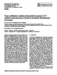
Figure 1. The coral animal (polyp) manipulates the chemistry of seawater sequestered in an isolated calcifying compartment to produce many millions of tiny calcium carbonate (aragonite) crystals that are assembled into a skeleton. In (A), each polyp (p) in the colony builds its own skeleton, the corallite (cl). The polyp sits atop of, and completely covers, the skeletal surface. The arrow points to the interface between the basal epithelial cells and the skeletal surface, where calcification occurs. In (B), the skeleton is composed of radiating arrays of needle-shaped crystals (f) that grow on aggregates of fine granular crystals (g). In (C), the granular crystals are accreted at night and in (D), the needle-shaped crystals are accreted mainly in the day. Scale bars: (A) 500 µm, (B) 10 µm, (C) and (D) 1 µm. (A) is adapted from Veron (1993)
in some cases the carbonate chemistry of the calcifying fluid, to enable CaCO3 precipitation to occur. Coccolithophores and gorgonians, for example, produce calcite and high-magnesium calcite intracellularly, the marine alga Halimeda accretes aggregations of acicular aragonite needles extracellularly within spaces created between cell membranes, and corals and mollusks accrete their CaCO3 crystals within compartments created between the tissue and the existing skeleton or shell. In addition, there is substantial evidence for the involvement of organic molecules in the biomineralization processes of many marine Anne L. Cohen (
[email protected]) is Research Specialist, Department of Geology and Geophysics, Woods Hole Oceanographic Institution, Woods Hole, MA, USA. Michael Holcomb is PhD Candidate, Joint Program, Woods Hole Oceanographic Institution, Woods Hole, MA, USA.
120
Oceanography
Vol.22, No.4
calcifiers. Organic molecules may play a role in reducing the surface free energy required for nucleation (e.g., Teng et al., 1998), in guiding site-specific nucleation that results in species-specific skeletal architectures, or in buffering the pH of the calcifying fluid against large fluctuations in internal CO2 concentrations caused by cycles in photosynthesis and respiration (Holcomb et al., 2009). In stony corals, skeletal formation is entirely external to the organism (extracellular) but the skeleton is not exposed to seawater. Corals either sit on top of or wrap themselves around their skeletons (Figure 1A) and the skeletal surface is completely enveloped by tissue, separated from the external seawater environment by four layers of cells. Data from a combination of geochemical, mineralogical, and tracer dye studies indicate that seawater is likely the starting fluid for calcification (Cohen et al., 2001, 2009; Braun and Erez, 2004; Gaetani and Cohen, 2006; Holcomb et al., 2009), and
it is transported to the site of calcification located between the base of the calicoblastic epithelium and the existing skeletal surface (Braun and Erez, 2004). The route by which seawater enters this space remains unclear, although calcein dye tracer studies rule out cross-membrane transport (Braun and Erez, 2004). One possibility is that the calicoblastic epithelium that loosely but directly overlies the skeletal surface (or reef substrate in the case of newly settled noncalcified polyps) pulsates, periodically lifting away from the skeletal surface to create small pockets that fill with seawater via paracellular channels or other “leaks” in the tissue membranes.
Abiogenic Ar agonite Crystals Provide a Window on the Cor al’s Calcifying Environment For reasons discussed above, bringing seawater into a calcifying compartment is not, by itself, adequate for producing
aragonite crystals, at least not at the rate that corals need to build their skeletons. Kinetic inhibition of aragonite nucleation and growth has to be overcome, and there are several ways this could be accomplished by the coral. The exact mechanisms involved have important implications for understanding and predicting the impact of ocean climate changes on coral; however, opportunities for scientists to directly observe or monitor the site of calcification in corals have been limited because of its small size and inaccessible location beneath the tissue. One approach that has proven fruitful is to compare the composition and morphology of coral aragonites with abiogenic aragonites precipitated experimentally from highly supersaturated seawater solution in a laboratory beaker (e.g., Gaetani and Cohen, 2003; Holcomb et al., 2009). Figure 1B–D shows the morphology and microstructural arrangement of crystals in a coral skeleton. All the different components of the skeleton seen in Figure 1A are built of bundles of fine, needle-shaped crystals that are several microns in length (f in Figure 1B and Figure 1D) and that radiate from discrete aggregations of tiny granular crystals, each several nanometers in diameter (g in Figure 1B, and Figure 1C). In many corals, there is a distinct diurnal cycle to the formation of these crystals: the granular crystals form at night, whereas the needle-shaped crystals form during the day (Holcomb, 2009). Each aggregate of granular crystals and its associated bundle of needles is called a sclerodermite (Wells, 1956). Sclerodermites are the basic building blocks or “bricks” of the coral skeleton. Aragonite crystals precipitated in the laboratory from a highly supersaturated seawater solution in the absence of a
coral (i.e., abiogenic aragonites) bear remarkable resemblance to the coral sclerodermites (Figure 2). Nucleation, which occurs at very high aragonite saturation states (Ω ar > 20), produces aggregates of submicron-sized granular crystals. At lower aragonite saturation states (Ω ar ~ 6–19), crystal growth is favored over nucleation. The growth phase produces bundles of fine, needleshaped crystals. Each aggregate of granular crystals and its associated needle-shaped crystals is called a “spherulite” (Figure 2B; Holcomb et al., 2009). Aragonitic spherulites precipitated from a supersaturated seawater solution are compositionally (i.e., chemically) and morphologically similar to coral “sclerodermites,” as evident in Figure 2. Based on this similarity, it is feasible that one mechanism corals may employ to achieve aragonite nucleation and growth is by elevating the saturation state of seawater trapped in an isolated or semiisolated calcifying compartment (Cohen and McConnaughey, 2003). Abiogenic aragonites grown experimentally from seawater over a range of aragonite saturation states show systemmatic and progressive changes in crystal morphology caused by changes in crystal growth rate (Figure 3). Crystals grown experimentally at relatively low aragonite saturation states (Ω ar ~ 6) are short, wide, and highly faceted, consistent with low crystal growth rates (e.g., Lofgren, 1974, 1981; Figure 3C). When the saturation state of the experimental seawater is increased during growth, the crystals produced are longer and thinner, more bladelike in appearance, and less faceted, consistent with a progressive increase in crystal growth rate (Figure 3A,B). This change in crystal morphology with increasing seawater saturation state can be quantified using the crystal aspect
ratio or the ratio of its length to its width. In Figure 3D, the aspect ratio of aragonite crystals grown under experimental conditions is plotted against the saturation state of the seawater in which they grew. The crystal aspect ratio is linearly related to aragonite saturation state. The lower the seawater saturation state, the lower the crystal growth rate, and thus the lower the crystal aspect ratio. The relationship between seawater saturation state and crystal aspect ratio in these experiments can be described by the following equation: Ω ar = 0.93 (± 0.06) x crystal aspect ratio (µm) + 0.20(± 0.89); r2 = 0.94. This relationship between crystal aspect ratio and aragonite saturation state can be applied to coral skeletons to
Figure 2. Aragonite crystals produced by corals (B) and aragonite crystals produced experimentally from a highly supersaturated seawater solution (A) share many similarities that allow us to conclude that corals expend energy to elevate the saturation state of seawater in order to calcify. In both (A) and (B), bundles of aragonite fibers radiate out from a central region occupied by aggregates of submicronsized granular crystals. Scale bars are 1 µm in both. Images from Cohen and McConnaughey (2003) and Holcomb et al. (2009)
Oceanography
December 2009
121
constrain the aragonite saturation state of the calcifying fluid of corals reared under different conditions. Aspect ratios estimated from measurements of crystals in wild coral skeletons using scanning electron microscope (SEM) images can range from ~ 11 in the ahermatypic coral Astrangia to 20 in the septa of a tropical Porites coral. Using the equation above, we can estimate the saturation state of the coral’s calcifying fluid during active calcification. Doing so reveals that conditions within the calcifying space during crystal growth are significantly elevated above that of the external seawater, with Ω values between 10 and 19.5. Note, however, as we are able to nucleate these crystals in the laboratory only at Ω ar
A
values of 20 and higher. Thus, the nucleation phase, which results in formation of aggregates of submicron-sized granular crystals, likely occurs at even higher aragonite saturation states. Al-Horani et al. (2003) placed micro-pH electrodes within the calcifying space of the zooxanthellate coral Galaxea fascicularis while manipulating light levels in the laboratory. The pH of the fluid within the coral’s calcifying compartment cycle from ambient seawater values (~ 8.2) in the dark to values significantly elevated above ambient (> 9.0) in the light, when most of the calcification occurs. This rise in pH translates to an increase in the aragonite saturation state of the calcifying
B
C
D
Figure 3. Aragonites precipitated experimentally from seawater over a range of aragonite saturation states display systematic changes in crystal morphology (A–C) that can be quantified using the crystal aspect ratio (the ratio of crystal length to width). In (D), crystal aspect ratio is plotted against aragonite saturation state of the seawater in which they grew. This relationship is used in Figure 5 to estimate the saturation state of the coral’s calcifying fluid. Scale bars are 1 µm.
122
Oceanography
Vol.22, No.4
fluid that is significantly above that of the ambient seawater, potentially five to 10 times higher depending on DIC concentration. Al-Horani et al. (2003) also reported a small increase, ~ 10% above ambient, in the concentration of calcium ions at the calcifying site. This increase suggests that the elevation in fluid saturation state in the coral’s calcifying compartment may be driven largely by an increase in carbonate ion concentration and to a lesser extent by the change in calcium ion concentration.
Calcification Is Energetically Costly Thus, conditions within the coral’s calcifying region during active calcification are very different from those of the external seawater environment. Fluid with the pH, calcium, and carbonate ion concentration of the external seawater is brought into the calcifying space at the base of the polyp tissue, but does not stay that way for very long. Indeed, if Al-Horani et al.’s (2003) unique timeseries data of pH changes at the site of calcification reflect actual conditions and changes within the calcifying space over the diurnal cycle, then the saturation state of the calcifying fluid ramps up immediately following daybreak. How is this achieved? Cohen and McConnaughy (2003) proposed that rapid and effective elevation of calcifying fluid pH and saturation state could be achieved with a plasma membrane Ca2+-ATPase acting as a Ca2+/H+ exchanger (or antiporter), which removes two H+ ions from the calcifying fluid for every Ca2+ ion transported from the calicoblastic epithelial cells into the calcifying space. By this process, calcifying fluid saturation can be elevated significantly above ambient
seawater in a very short amount of time. Subsequent work by Zoccola et al. (2004) lends some support to this model, showing that Ca2+-ATPase expression is highly concentrated in the calicoblastic epithelial cells that overly the coral skeleton and are presumed to be involved in calcification. The potential role of Ca2+-ATPase, proton pumps and other enzymes in coral calcification highlights the energetic cost to the coral of producing aragonite crystals to build a skeleton. Under the Cohen and McConnaughey (2003) model, every µmol of CaCO3 produced by the coral requires the removal of two µmol protons (H+ ions) from the calcifying fluid, and the expenditure of 1 µmol of “energy” or ATP. For a coral producing 3.7 µmol CaCO3 per hour and respiring at a rate of 3.1 µmol CO2 per hour (e.g., Dennison and Barnes, 1988), and assuming 6 mol ATP per mol CO2 produced, calcification may consume up to 20% of the coral’s energy budget. When the energy required to produce organic matrix proteins that constitute 0.1–1% of the coral skeleton is included, up to 30% of the coral’s energy budget may be devoted to calcification (Allemand et al., in press). Although these estimates of the energetic cost of calcification are preliminary, it is clear from studies of corals reared under fed and starved conditions (e.g., Houlbreque et al., 2008) that calcification is an active, physiological process that requires significant amounts of energy to drive it.
a profound negative influence on skeletal growth (Figure 4; see Kleypas and Yates, 2009). This is most likely due to the lowering of seawater carbonate ion concentration [CO 32–] and, thus, the initial availability of ions required to build CaCO3 crystals. Cohen et al. (2009) found that corals reared in highly corrosive (i.e., undersaturated) conditions were able to accrete and maintain aragonite crystals beneath the polyp, testifying to the organism’s degree of control on the internal calcifying environment (Figure 4D). However, the skeleton accreted under these conditions was far from healthy, and SEM images of the aragonite crystals revealed that their morphology, organization, and packing is very different from those accreted by corals under ambient CO2 conditions. Figure 5 illustrates progressive changes in coral crystal morphology as a function of seawater saturation state in the experimental aquaria. Aragonite crystals accreted by corals reared under ambient CO2 are similar to those found
in coral skeletons collected in the field: acicular in shape, several µm in length, 0.5–1 µm wide, tightly packed into discrete bundles, and oriented with their c-axes aligned or in parallel with adjacent crystals in the bundle (Figure 5A). Crystals in coral skeletons accreted in seawater with decreasing saturation states show progressive changes in crystal morphology that are consistent with a slowdown in crystal growth rate (Figure 5B–D). Individual crystals within bundles become increasingly short, fat, and faceted as the saturation state in the aquarium seawater decreases. Near aragonite saturation (Ω ar ~ 1), the crystals are no longer organized into discrete, tightly packed bundles, and in the strongly undersaturated treatment (Ω ar ~ 0.2), the crystals are no longer oriented with their c-axes in parallel. In this treatment, crystal growth is so slow that crystal morphology assumes an orthorhombic shape characteristic of aragonite grown slowly at near equilibrium conditions (Figure 5D).
Figure 4. Ocean acidification negatively impacts the skeletal growth of eight-dayold corals reared from planula larvae in laboratory aquaria. In these images, the polyp has been removed to reveal the first skeleton or “primary corallite.” In A, Ωar of the aquarium seawater is ~ 3.7; in B, Ωar ~ 2.4; in C, Ωar ~ 1, and in D, Ωar ~ 0.2. A–D scale bars are ~ 100 µm. From Cohen et al., 2009
How Ocean Acidification Impacts Cor al Calcification Most experimental studies with corals reared under elevated CO2 conditions indicate that a decrease in Ω ar can have
Oceanography
December 2009
123
In Figure 5E, we plot the aspect ratio of crystals accreted by corals reared in seawater with Ωar ranging from ambient (3.7) to strongly undersaturated (0.2). Note that the aspect ratio of the crystals decreases as the Ωar of the aquarium seawater decreases. Applying the relationship between crystal aspect ratio and fluid saturation state established for experimental abiogenic aragonites, we can show that the saturation state of the coral’s internal calcifying fluid is maintained significantly above that of the external seawater in all experimental conditions (Figure 5E). However, the saturation state of the coral’s internal calcifying fluid decreases as that of the external seawater decreases. When the Ωar of the external seawater is ~ 3.7 (i.e., at ambient levels), the coral pumps enough protons from the initial calcifying “intake” water to maintain an internal calcifying fluid saturation state
significantly higher than it is outside (Ωar ~ 19). At this level of aragonite supersaturation, crystals growth is very fast, allowing the coral to quickly build a massive and dense skeleton. When the Ωar of the external seawater is ~ 2.4, the coral works to maintain the calcifying fluid Ωar at around 15. At this level of aragonite supersaturation, crystal growth slows enough to cause the rate of skeletal formation to slow down as well. When the Ωar of the external seawater is ~ 1, the coral works to maintain the calcifying fluid Ωar at ~ 7. At this level of aragonite supersaturation, the rate of crystal growth slows dramatically and building a normal skeleton becomes almost impossible. When reared in strongly undersaturated seawater, the coral is able to maintain the fluid saturation state just above that at which its minute skeleton will dissolve (Ωar ~ 2). Thus, coral calcification
declines with declining seawater saturation state because the saturation state of the internal calcifying fluid declines in concert with it. And, as the saturation state of the internal calcifying fluid declines, the rate of CaCO3 crystal growth becomes too slow to sustain growth of a normal skeleton.
Why Don’t Cor als Simply Pump More Protons? If corals grow aragonite from seawater by pumping protons to raise the saturation state within a calcifying compartment, why don’t corals reared under elevated CO2 (ocean acidification) conditions simply pump more protons in an effort to build a normal skeleton? After all, under elevated CO2, there is even more carbon around, in the form of bicarbonate. And, removing protons from the calcifying fluid would turn that excess bicarbonate into even more carbonate
E
Figure 5. In (A) aragonite crystals accreted by corals (shown in Figure 4) reared in seawater with a range of aragonite saturation states, display systematic and progressive changes in crystal morphology (A–D). In (E), these changes can be quantified using the crystal aspect ratio (the ratio of crystal length to width) and used to estimate changes in the saturation state of the coral’s internal calcifying fluid in response to ocean acidification. Scale bars are 1 µm. From Cohen et al., 2009
124
Oceanography
Vol.22, No.4
ions for calcification. The answer might lie in the fact that calcification is energetically costly and the coral’s energy budget is not that flexible. A coral building a normal skeleton under elevated CO2 conditions would have to divert more energy to pumping protons from the calcifying fluid than a coral building a normal skeleton under ambient CO2 conditions. For example, a coral reared in seawater with an aragonite saturation state of ~ 3.7 (pCO2 ~ 400 ppm) would need to remove ~ 4500 nmol protons per ml of fluid to raise the Ω ar of the calcifying fluid to 19 (Figure 6). By comparison, a coral reared in seawater with a saturation state of ~ 2.4 (pCO2 ~ 740 ppm) would have to remove > 8000 nmol protons per ml of calcifying fluid to reach Ω ar of 19. Conversely, if the energy budget for calcification were fixed and the coral could expend only enough energy to remove 4500 nmol protons from the calcifying fluid independent of the saturation state of the “intake” water, then our calculations show that a coral reared at Ω ar ~ 2.4 would be able to elevate the internal saturation state from 2.4 to about 15 (Figure 6). And a coral reared in seawater with Ω ar of 1.5 would elevate the internal saturation state to approximately 10 by removing the same number of protons. These numbers are consistent with our estimate of actual calcifying fluid saturation state based on crystal aspect ratios (Figure 5E), and suggest that corals reared under high-CO2 conditions may have worked as hard, and pumped as many protons from their calcifying fluid, as corals in the ambient treatment. However, in our experimental aquaria, there appears to be a limit to the amount of work the corals will do even at the expense of building the healthy skeleton
Figure 6. Calcification achieved by removing protons from the internal calcifying fluid is energetically expensive. The energy expended in calcification is represented here by the number of protons pumped from each milliliter of calcifying fluid. Corals reared in seawater with a range of aragonite saturation states (depicted by different colors) remove the same number of protons from that seawater (4500 nmol ml-1) but achieve different calcifying saturation states (shown by grey bar). This suggests that corals reared in acidified seawater did not divert more energy to proton removal even at the expense of building a normal skeleton. In this CO2Sys calculation, the initial calcifying fluid is seawater with an initial alkalinity of 2470 µmol kg-1 in equilibrium with the specified pCO2 (400, 740, or 1340 µatm). The fluid is isolated from the surrounding seawater; proton pumping elevates the alkalinity and saturation state within the calcifying space while CO2 diffuses through the calicoblastic epithelium to maintain equilibrium pCO2. Calculations were made with a Matlab implementation of CO2Sys using constants of Mehrbach et al. (1973) refit by Dickson and Millero (1987) for carbonate, and Dickson (1990) for sulfate; input conditions are as follows: S = 30, T = 25, atmospheric pressure = 1atm. Concentrations of silicate, phosphate, ammonia and HS were set at 0. Ca and B concentrations were calculated from salinity.
that is so critical to recruitment success. This result suggests that the coral’s energy budget for calcification might be limited by the availability of nutrients or food.
The Potential Role of Nutrients and Food in Modulating Cor al Response to Ocean Acidification If it is true that the coral’s energy budget for calcification is limited by the availability of nutrients, then elevating the nutritional status of a coral calcifying in low-saturation-state seawater might provide the additional fuel it needs to maintain healthy calcification rates. This could be done by enhanced heterotrophic feeding or by adding moderate amounts of inorganic nutrients, such as nitrates and phosphates, to stimulate zooxanthellate photosynthesis, providing the coral with carbohydraterich photosynthate to fuel calcification.
Normally, nutrient addition stimulates zooxanthellate photosynthesis, which, under ambient CO2 conditions, can lead to CO2 limitation and a decline in calcification (e.g., Marubini and Davies, 1996). However, under elevated CO2 conditions, nutrient addition can support increased zooxanthellate photosynthesis without reducing the total amount of carbon available for calcification. Increased photosynthesis means increased photosynthate and more energy for calcification. Therefore, combining elevated nutrients with elevated CO2 could help to offset the negative impact on calcification of elevated CO2 alone. In at least four separate experiments conducted to date, five different species of coral were reared under significantly elevated CO2 conditions (780–1200 ppm, Ω ~ 1.5–2) with the simultaneous addition of food or inorganic nutrients. Calcification rates under these
Oceanography
December 2009
125
conditions were maintained between 75 and 100% of those attained by corals reared under ambient CO2 conditions (Langdon and Atkinson, 2005; Holcomb et al., in press; Ries et al., 2009; Cohen et al., 2010; Figure 7). These experimental results are consistent with the earlier findings of Atkinson et al. (1995), which show that nutritionally replete zooxanthellate corals in naturally lowsaturation-state seawaters are capable of accreting skeletons at rates comparable to those achieved by conspecifics in high-saturation-state seawaters. Today, several reefs, including Galàpagos, areas of Pacific Panama, and Jarvis (southern Line Islands), experience levels of aragonite saturation equivalent to that predicted for the open ocean under two times and three times pre-industrial CO2 levels (Manzello et al., 2008; Kathryn Shamberger [PMEL/NOAA] and colleagues, pers. comm., August 2009). Available data on coral colony growth rates on these
reefs, albeit limited, suggest that they are equivalent to and sometimes even rival those of conspecifics in areas where aragonite saturation states are naturally high, such as the western Pacific warm pool. Although no comparative data exist regarding the nutritional status of corals on these high-CO2 reefs, surface ocean nutrient concentrations and rates of primary and secondary production around the islands are significantly elevated compared with oligotrophic reefs. Thus, it appears likely that naturally elevated levels of inorganic nutrients and, consequently, high levels of primary and secondary production, may already be facilitating high coral calcification rates in regions with naturally high dissolved CO2 levels. If, as these experimental and field data suggest, nutrients or food can potentially lessen the severity of the impact of ocean acidification on coral calcification, does this observation change the outlook for corals and coral reefs as the ocean
Figure 7. Moderate elevation of food or inorganic nutrients can help corals in laboratory experiments to cope with ocean acidification. Here, calcification by five species of corals reared in four separate ocean acidification experiments (Ωar ~ 1.5) is expressed as a percent of calcification by conspecifics reared under ambient CO2 conditions. When corals are reared under elevated CO2 (acidified conditions) with slightly elevated inorganic nutrients and/or food (solid bars), calcification can be maintained at 75–100% of ambient (normal) rates, probably because corals have more energy to divert to proton removal. By comparison, corals reared under elevated CO2 without additional nutrient and/or food usually exhibit significantly reduced calcification rates (graded bars). Data are from Holcomb et al. (in press: Astrangia), Ries et al. (2009: Oculina), Langdon and Atkinson (2005: Porites and Montipora); unpublished data of author Cohen (Favia). The unfed Oculina response (graded blue bar) is estimated from data generated by Holcomb et al. (2009).
126
Oceanography
Vol.22, No.4
becomes more acidic over this century? The answer is, probably not. Seawater saturation states across the global tropics are predicted to drop to an average of around 2.5–3 by the year 2100 (Feely et al., 2009). However, several models (e.g., Boyd and Doney, 2002) predict that this decline will not be accompanied by an elevation in surface ocean nutrient concentrations, at least in the open ocean. Enhanced stratification, reduced mixed-layer depth, and slowed circulation caused by greenhouse-gas-induced global warming will likely cause a significant reduction in surface nutrient concentrations across much of the global tropics and subtropics. A recent study showed that spawning female corals are significantly more susceptible to the negative effects of ocean acidification than spawning male corals (Holcomb et al., 2010). The apparent gender discrimination is likely due to the energetically costly process of egg production, leaving little energy available to the coral to temper the effects of acidification on calcification. This example highlights the compounding difficulties that corals will face as they strive to build skeletons in a high-CO2 world. Climate changes that simultaneously reduce the availability of food while making it harder for corals to calcify will impact not only rates of skeletal growth but sexual reproduction, genetic diversity, and rates of recolonization, rendering coral reef ecosystems even more vulnerable to CO2-induced climate changes than they would be to ocean acidification alone.
Acknowledgements The authors extend gratitude to Dan McCorkle (Woods Hole Oceanographic Institution [WHOI]), Samantha de
Putron (Bermuda Institute for Ocean Sciences [BIOS]), Justin Ries (University of North Carolina), Glenn Gaetani (WHOI), Rinat Gabitov (California Institute of Technology), Louis Kerr (Marine Biological Laboratory). Kathryn Rose (WHOI) and Elizabeth Drenkard (WHOI) provided the opening spread photo. BIOS, in part, supported our larval coral work. Anne L. Cohen acknowledges support from the WHOI Directorate for our Marine Calcification and Culture Labs, from WHOI’s Ocean Life and Tropical Research Institutes, and from NSF CO-0648157. Michael Holcomb’s graduate research was supported in part by an NSF graduate student fellowship, an MIT Presidential Award, and an International Coral Reef Society fellowship.
References Al-Horani, F.A., S.M. Al-Moghrabi, and D. de Beer. 2003. The mechanism of calcification and its relation to photosynthesis and respiration in the scleractinian coral Galaxea fascicularis. Marine Biology 142:419–426. Allemand, D., É. Tambutté, D. Zoccola, and S. Tambutté. In press. Coral calcification, cells to reefs. In Coral and Coral Reefs. Z. Dubinsky, ed., Springer. Atkinson, M.J., B. Carlson, and J.B. Crowe. 1995. Coral growth in high-nutrient, low pH seawater: A case study in coral growth at the Waikiki aquarium. Coral Reefs 14:215–233. Boyd, P., and S.C. Doney. 2002. The impact of climate change and feedback processes on the ocean carbon cycle. Chapter 7 in Ocean Biogeochemistry: The Role of the Ocean Carbon Cycle in Global Change. International Joint Global Ocean Flux Study Synthesis, M. Fasham, ed., Springer. Braun, A., and J. Erez. 2004. Preliminary observations on sea water utilization during calcification in scleractinian corals. Paper presented at American Geophysical Union Fall Meeting, December 13–17, San Francisco. Abstract #B14B-04 available online at: http://adsabs.harvard.edu/abs/2004AGUFM. B14B..04B (accessed December 2, 2009). Cesar, H., L. Burke, and L. Pet-Soede. 2003. The Economics of Worldwide Coral Reef Degradation. Cesar Environmental Economics Consulting, Arnhem, The Netherlands.
Cohen A.L., G.D. Layne, S.R. Hart, and P.S. Lobel. 2001. Kinetic control of skeletal Sr/Ca in a symbiotic coral: Implications for the paleotemperature proxy. Paleoceanography 16(1):20–26. Cohen, A.L., and T.A. McConnaughey. 2003. Geochemical perspectives on coral mineralization. Pp. 151–187 in Biomineralization. P.M. Dove, S. Weiner, and J.J. deYoreo, eds., Reviews in Mineralogy and Geochemistry, vol. 54, doi:10.2113/0540151, The Mineralogical Society of America, Washington, DC. Cohen, A.L., D.C. McCorkle, S. de Putron, G.A. Gaetani, and K.A. Rose. 2009. Morphological and compositional changes in the skeletons of new coral recruits reared in acidified seawater: Insights into the biomineralization response to ocean acidification. Geochemistry, Geophysics, Geosystems 10, Q07005, doi:10.1029/2009GC002411. Cohen, A.L., D.C. McCorkle, S.J. de Putron, and K.A. Rose. 2010. Why corals care about ocean acidification: The role of nutrition. 2010 Ocean Sciences Meeting, Portland, OR (abstract). Dennison, W.C., and D.J. Barnes. 1988. Effect of water motion on coral photosynthesis and calcification. Journal of Experimental Marine Biology and Ecology 115:67–77. Feeley, R.A., S.C. Doney, and S.R. Cooley. 2009. Ocean acidification: Present conditions and future changes in a high-CO2 world. Oceanography 22(4):36–47. Garrels, R.M., and M.E. Thompson. 1962. A chemical model for seawater at 25°C and one atmosphere total pressure. American Journal of Science 260:57–66. Gaetani, G.A., and A.L. Cohen. 2006. Element partitioning during precipitation of aragonite from seawater: A framework for understanding paleoproxies. Geochemica et Cosmochimica Acta 70:4,617–4,634. Holcomb, M. 2009. Coral Calcification: Insights From Inorganic Experiments and Coral Responses to Environmental Variables. PhD Thesis, Massachusetts Institute of Technology/Woods Hole Oceanographic Institution Joint Program in Oceanography/Applied Ocean Science and Engineering, 227 pp. Holcomb, M., A. Cohen, R. Gabitov, and J. Hutter. 2009. Compositional and morphological features of aragonite precipitated experimentally from seawater and biogenically by corals. Geochimica et Cosmochimica Acta 73:4,166–4,179, doi:10.1016/ j.gca.2009.04.015. Holcomb, M.C., A.L. Cohen, and D.C. McCorkle. 2010. Gender bias in the coral response to ocean acidification. 2010 Ocean Sciences Meeting, Portland, OR (abstract). Holcomb, M.C., D.C. McCorkle, and A.L. Cohen. In press. Long-term effects of nutrient and CO2 enrichment on the temperate coral Astrangia poculata (Ellis and Solander, 1786). Journal of Experimental Marine Biology and Ecology. Houlbrèque, F., and C. Ferrier-Pagès. 2008. Heterotrophy in tropical scleractinian corals. Biological Reviews 84:1–17, doi:10.1111/j.1469185X.2008.00058.
Kastner, M. 1984. Control of dolomite formation. Nature 311:410–411. Kleypas, J.A., and K.K. Yates. 2009. Coral reefs and ocean acidification. Oceanography 22(4):108–117. Langdon, C., and M.J. Atkinson. 2005. Effect of elevated pCO2 on photosynthesis and calcification of corals and interactions with seasonal change in temperature/irradiance and nutrient enrichment. Journal of Geophysical Research 110, C09S07, doi:10.1029/2004JC002576. Lippmann, F. 1973. Sedimentary carbonate minerals. Springer-Verlag, Berlin, 228 pp. Lofgren, G. 1974. An experimental study of plagioclase crystal morphology: Isothermal crystallization. American Journal of Science 274:243–273. Lofgren, G. 1980. Experimental studies on the dynamic crystallization of silicate melts. Pp. 487–565 in Physics of Magmatic Processes. R.B. Hargraves, ed., Princeton University Press, Princeton, NJ. Manzello, D.P., J.A. Kleypas, D. Budd, C.M. Eakin, P.W. Glynn, and C. Langdon. 2008. Poorly cemented coral reefs of the eastern tropical Pacific: Possible insights into reef development in a highCO2 world. Proceedings of the National Academy of Sciences of the United States of America 105, doi:10.1073/pnas.0712167105. Marubini, F., and P.S. Davies. 1996. Nitrate increases zooxanthellae population density and reduces skeletogenesis in corals. Marine Biology 127:319–328. Ries, J.B., A.L. Cohen, and D.C. McCorkle. 2009. Marine calcifiers exhibit mixed responses to CO2-induced ocean acidification. Geology 37(12):1,131–1,134; doi:10.1130/G30210A. Teng, H.H., P.M. Dove, C.A. Orme, and J.J. de Yoreo. 1998. Thermodynamics of calcite growth: Baseline for understanding biomineral formation. Science 282(5389):724–727, doi:10.1126/ science.282.5389.724. Usdowski, H.-E. 1968. The formation of dolomite in sediments. Pp. 21–32 in Recent Developments in Carbonate Sedimentology in Central Europe. G. Müller and G.M. Friedman, eds., SpringerVerlag, Berlin. van Heuven, S., D. Pierrot, E. Lewis, and D.W.R. Wallace. 2009. MATLAB Program Developed for CO2 System Calculations. ORNL/CDIAC-105b. Carbon Dioxide Information Analysis Center, Oak Ridge National Laboratory, US Department of Energy, Oak Ridge, TN, http://cdiac.ornl.gov/ oceans/co2rprt.html. Veron, J.E.N. 1993. Corals of Australia and the IndoPacific. University of Hawaii Press, Honolulu, Hi, 656 pp. Wells, J.W. 1956. Scleractinia. Pp. F328–F440 in Treatise on Invertebrate Paleontology Part F: Coelenterata. R.C. Moore, ed., Geological Society of America and University of Kansas Press, Lawrence, KS. Zoccola, D., É. Tambutté, E. Kulhanek, S. Puverel, J.-C. Scimeca, and D. Allemand, and S. Tambutté. 2004. Molecular cloning and localization of a PMCA P-type calcium ATPase from the coral Stylophorapistillata pistillata. Biochimica et Biophysica Acta 1663:117–126.
Oceanography
December 2009
127