Wireless Telemetry Performance of Transplanted Organ Monitoring at Ultra Wideband Range Considering Respiration-Induced Organ Movement Pongphan Leelatien, Koichi Ito, Kazuyuki Saito Medical System Engineering Chiba University Chiba, Japan
[email protected],
[email protected],
[email protected]
Akram Alomainy, Yang Hao
Manmohan Sharma
Electronic Engineering & Computer Science Queen Mary University of London London, United Kingdom
[email protected],
[email protected]
Electrical & Electronic Engineering Nanyang Technological University Singapore
[email protected]
Abstract—In this paper, we aim to assess the performance of a transplanted organ monitoring system by considering the effect of respiration-induced organ movements and locations of the onbody antenna. A heterogeneous human model was used in our numerical investigations. The performance of an implanted antenna and its in-body channels around the liver area are studied using ultra wideband (UWB) technology. S-parameter results for various separating distances between the implanted antenna and the on-body antenna were acquired and investigated. The results demonstrate the influence of organ movement as well as the surrounding tissues on the level of signal attenuation. Our analysis indicates that UWB channel has the potential to be successfully utilized for the transplanted organ monitoring in this in-body scenario.
communications. Simulations and measurements using simplified human phantom model were performed in our previous investigations of this wireless implanted monitoring system [3]. As part of our further investigations, the effect of movement of upper abdominal organs due to respiration as well as location of the on-body antenna on the level of signal attenuation is considered in this paper.
Keywords—antenna; propagation; simulation; ultra wideband; in-body communication
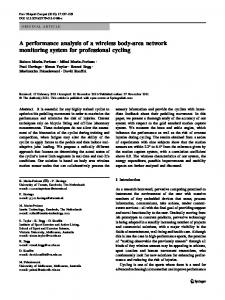
Fig. 1. Overview of the transplanted organ monitoring system.
I. INTRODUCTION Wireless implanted monitoring applications for transplanted organs are considered as promising solutions for increasing the chances of successful organ transplantation. In particular, the technical failure rate of liver transplantation is reportedly highest during the first two week period after surgery [1]. This application can provide a real-time monitoring for reporting the medical status of the organ. This will allow early detection and timely intervention before any critical damage occurs, thus preventing the failure of organ transplantation [1]. Fig. 1 presents an example scenario for a wireless implanted monitoring system. A wireless implanted device placed on the surface of the liver transmits organ parameters to a body-worn device operating as a transmission hub, before wirelessly relaying this data to the healthcare personnel. One critical parameter that needs to be considered for such a kind of system is the operating frequency. Ultrawideband (UWB) technology is a promising candidate for wireless implanted applications because of its wide available bandwidth and low power consumption [2]. However, UWB signals suffer from high attenuation inside the human body, leading to difficulty in the implementation of in-body wireless
II. ANTENNA AND SIMULATION SETUP Simulations for this study were carried out using CST Microwave Studio version 2016.6. The antenna model used in the simulations was a compact tapered-slot UWB antenna which was developed in the Antenna Measurement Laboratory at Queen Mary University of London (QMUL) [4]. The antenna model was inserted in a square-shaped case and to improve the impedance matching characteristics of the implanted antenna, part of the antenna was inserted in the material mimicking liver tissue. The case dimensions were 10.7 × 19.38 × 7.05 mm3. The anatomical model of a Japanese Male named TARO [5] was used in our investigation. The artificial skin and liver tissues as shown in Fig. 2 and Fig. 3 were used to establish a flat surface between the antenna case and the tissues. Numerical S-parameter results were then recorded at various distances between the two antennas. For this, the in-body antenna was kept fixed on the artificial liver tissue and the on-body antenna (mounted on the artificial skin tissue) was moved horizontally along the reference line in steps of 20 mm up to a distance of 80 mm. The average liver movement during normal respiration has been reported in literature to be around 21 mm [6]. Hence, we repeated the same steps at 20 mm vertically above and below the reference line to
consider the effect of organ movement. All positions of the onbody antenna are presented in Fig. 3.
III. RESULTS Fig. 4 shows the return loss curves of the on-body antenna and the in-body antenna within the frequency range of 3.5-5.5 GHz. It is observed that both the antennas give good performance. Numerical S21 results were collected for various locations of the on-body antenna as presented in Fig. 5. The variation of S21 along the vertical axis also suggests the influence of organ movement and the tissues between the two antennas on the level of signal attenuation inside the human body. IV. CONCLUSION AND FUTURE WORK
Fig. 2. Cross-section picture of the human voxel model showing locations of the on-body and in-body antennas in the simulation setup.
Fig. 3. Front-view picture of the human voxel model showing all positions of the on-body antenna. The on-body antenna was moved horizontally from its initial position in steps of 20 mm up to 80 mm and its vertical positions were moved by 20 mm above and below the reference line.
The performance of an implanted antenna and its in-body communication channels around liver location at UWB range were studied in this paper by considering respiration-induced organ movement and the positions of the on-body antenna. An inhomogeneous human male model was used for these investigations. Numerical S-parameter results for various separation gaps between the two antennas are presented within a frequency range of 3.5-5.5 GHz. The attenuation variations due to organ movements were observed in the simulated results. This information can be useful to estimate the deviation of attenuations and consider the worst case scenario. Our results have indicated that it is feasible to establish in-body wireless communications and the wireless implanted monitoring applications using UWB channel even in the worst case scenario. For future work, we will perform simulations using different human voxel models to assess the effect of various compositions on the wireless implanted system’s performance. ACKNOWLEDGMENT The authors would like to thank AET, Inc. for their support with CST STUDIO SUITE. REFERENCES [1]
[2]
Fig. 4. Return loss results of the on-body and in-body antennas within the 3.55.5 GHz band.
[3]
[4]
[5]
[6] Fig. 5. S21 results vs horizontal distance of the on-body antenna from the initial location at different vertical positions.
T. J. Akl, M. A. Wilson, M. N. Ericson, E. Farquhar, and G. L. Coté, “Wireless Monitoring of Liver Hemodynamics In Vivo,” PLoS ONE, vol. 9, no. 7, p. e102396, Jul. 2014. A. Khaleghi, R. Chavez-Santiago, and I. Balasingham. “An improved ultra wideband channel model including the frequency-dependent attenuation for in-body communications,” In 2012 Annual International Conference of the IEEE Engineering in Medicine and Biology Society, pp. 1631-1634. IEEE, 2012. P. Leelatien, K. Ito, K. Saito, A. Alomainy, M. Sharma, Y. Hao, “Radio Telemetry Performance of Liver Implanted Ultra Wideband Antenna,” 11th EuCAP, 19-24 March 2017, Paris, France. M. Sharma, C. G. Parini, and A. Alomainy, “Time domain analysis of a miniature tapered-slot UWB antenna." In Antenna Technology,” Small Antennas, Novel EM Structures and Materials, and Applications"(iWAT), 2014 International Workshop on, pp. 318-321, IEEE, 2014. T. Nagaoka, S. Watanabe, K. Sakurai, E. Kunieda, S. Watanabe, et al, “Development of realistic high-resolution whole-body voxel models of Japanese adult males and females of average height and weight, and application of models to radio-frequency electromagnetic-field dosimetry, ” Phys. Med. Biol. vol. 49, no. 1, pp. 1-15, Dec. 2003. S. Shimizu, H Shirato, B. Xo, K. Kagei, T. Nishioka, et al, “Threedimensional movement of a liver tumor detected by high-speed magnetic resonance imaging,” Radiother Oncol. vol.50, no.3, pp. 367370, Mar. 1999.