Cell, Tumor, and Stem Cell Biology
Wnt Signaling Stimulates Transcriptional Outcome of the Hedgehog Pathway by Stabilizing GLI1 mRNA Felicite K. Noubissi,1 Srikanta Goswami,1 Nicholas A. Sanek,2 Kazuyuki Kawakami,5 Toshinari Minamoto,5 Amy Moser,3,4 Yevgenya Grinblat,2 and Vladimir S. Spiegelman1,4 Departments of 1Dermatology, 2Zoology and Anatomy, and 3Human Oncology and 4Paul P. Carbone Comprehensive Cancer Center, University of Wisconsin School of Medicine and Public Health, Madison, Wisconsin and 5Cancer Research Institute, Kanazawa University, Kanazawa, Japan
Abstract Wnt and Hedgehog signaling pathways play central roles in embryogenesis, stem cell maintenance, and tumorigenesis. However, the mechanisms by which these two pathways interact are not well understood. Here, we identified a novel mechanism by which Wnt signaling pathway stimulates the transcriptional output of Hedgehog signaling. Wnt/β-catenin signaling induces expression of an RNA-binding protein, CRD-BP, which in turn binds and stabilizes GLI1 mRNA, causing an elevation of GLI1 expression and transcriptional activity. The newly described mode of regulation of GLI1 seems to be important to several functions of Wnt, including survival and proliferation of colorectal cancer cells. [Cancer Res 2009;69(22):8572–8]
Introduction Wnt and Hedgehog (Hh) are two major pathways that are critical in embryonic development, stem cell maintenance, and tumorigenesis. Both signaling pathways play critical roles in patterning, morphogenesis, and proliferation during embryogenesis and in tumorigenesis. β-Catenin is a pivotal player in the canonical signaling pathway initiated by Wnt proteins. This pathway has been shown to control the establishment of the body axis at the very early stages of embryogenesis and the development of many organs and tissues, including brain, limbs, kidney, reproductive tract, teeth, and mammary glands (reviewed in ref. 1). In the absence of Wnt signaling, βcatenin (contained within a multiprotein complex of axin, APC, and GSK3β) is phosphorylated by GSK3β and subsequently degraded by ubiquitin-dependent proteolysis. Following the binding of Wnt proteins to receptors of the Frizzled and LRP families on the cell surface, GSK3β is inactivated and unphosphorylated β-catenin is released from the complex. It is subsequently translocated into the nucleus, where it forms a complex with Tcf/Lef, resulting in the activation of Wnt target genes. Mutational loss of APC, stabilizing mutations of β-catenin, or mutations in axin cause constitutive activation of the Wnt signaling pathway and lead to colorectal cancers (reviewed in ref. 2).
Note: Supplementary data for this article are available at Cancer Research Online (http://cancerres.aacrjournals.org/). Requests for reprints: Vladimir S. Spiegelman, Department of Dermatology, University of Wisconsin School of Medicine and Public Health, 1300 University Avenue, MSC 417, Madison, WI 53706. Phone: 608-265-8197; Fax: 608-263-5223; E-mail:
[email protected]. ©2009 American Association for Cancer Research. doi:10.1158/0008-5472.CAN-09-1500
Cancer Res 2009; 69: (22). November 15, 2009
The Hh signaling pathway is also crucial for growth, patterning, and morphogenesis of many organs. This pathway is mediated by the Ci/GLI family of zinc finger transcription factors. In the absence of the Hh ligand, its transmembrane receptor Patched (Ptch) inhibits the activity of another transmembrane protein, Smoothened (Smo), resulting in inactivation of Hh signaling. Binding of the Hh ligand to Ptch abrogates the inhibitory effect of Ptch on Smo, thereby activating the transcription factor Ci/GLI. In vertebrates, three GLI genes have been identified, with GLI1 being predominantly a transcriptional activator and GLI2 and GLI3 acting as both activators and repressors. Aberrant regulation of the Hh pathway contributes to the development of many human cancers. Activating mutations of Smo or suppressing mutations of Ptch have been shown to constitutively activate the Hh signaling pathway (reviewed in ref. 3). The Wnt and Hh signaling pathways, being fundamental in the coordination of developmental transitions, have been postulated to interact or cross-regulate at multiple levels; however, the mechanisms of these interactions are not clear. Some studies have suggested an antagonistic role of Hh signaling toward Wnt signaling. This antagonism has been reported during patterning of the dorsal somite in chick (4), in the mouse somitic mesoderm, possibly through upregulation of SFRP2 (5), and in colonic epithelial cell differentiation and colorectal cancers, probably through a GLI1-mediated mechanism (6, 7). Conversely, a Gli-dependent activation of Wnt signaling has been shown during ventro-posterior morphogenesis in Xenopus embryos (8) and during epithelial transformation, likely through Snail activation and E-cadherin inhibition (9). Active canonical Wnt signaling pathway has also been shown to be required for Hh pathway–driven development of basal cell carcinomas (10). Several reports have suggested that Hh signaling is controlled by Wnt signaling during embryogenesis (11, 12) and in development of colorectal cancers (13–15). The mechanisms of cross-regulation between Wnt and Hh signaling pathways are not well understood. In this study, we identify a novel mechanism by which Wnt signaling regulates the transcriptional outcome of Hh signaling pathway. We show that this mechanism uses GLI1 mRNA stabilization by the RNA-binding protein CRD-BP, a direct target of the Wnt signaling pathway, and show its importance for colorectal tumorigenesis.
Materials and Methods Expression vectors. The full-length GLI1 subcloned into pOTB7 [American Type Culture Collection (ATCC)] was amplified by PCR using Pfu Turbo DNA polymerase (Stratagene) and cloned into two vectors: pTRETight (Clontech) under the control of TRE promoter and pcDNA3.1 (Invitrogen) downstream of the T7 promoter.
8572
www.aacrjournals.org
Wnt Signaling Stabilizes GLI1 mRNA The expression vectors for Flag-CRD-BP were a kind gift of Dr. J. Ross (McArdle Laboratory for Cancer Research, University of Wisconsin, Madison, WI). CRD-BP shRNA was described previously (16). In brief, we used the siRNA Target Finder and Design Tool6 to select siRNA sequences. The annealed shRNA inserts were cloned into the pSilencer 1.0-U6 siRNA expression vector in accordance with the recommendations of Ambion. βCateninS33Y and pHR-hTcf4 (wild-type) were a gift from Drs. K. Kinzler (The Sol Goldman Pancreatic Cancer Research Center, The Ludwig Center and The Howard Hughes Medical Institute at the Johns Hopkins Kimmel Cancer Center, Baltimore, MD) and B. Vogelstein (The Sol Goldman Pancreatic Cancer Research Center, The Ludwig Center and The Howard Hughes Medical Institute at the Johns Hopkins Kimmel Cancer Center, Baltimore, MD). GLI1 shRNAs were kindly provided by Dr. F. Aberger (Department of Molecular Biology, University of Salzburg, Salzburg, Austria), and 8×3′Gli BS-LucII (eight directly repeated copies of 3′Gli binding site from HNF3β floor plate enhancer cloned into pδ51LucII) was a gift of Dr. H. Sasaki (Laboratory of Developmental Biology, Institute for Molecular and Cellular Biology, Osaka University, Osaka, Japan). Plasmids for expression of β-galactosidase (pSV-40 β-galactosidase) were purchased from Promega. Tissue culture and transfections. 293T cells were obtained from ATCC; they were maintained in DMEM medium with 10% fetal bovine serum (FBS) and transfected with the calcium phosphate method (16). The cell lines HeLa, NIH3T3, HCT116, DLD1, HT-29, RKO, SW48, SW480, SW620, and CCD-841/CoTr were also obtained from ATCC and maintained in accordance with the manufacturer's recommendations. The cancer cell lines DLD1D7Δ15 and LS174 T-L8 were gifts from Dr. H. Clevers (Hubrecht Laboratory and Utrecht University, Utrecht, the Netherlands). These cell lines are characterized by constitutive activation of the β-catenin/Tcf signaling. DLD1D7Δ15 cells carry a mutation in APC, whereas LS174 T-L8 cells have their mutation in β-catenin. Additionally, these cells carry a doxycyclineinducible dnTcf4 (17). They were maintained in RPMI medium with 5% FBS. All our tissue culture media contained 1% penicillin and streptomycin. NIH 3T3, DLD1D7Δ15, and LS174 T-L8 cells were transfected with Lipofectamine 2000 (Invitrogen) in accordance with the manufacturer's recommendations. The amount of DNA in each transfection was kept constant by the addition of an appropriate amount of empty expression vector. The chlorobenzothiophene-containing Hh pathway agonist (SAG) obtained from Alexis Biochemicals was diluted in DMSO and used to treat DLD1D7Δ15 cells at the concentration of 3 nmol/L for 30 h (18). Cyclopamine (Biomol) was also diluted in DMSO and used at the concentration of 10 μmol/L (19) to treat DLD1D7Δ15 cells for 24 h. Antibodies and immunotechniques. Antibodies against GLI1, β-actin (Santa Cruz Biotechnology), and Flag (Sigma-Aldrich) were purchased, as well as the secondary antibody conjugated with horseradish peroxidase (Chemicon). Antibody against CRD-BP was generous gift of Dr. J. Ross (20). Immunoblotting procedures were performed as described previously (21). Whole cell extracts. To obtain the whole cell lysate for Western blot analysis, cells were lysed using a denaturing radioimmunoprecipitation assay buffer containing PBS (pH 7.4), 0.5% sodium deoxycholate, 0.1% SDS, 1% (v/v) IGEPAL, 100 mmol/L sodium orthovanadate, and proteinase inhibitor cocktail (Sigma). For UV cross-linking reactions, protein extracts were made in nondenaturing lysis buffer containing 10 mmol/L HEPES (pH 7.6), 3 mmol/L MgCl2, 40 mmol/L KCl, 2 mmol/L DTT, 5% (v/v) glycerol, 0.5% (v/v) IGEPAL, and proteinase inhibitor cocktail (Sigma). GLI1 mRNA degradation in vivo. 293T cells were transfected by the calcium phosphate method with 5 μg of Tet-off plasmid (Clontech), 3 μg of pTRE-GLI1 and β-cateninS33Y/Tcf4 expression vectors (3 μg each), or 4 μg of Flag-CRD-BP per 100-mm plate. At 48 h after transfection, the cells were treated with doxycycline (1 μg/mL) and harvested at different time points after treatment. The levels of GLI1 mRNA were analyzed by Northern blot analysis with a GLI1-specific probe.
6
http://www.ambion.com/techlib/misc/siRNA_finder.html
www.aacrjournals.org
Luciferase reporter assays. NIH 3T3 cells, DLD1D7Δ15 cells, and 293T cells were transfected by lipofection with 8×3′GLI BS-LucII reporter plasmid, pSV-40 β-galactosidase (Promega), and different additional plasmids as indicated in the figures. Luciferase activity was estimated 48 h after transfection using luciferase reporter assay reagent (Promega). β-Galactosidase used for normalization was estimated by the β-galactosidase assay reagent (Pierce). In vitro transcription of labeled RNA with α-32P. The fragments encompassing nucleotides 41-990, 973-1824, 1808-2730, and 2713-3600 of GLI1 cDNA were amplified by PCR downstream of the SP6 promoter using Pfu Turbo DNA polymerase (Stratagene). Primer sequences for PCR amplification are in Supplementary Table S1. The full-length GLI1 in the pOTB7 vector was linearized immediately 3′ to the target DNA insert, and in vitro transcription was performed for the full-length mRNA as well as for the fragments using SP6 DNA–directed RNA polymerase to produce 5′-capped, uniformly labeled mRNAs according to the kit Riboprobe in vitro Transcription Systems (Promega). Radiolabeled probes were prepared using [α-32P] UTP (800 Ci/mmol; Amersham). Following transcription, RNase-free DNase (Promega) was added to the mixture to remove the template DNA and the probe was precipitated with 0.5 volume of 7.5 mol/L ammonium acetate in 2.5 volumes of ethanol and resuspended in RNase-free water. UV cross-linking. Twenty micrograms of protein from the whole cell extract of 293T cells transfected with either Flag-CRD-BP expression vector or pcDNA3.1 plasmid were incubated with 1.5 × 10⁶ cpm of each RNA probe in 96-well plate at room temperature for 20 min in 20 μL of binding buffer [10 mmol/L HEPES (pH 7.6), 3 mmol/L MgCl2, 40 mmol/L KCl, 2 mmol/L DTT, 5% (v/v) glycerol, 0.5% (v/v) IGEPAL]. Heparin and yeast tRNA were added to final concentrations of 2.5 μg/μL and 50 ng/μL, respectively, for an additional 10 min. The 96-well plate was then placed on ice and irradiated at 254 nm UV light in a Stratalinker (Stratagen) for 30 min at a distance of 5 cm from the light source. RNA not associated with protein was digested with 100 units of RNase T1 (Ambion) for 20 min at room temperature and further digested with 25 μg of RNase A (Sigma Aldrich) at 37°C for 15 min. The remaining RNA-protein complexes were incubated overnight with 2 μg of anti-Flag antibody and 25 μL of protein A/G-plus agarose beads (Santa Cruz Biotechnology). Immunoprecipitates were washed six times in lysis buffer, boiled with 20 μL of sample buffer, and separated in 8% SDS-PAGE. The gel was dried and exposed to X-ray film for a week. Real-time PCR. Real-time PCR for quantitative measurements in zebrafish embryos, primary human tissue samples (16), mouse tissue samples, and cell lines was done using SYBR Green PCR Core reagents (Applied Biosystems). Primer sequences for Gli1, Gli2, Gli3, Axin2, Tcf1 (22), cyclin D1 (23), c-myc (24), and CRD-BP are in Supplementary Table S2. α-Tubulin was used as a reference gene for zebrafish (25) and GAPDH as a reference gene for human (16) and mouse samples. Zebrafish strains and embryo culture. Adult zebrafish were maintained according to established methods (26). Embryos were obtained from natural matings and staged according to Kimmel and colleagues (27). Heterozygous Tg(hs:Gfptcf) embryos (28) at shield stage (6 hpf) were heat shocked at 37°C for 30 or 45 min, then incubated at 29°C until they reached tailbud stage (10 hpf). Embryos expressing GfpTcf were identified by green fluorescent protein (GFP) using a Leica MZFLIII stereoscope, then lysed for RNA extraction with Trizol (Invitrogen).
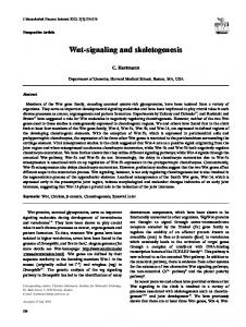
Results and Discussion Wnt/β-catenin signaling induces expression of GLI1. To analyze the effect of Wnt/β-catenin signaling on the modulation of the Hh pathway, we studied the expression of the GLI family transcription factors in different cell types after Wnt/β-catenin signaling was either activated or inhibited. We observed an upregulation of GLI1 mRNA levels in HeLa cells treated with recombinant Wnt3A protein (Fig. 1A). Transfection of 293T cells with β-catenin and Tcf4 resulted in elevated expression of GLI1 mRNA and protein (Fig. 1B). Colorectal cancer cells DLD1D7Δ15 and LS174 T-L8, characterized by constitutive activation of the β-catenin/Tcf4
8573
Cancer Res 2009; 69: (22). November 15, 2009
Cancer Research
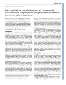
signaling, carry doxycycline-inducible dominant-negative Tcf4 mutant (dnTcf4). Both of these cell lines showed a significant reduction in GLI1 expression in the presence of doxycycline at the RNA and protein levels (Fig. 1C). Similarly, blocking canonical Wnt signaling with dnTCF in zebrafish embryos resulted in reduction of gli1 mRNA levels (Fig. 1D). These results show that Wnt/β-catenin signaling induces GLI1 expression to the extent similar to other Wnt-regulated genes (axin2 and Tcf1; Supplementary Fig. S1A–C) and this induction is evolutionarily conserved and not cell type restricted. Our findings corroborate a recent study that suggested enhancement of GLI transcriptional activity by β-catenin in different human cancer cells (14). Our results also show that GLI1 induction is Tcf dependent, as overexpression of the dominant-negative form of Tcf inhibited GLI1 expression in DLD1D7Δ15 and LS174 T-L8 cells and in zebrafish embryos. CRD-BP binds to the coding region of GLI1 mRNA and stabilizes it. To investigate the mechanism of GLI1 regulation by Wnt/β-catenin signaling, 293T cells transfected with β-catenin/ Tcf were treated with actinomycin D to inhibit transcription, and GLI1 mRNA expression was determined. In contrast to the regulation of the Wnt transcriptional target axin2 (29–31), this treatment did not prevent β-catenin/Tcf–dependent GLI1 induction in 293T cells (Supplementary Figs. S2A and B), suggesting that upregulation of GLI1 is posttranscriptional. Further studies using doxycycline-regulated expression of GLI1 showed that Wnt signaling stabilizes GLI1 mRNA in cells (Fig. 2A). We have previously reported that Wnt/β-catenin signaling induces the expression of
mRNA binding protein, CRD-BP (16). This protein was shown to bind and stabilize different mRNAs, including the mRNA of the proto-oncogene c-myc (32), the mRNA of the β-TrCP1 ubiquitin ligase receptor (16, 33), and the mRNA of MDR1, the multidrug resistance P-glycoprotein gene (34). We sought to determine whether GLI1 mRNA expression could be affected by CRD-BP as well. We found that GLI1 mRNA half-life was drastically increased when CRD-BP was overexpressed (Fig. 2A). Overexpression of CRD-BP also upregulated steady-state levels of GLI1 mRNA and protein (Fig. 2B). Because CRD-BP is an mRNA binding protein, we hypothesized that it might directly bind to GLI1 mRNA and induce its stabilization. Indeed, CRD-BP interacted directly with GLI1 mRNA, with the strongest binding observed within the first ∼900 bases (41–990) of the coding region of GLI1 mRNA (Fig. 2C). Overall, these results suggest that, upon Wnt/β-catenin signaling activation, CRD-BP is upregulated and then binds to the GLI1 mRNA and stabilizes it. CRD-BP did not target GLI2 and GLI3 mRNAs. These data are anticipated because β-catenin/Tcf4 that upregulates CRD-BP expression (16) could not induce GLI2 and GLI3 mRNA expression (Supplementary Fig. S2C). Wnt/β-catenin signaling induces the expression and transcriptional activity of GLI1 in a CRD-BP–dependent manner. CRD-BP knockdown largely prevented β-catenin/Tcf–dependent GLI1 upregulation in 293T cells, as well as in NIH3T3 cells (Fig. 2D and Supplementary Fig. S3). Moreover, downregulation of CRD-BP by shRNA prevented the induction of Gli1 transcriptional activity by the Wnt/β-catenin signaling (Fig. 3A). Doxycycline-induced
Figure 1. Wnt/β-catenin signaling induces the expression of GLI1. A, Northern blot (top) analysis of GLI1 mRNA levels in HeLa cells either untreated or treated with the recombinant mouse Wnt-3A (100 ng/mL) for 15 h. Immunoblot (bottom) analysis of CRD-BP expression levels in the corresponding cells. B, Northern blot (top) analysis of GLI1 mRNA levels in 293T cells transfected with pcDNA3.1 or β-catenin/Tcf4. Immunoblot (bottom) analyses of GLI1 and CRD-BP expression levels in the corresponding cells. C, Northern blot (top) analysis of GLI1 mRNA levels in DLD1D7Δ15 and LS174 T-L8 cells untreated or treated with doxycycline (Dox, 1 μg/mL) for 24 h. Immunoblot (bottom) analyses of GLI1 and CRD-BP expression levels in the corresponding cells. 7S was used as internal control for Northern blot analyses and β-actin as internal control for immunoblot analyses. Numbers in the A, B, and C represent relative densitometry measurements. D, the levels of gli1 mRNA were determined by quantitative reverse transcriptase-PCR (RT-PCR) in zebrafish transgenic Tg(HS:dTCF-GFP) embryos bearing heat shock–inducible dnTCF fused to GFP. GFP-positive embryos (dTCF), which expressed dnTCF, were selected by fluorescence and analyzed separately from their GFP-negative siblings (control).
Cancer Res 2009; 69: (22). November 15, 2009
8574
www.aacrjournals.org
Wnt Signaling Stabilizes GLI1 mRNA
Figure 2. Wnt/β-catenin signaling modulates Hh signaling by upregulating CRD-BP, which stabilizes GLI1 mRNA. A, 293T cells were cotransfected with Tet-off, pTRE-Tight-GLI1, and either pcDNA3.1, β-catenin/Tcf4, or Flag-CRD-BP. Forty-eight hours after transfection, transcription was stopped by treatment with doxycycline (1 μg/mL) for the indicated durations, and the stability of GLI1 mRNA was analyzed by Northern blotting (top) and presented graphically (bottom). B, expression levels of GLI1 mRNA determined by Northern blot (top) analysis in 293T cells transfected with pcDNA3.1 or Flag-CRD-BP. Protein expression of GLI1 and CRD-BP was determined by immunoblot (bottom) analyses in the corresponding cells. C, Flag immunoprecipitation of UV cross-linked complexes of the 41-990, 973-1824, 1808-2730, and 2713-3600 bp fragments of GLI1 mRNA as well as the full-length form and proteins from 293T cells transfected with Flag-CRD-BP or pcDNA3.1 plasmid. The diagram illustrates the position of each fragment on GLI1 mRNA. D, Northern blot (top) analysis of GLI1 expression in 293T cells and immunoblot (bottom) analyses of GLI1 and CRD-BP expression in 293T and NIH3T3 cells cotransfected with pcDNA3.1 and irrelevant shRNA, β-catenin/Tcf4, and irrelevant shRNA or β-catenin/Tcf4 and CRD-BP shRNA. 7S was used as internal control for Northern blot analyses and β-actin as internal control for immunoblot analyses. Numbers in B and D represent relative densitometry measurements.
inhibition of β-catenin/Tcf4 signaling resulted in downregulation of GLI1-dependent transcription, whereas CRD-BP was able to upregulate GLI1-dependent transcriptional activity in DLD1D7Δ15 colorectal cancer cells, regardless of the status of Wnt/β-catenin signaling (Fig. 3B). Knockdown of GLI1 abrogated CRD-BP– controlled regulation of GLI-dependent transcription (Fig. 3C), confirming the specificity of the CRD-BP role in GLI1 regulation. These results show that control of GLI1 expression and activity by the Wnt/β-catenin signaling depends on CRD-BP.
www.aacrjournals.org
Interestingly, this regulation of GLI-dependent transcriptional activity seems to be independent of upstream Hh signaling, as neither inhibitor (cyclopamine) nor activator (SAG) of SMO had any effect on either basal or Wnt- and CRD-BP–regulated GLI-luciferase (Fig. 3B). These data, whereas confirming a previous observation that upstream Hh pathway inhibitors do not affect GLI transcriptional activity in colorectal cancer cells (19), suggest a novel, SMO-independent, mode of regulation of GLI transcriptional outcome by Wnt signaling pathway.
8575
Cancer Res 2009; 69: (22). November 15, 2009
Cancer Research
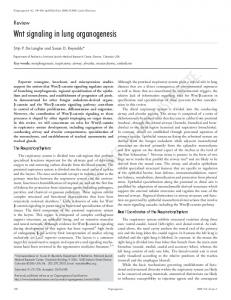
GLI1 contributes to β-catenin and CRD-BP–dependent proliferation of colorectal cancer cells. Several reports point to the involvement of Hh signaling in the genesis of colorectal cancers (13, 15), where the role of Wnt signaling is well established. Enhancement of GLI transcriptional activity by β-catenin has been shown in different human cancer cells (14), although the mechanism of this activation is not clear. Interestingly, a recent study reported the absence of canonical Hh signaling in a variety of epithelial (including colorectal) cancer cells (19). However, GLI1 protein was also found to be overexpressed in some colorectal cancers independently of Shh signaling (35, 36). Taken together, these studies imply that Wnt signaling controls Hh signaling in colorectal cancers. In cancer cells, Hh is primarily a proliferative stimulus (37) and GLI1 is a transcriptional mediator of Hh signaling (38). Because GLI1 is also upregulated by the Wnt/β-catenin signaling, we sought to investigate whether GLI1 contributes to Wnt/βcatenin–dependent proliferation of human colorectal cancer cells. To assess this function of Wnt/β-catenin–mediated upregulation of GLI1, we tested whether GLI1 coexpression could rescue Wnt/β-catenin–dependent colony formation when this pathway is inhibited. GLI1 could indeed partially rescue the ability of DLD1D7Δ15 and LS174 T-L8 cells to form colonies when Wnt/βcatenin signaling was inhibited by doxycycline treatment–induced expression of dnTcf4 (Fig. 4A). Interestingly, ectopic expression of GLI1 failed to induce cyclin D1 and c-myc mRNA expression in the
absence of Wnt signaling (Supplementary Fig. S4A), suggesting that the partial rescue of the ability of DLD1D7Δ15 and LS174 T-L8 cells to form colonies by GLI1 is probably independent of cyclin D1 and c-myc. In addition, although the knockdown of CRD-BP may affect the proliferation of colorectal cancer cells through multiple mechanisms, our data showed that overexpression of GLI1 resulted in attenuation of the inhibitory effect of CRD-BP shRNA in colony formation in colorectal cancer cells DLD1D7Δ15 and LS174 T-L8 (Fig. 4B). This indicates that GLI1 contributes to Wnt- and CRDBP–dependent proliferation of DLD1D7Δ15 and LS174 T-L8 cells. GLI1 did not affect the expression of cyclin D1 and c-myc mRNA in these experiments either (Supplementary Fig. S4B), which also suggests that other target genes are involved in partial rescue of these cells to form colonies by GLI1. GLI1 contribution to the growth of colorectal cancer cells was not limited to DLD1D7Δ15 and LS174 T-L8 cells: Knockdown of GLI1 (using two independent GLI1-targeting shRNA constructs) drastically decreased the ability of SW480, SW620, and HCT116 cells to form colonies (Supplementary Fig. S5A and B), indicating a requirement for GLI1 in proliferation and survival of colorectal cancer cells. Our data support previous findings that showed the involvement of GLI1 in colorectal cancer cell proliferation (13, 15). In contrast, another study showed that GLI1 overexpression suppressed proliferation of SW480 and HCT116 colorectal cancer cells with activated Wnt signaling (7). These cells exhibit high levels of endogenous GLI1 (Supplementary
Figure 3. Wnt/β-catenin signaling induces the expression of GLI1 in a CRD-BP–dependent manner. A, NIH 3T3 cells were grown in six-well plates and cotransfected with 8×3′GLI BS-LucII reporter plasmid and pSV-40-galactosidase, and irrelevant shRNA or CRD-BP shRNA plasmid, and then treated with Wnt-3A (100 ng/mL) for 15 h as indicated. Forty-eight hours after transfection, luciferase activity was estimated using luciferase reporter assay reagent (Promega). β-Galactosidase was used for normalization and estimated using the β-galactosidase assay reagent (Pierce). B, DLD1D7Δ15 cells were grown in a six-well plate and cotransfected with 8×3′GLI BS-LucII reporter plasmid, pSV-40 β-galactosidase, and Flag-CRD-BP or empty plasmid and then treated with doxycycline (1 μg/mL) for 24 h, cyclopamine (10 μmol/L) for 24 h, or SAG (3 nmol/L) for 30 h as indicated. Luciferase activity was estimated as in A. C, 293T cells grown in a six-well plate were cotransfected with 8×3′GLI BS-LucII reporter plasmid, pSV-40 β-galactosidase, and Flag-CRD-BP or empty plasmid and irrelevant shRNA or GLI1 shRNA (shGLI1-C). Luciferase activity was estimated as in A. *, P < 0.01, compared with controls in Student's t test.
Cancer Res 2009; 69: (22). November 15, 2009
8576
www.aacrjournals.org
Wnt Signaling Stabilizes GLI1 mRNA
Figure 4. GLI1 contributes to β-catenin and CRD-BP–dependent proliferation of colorectal cancer cells. A, DLD1D7Δ15 and LS174 T-L8 cells were grown in a 100-mm dish and cotransfected with pTK-puro plasmid and either the empty vector or pcDNA3.1-GLI1 as indicated. Forty-eight hours after transfection, cells from each plate were seeded in five 100-mm plates and the cells were treated with doxycycline (1 μg/mL) for 72 h and puromycin (8 μg/mL) for 10 d. The colonies formed were counted under a microscope. B, DLD1D7Δ15 and LS174 T-L8 cells grown in 100-mm plates were cotransfected with pTK-puro plasmid and irrelevant shRNA, CRD-BP shRNA, irrelevant shRNA and pcDNA3.1-GLI1, or CRD-BP shRNA and pcDNA3.1-GLI1 as indicated. Forty-eight hours after transfection, cells from each plate were seeded in five 100-mm plates and treated with puromycin (8 μg/mL) for 10 d. The colonies were counted under a light microscope. C, levels of GLI1 mRNA in primary colorectal normal and tumor human tissue samples determined by quantitative RT-PCR. D, immunoblot analysis of proteins isolated from normal (N) and tumor (T) samples of ApcMin−/+ mouse small intestine and colon for Gli1, CRD-BP, and β-catenin. β-Actin was used as internal control.
Fig. S5C), and, perhaps, overexpression of GLI1 does not represent relevant pathophysiologic conditions. On the other hand, knockdown of GLI1 expression in a variety of colorectal cancer cells, including SW480 and HCT116, resulted in dramatic inhibition of colony formation (Supplementary Fig. S5A), further supporting the importance of GLI1 for the proliferation of colorectal cancer cells. The Wnt/β-catenin pathway is an important player in colorectal carcinogenesis. We next assessed the expression of GLI1 in primary human colorectal tumor samples and colorectal cancer cell lines. We found that all human colorectal tumor samples previously characterized by an activation of β-catenin and high levels of CRD-BP (16, 39) overexpressed GLI1 mRNA to different extents (Fig. 4C). Similarly, we have found a positive correlation between activation of β-catenin signaling and expression of CRD-BP and GLI1 in a panel of established colorectal cancer cell lines (Supplementary Fig. S5C). We also analyzed ApcMin+/− mice, heterozygous for a nonsense mutation in the APC locus that predisposes them to the development of multiple adenomas throughout the entire intestinal tract (40), for expression of Gli1. These mice overexpressed Gli1 in their intestinal tumors but not in the normal intestine
www.aacrjournals.org
(Fig. 4D and Supplementary Fig. S6). These findings further support both a role for GLI1 in colorectal cancer formation and its regulation by Wnt/β-catenin signaling. Increased expression of GLI1 mRNA was previously reported in human colonic adenocarcinomas (13); however, its association with Wnt/β-catenin signaling was not studied. Two other studies observed that the upregulation of GLI1 expression in colorectal carcinomas was not always consistent with the expression pattern of Shh, suggesting that the Hh pathway might be activated by other regulatory mechanisms in colorectal carcinomas (35, 36). Another study reported that Indian hedgehog (Ihh) signaling stimulates colonic epithelial differentiation and inhibits proliferation by antagonizing Wnt signaling (6); however, Yauch and colleagues have recently shown that Hh ligands failed to activate canonical Hh signaling in tumor epithelial cells (19), suggesting that the observed upregulation of GLI1 in colorectal cancer cells may not be a result of activation of upstream Hh signaling. A body of evidence points to a regulatory role of Wnt and Hh pathways in stem cell development in epithelia, including those of the intestine (reviewed in ref. 37). Additionally, recent studies suggest that colorectal tumors might arise from intestinal stem cells (41). Taken together, it is likely that cross-talk between
8577
Cancer Res 2009; 69: (22). November 15, 2009
Cancer Research
the Wnt and Hh pathways exists in the development of colorectal cancers and one proposed mechanism of this interaction involves control of GLI1 expression by both pathways. Wnt and Hh are pivotal pathways similar to each other in several respects (42, 43). These pathways interact at multiple levels in embryonic organ patterning as well as in the development of some cancers. It is likely that different mechanisms are used for this cross-talk in different contexts. In this study, we identify a novel mechanism by which Wnt signaling regulates Hh signaling and show that this mechanism uses GLI1 mRNA stabilization. To the best of our knowledge, this is the first study that shows posttranscriptional regulation of the Hh signaling by the Wnt/β-catenin signaling pathway. It is also the first study to identify CRD-BP as a mechanistic link between the two pathways. Altogether, this work sheds light on the regulatory role of Wnt signaling in controlling Hh signaling and offers significant new insight on the involve-
References 1. Smalley MJ, Dale TC. Wnt signalling in mammalian development and cancer. Cancer Metastasis Rev 1999; 18:215–30. 2. Nusse R. Wnt signaling in disease and in development. Cell Res 2005;15:28–32. 3. Wicking C, Smyth I, Bale A. The hedgehog signalling pathway in tumorigenesis and development. Oncogene 1999;18:7844–51. 4. Marcelle C, Stark MR, Bronner-Fraser M. Coordinate actions of BMPs, Wnts, Shh and noggin mediate patterning of the dorsal somite. Development 1997;124: 3955–63. 5. Lee CS, Buttitta LA, May NR, Kispert A, Fan CM. SHH-N upregulates Sfrp2 to mediate its competitive interaction with WNT1 and WNT4 in the somitic mesoderm. Deve‐ lopment 2000;127:109–18. 6. van den Brink GR, Bleuming SA, Hardwick JC, et al. Indian Hedgehog is an antagonist of Wnt signaling in colonic epithelial cell differentiation. Nat Genet 2004; 36:277–82. 7. Akiyoshi T, Nakamura M, Koga K, et al. Gli1, down‐ regulated in colorectal cancers, inhibits proliferation of colon cancer cells involving Wnt signalling activation. Gut 2006;55:991–9. 8. Mullor JL, Dahmane N, Sun T, Ruiz i Altaba A. Wnt signals are targets and mediators of Gli function. Curr Biol 2001;11:769–73. 9. Li X, Deng W, Lobo-Ruppert SM, Ruppert JM. Gli1 acts through Snail and E-cadherin to promote nuclear signaling by β-catenin. Oncogene 2007;26:4489–98. 10. Yang SH, Andl T, Grachtchouk V, et al. Pathological responses to oncogenic Hedgehog signaling in skin are dependent on canonical Wnt/β-catenin signaling. Nat Genet 2008;40:1130–5. 11. Borycki A, Brown AM, Emerson CP, Jr. Shh and Wnt signaling pathways converge to control Gli gene activation in avian somites. Development 2000;127: 2075–87. 12. Iwatsuki K, Liu HX, Gronder A, et al. Wnt signaling interacts with Shh to regulate taste papilla development. Proc Natl Acad Sci U S A 2007;104:2253–8. 13. Douard R, Moutereau S, Pernet P, et al. Sonic Hedgehogdependent proliferation in a series of patients with colorectal cancer. Surgery 2006;139:665–70. 14. Maeda O, Kondo M, Fujita T, et al. Enhancement of GLI1-transcriptional activity by β-catenin in human cancer cells. Oncol Rep 2006;16:91–6. 15. Qualtrough D, Buda A, Gaffield W, Williams AC, Paraskeva C. Hedgehog signalling in colorectal tumour
ment of the two pathways in colorectal cancer development. These findings suggest CRD-BP as a valuable candidate drug target for therapy in cancers associated with deregulation of Wnt/β-catenin and Hh signaling pathways.
Disclosure of Potential Conflicts of Interest No potential conflicts of interest were disclosed.
Acknowledgments Received 4/23/09; revised 8/25/09; accepted 9/18/09; published OnlineFirst 11/3/09. Grant support: grant (V.S. Spiegelman) and grant (Y. Grinblat). The costs of publication of this article were defrayed in part by the payment of page charges. This article must therefore be hereby marked advertisement in accordance with 18 U.S.C. Section 1734 solely to indicate this fact. We thank Drs. F. Aberger, H. Clevers, K. Kinzler, J. Ross, H. Sasaki, and B. Vogelstein for their generous gifts of reagents; Dr. S. Fuchs for critical reading of the manuscript; and Dr. K. Spiegelman for help with the manuscript preparation.
cells: induction of apoptosis with cyclopamine treatment. Int J Cancer 2004;110:831–7. 16. Noubissi FK, Elcheva I, Bhatia N, et al. CRD-BP mediates stabilization of βTrCP1 and c-myc mRNA in response to β-catenin signalling. Nature 2006;441: 898–901. 17. van de Wetering M, Sancho E, Verweij C, et al. The βcatenin/TCF-4 complex imposes a crypt progenitor phenotype on colorectal cancer cells. Cell 2002;111: 241–50. 18. Chen JK, Taipale J, Young KE, Maiti T, Beachy PA. Small molecule modulation of Smoothened activity. Proc Natl Acad Sci U S A 2002;99:14071–6. 19. Yauch RL, Gould SE, Scales SJ, et al. A paracrine requirement for hedgehog signalling in cancer. Nature 2008;455:406–10. 20. Ross J, Lemm I, Berberet B. Overexpression of an mRNA-binding protein in human colorectal cancer. Oncogene 2001;20:6544–50. 21. Spiegelman VS, Slaga TJ, Pagano M, Minamoto T, Ronai Z, Fuchs SY. Wnt/β-catenin signaling induces the expression and activity of βTrCP ubiquitin ligase receptor. Mol Cell 2000;5:877–82. 22. Willinger T, Freeman T, Herbert M, Hasegawa H, McMichael AJ, Callan MF. Human naive CD8 T cells down-regulate expression of the WNT pathway transcription factors lymphoid enhancer binding factor 1 and transcription factor 7 (T cell factor-1) following antigen encounter in vitro and in vivo. J Immunol 2006;176: 1439–46. 23. Medeiros LJ, Hai S, Thomazy VA, Estalilla OC, Romaguera J, Luthra R. Real-time RT-PCR assay for quantifying cyclin D1 mRNA in B-cell non-Hodgkin's lymphomas. Mod Pathol 2002;15:556–64. 24. Fields WR, Desiderio JG, Putnam KP, Bombick DW, Doolittle DJ. Quantification of changes in c-myc mRNA levels in normal human bronchial epithelial (NHBE) and lung adenocarcinoma (A549) cells following chemical treatment. Toxicol Sci 2001;63:107–14. 25. Grinblat Y, Gamse J, Patel M, Sive H. Determination of the zebrafish forebrain: induction and patterning. Development 1998;125:4403–16. 26. Westerfield M. The zebrafish book: a guide for the laboratory use of zebrafish. Oregon: University of Oregon Press; 1995. 27. Kimmel CB, Ballard WW, Kimmel SR, Ullmann B, Schilling TF. Stages of embryonic development of the zebrafish. Dev Dyn 1995;203:253–310. 28. Lewis JL, Bonner J, Modrell M, et al. Reiterated Wnt signaling during zebrafish neural crest development. Development 2004;131:1299–308.
Cancer Res 2009; 69: (22). November 15, 2009
8578
29. Jho EH, Zhang T, Domon C, Joo CK, Freund JN, Costantini F. Wnt/β-catenin/Tcf signaling induces the transcription of Axin2, a negative regulator of the signaling pathway. Mol Cell Biol 2002;22:1172–83. 30. Lustig B, Jerchow B, Sachs M, et al. Negative feedback loop of Wnt signaling through upregulation of conductin/axin2 in colorectal and liver tumors. Mol Cell Biol 2002;22:1184–93. 31. Yan D, Wiesmann M, Rohan M, et al. Elevated expression of axin2 and hnkd mRNA provides evidence that Wnt/β-catenin signaling is activated in human colon tumors. Proc Natl Acad Sci U S A 2001;98:14973–8. 32. Prokipcak RD, Herrick DJ, Ross J. Purification and properties of a protein that binds to the C-terminal coding region of human c-myc mRNA. J Biol Chem 1994; 269:9261–9. 33. Elcheva I, Goswami S, Noubissi FK, Spiegelman VS. CRD-BP protects the coding region of βTrCP1 mRNA from miR-183-mediated degradation. Mol Cell 2009;35: 240–6. 34. Sparanese D, Lee CH. CRD-BP shields c-myc and MDR-1 RNA from endonucleolytic attack by a mammalian endoribonuclease. Nucleic Acids Res 2007;35: 1209–21. 35. Bian YH, Huang SH, Yang L, Ma XL, Xie JW, Zhang HW. Sonic hedgehog-Gli1 pathway in colorectal adenocarcinomas. World J Gastroenterol 2007;13:1659–65. 36. Yoshizaki A, Nakayama T, Naito S, Wen CY, Sekine I. Expressions of sonic hedgehog, patched, smoothened and Gli-1 in human intestinal stromal tumors and their correlation with prognosis. World J Gastroenterol 2006; 12:5687–91. 37. Taipale J, Beachy PA. The Hedgehog and Wnt signalling pathways in cancer. Nature 2001;411:349–54. 38. Matise MP, Joyner AL. Gli genes in development and cancer. Oncogene 1999;18:7852–9. 39. Ougolkov A, Zhang B, Yamashita K, et al. Associations among β-TrCP, an E3 ubiquitin ligase receptor, β-catenin, and NF-κB in colorectal cancer. J Natl Cancer Inst 2004;96:1161–70. 40. Moser AR, Pitot HC, Dove WF. A dominant mutation that predisposes to multiple intestinal neoplasia in the mouse. Science 1990;247:322–4. 41. O'Brien CA, Pollett A, Gallinger S, Dick JE. A human colon cancer cell capable of initiating tumour growth in immunodeficient mice. Nature 2007;445:106–10. 42. Kalderon D. Similarities between the Hedgehog and Wnt signaling pathways. Trends Cell Biol 2002;12:523–31. 43. Nusse R. Wnts and Hedgehogs: lipid-modified proteins and similarities in signaling mechanisms at the cell surface. Development 2003;130:5297–305.
www.aacrjournals.org