Plant Physiology, July 1999, Vol. 120, pp. 727–737, www.plantphysiol.org © 1999 American Society of Plant Physiologists
Xanthophyll Cycle Pigment Localization and Dynamics during Exposure to Low Temperatures and Light Stress in Vinca major1 Amy S. Verhoeven2*, William W. Adams III, Barbara Demmig-Adams, Roberta Croce, and Roberto Bassi Department of Environmental, Population, and Organismic Biology, University of Colorado, Boulder, Colorado 80309–0334 (A.S.V., W.W.A., B.D.-A.); and Universita di Verona, Facolta di Scienze Matematiche, Fisiche e Naturali, Biotecnologie Vegetali, Strada Le Grazie, 37134 Verona, Italy (R.C., R.B.) found to be retained in leaves for extended periods after darkening (Demmig et al., 1988; Adams et al., 1995; Verhoeven et al., 1996; Demmig-Adams et al., 1998). The xanthophyll cycle pigments Z and A are formed from V when light is excessive, and they are involved in a photoprotective process whereby excess absorbed excitation energy is dissipated thermally in the light-harvesting antennae of PSII (Demmig-Adams and Adams, 1996; Eskling et al., 1997; Gilmore, 1997). The retention of Z plus A in photoinhibited leaves often correlates closely with sustained low PSII efficiencies measured as the Fv/Fm (Adams et al., 1995; Verhoeven et al., 1996; Demmig-Adams et al., 1998, and refs. therein). Such correlations have led to the suggestion that Z plus A may be engaged for thermal energy dissipation under these conditions and may therefore be involved in the reduced PSII efficiencies observed. To influence Chl fluorescence yield xanthophylls must be localized in close proximity to the pigment-protein complexes of the thylakoid membrane, and knowledge of their precise organization is important to understand the mechanism of (Z plus A)-dependent energy dissipation. Several studies have demonstrated that the xanthophyll cycle pigments (VAZ) are associated with all light-harvesting components, including LHCI (Thayer and Bjo¨rkman, 1992; Lee and Thornber, 1995). Among LHCII components, VAZ has been reported to be enriched in the minor CPs (CP29, CP26, and CP24) relative to the major LHC of PSII, LHCII (Bassi et al., 1993; Ruban et al., 1994; Lee and Thornber, 1995; Goss et al., 1997), suggesting an important role for the minor CPs in photoprotection (Bassi et al., 1993; Gilmore, 1997). Upon illumination, V is apparently converted to Z in all complexes (Ruban et al., 1994; Lee and Thornber, 1995; Phillip and Young, 1995; Fa¨rber et al., 1997; Zhu et al., 1997), with the degree of epoxidation varying among the different LHCs (Ruban et al., 1994; Croce et al., 1996a). Although several studies have been conducted to examine the distribution of the xanthophyll cycle pigments among pigment-protein complexes isolated from unstressed leaves, very little is know about their distribution
The distribution of xanthophyll cycle pigments (violaxanthin plus antheraxanthin plus zeaxanthin [VAZ]) among photosynthetic pigment-protein complexes was examined in Vinca major before, during, and subsequent to a photoinhibitory treatment at low temperature. Four pigment-protein complexes were isolated: the core of photosystem (PS) II, the major light-harvesting complex (LHC) protein of PSII (LHCII), the minor light-harvesting proteins (CPs) of PSII (CP29, CP26, and CP24), and PSI with its LHC proteins (PSILHCI). In isolated thylakoids 80% of VAZ was bound to protein independently of the de-epoxidation state and was found in all complexes. Plants grown outside in natural sunlight had higher levels of VAZ (expressed per chlorophyll), compared with plants grown in low light in the laboratory, and the additional VAZ was mainly bound to the major LHCII complex, apparently in an acidlabile site. The extent of de-epoxidation of VAZ in high light and the rate of reconversion of Z plus A to V following 2.5 h of recovery were greatest in the free-pigment fraction and varied among the pigment-protein complexes. Photoinhibition caused increases in VAZ, particularly in low-light-acclimated leaves. The data suggest that the photoinhibitory treatment caused an enrichment in VAZ bound to the minor CPs caused by de novo synthesis of the pigments and/or a redistribution of VAZ from the major LHCII complex.
Photoinhibition refers to a condition in which a persistent decrease in the efficiency of photosynthetic energy conversion in leaves is observed. Photoinhibition occurs in the field in plants exposed to conditions of high light in combination with environmental stress, such as cold temperatures, but can also be induced by exposure of shadeacclimated leaves to high light (Krause, 1994; Osmond, 1994). Under various photoinhibitory conditions large quantities of the xanthophyll cycle pigments Z and A have been 1
This work was supported by the U.S. National Science Foundation (award no. IBN-9631064 to W.W.A. and B.D.-A.), a fellowship from the David and Lucile Packard Foundation to B.D.-A., grants from the Ministry of University and Scientific Research, and the Biotechnology Project of the Italian National Research Council to R.B. 2 Present address: University of St. Thomas, Department of Biology, 2115 Summit Avenue, St. Paul, MN 55105–1096. * Corresponding author; e-mail
[email protected]; fax (Hawaii) 1– 808 –956 –3542; fax (Minnesota) 1– 651–962–5209.
Abbreviations: A, antheraxanthin; Chl, chlorophyll; CP, Chlbinding protein; ELIP, early-light-inducible protein; Fv/Fm, ratio of variable to maximal Chl fluorescence; GC, growth chamber; LHCI or II, light-harvesting complex I or II; OD, outdoors; V, violaxanthin; VAZ, V plus A plus Z; Z, zeaxanthin. 727
728
Verhoeven et al.
under photoinhibitory conditions when the rate of Z epoxidation is severely slowed following leaf darkening. The major goal of this study was to assess whether changes occur in the levels or distribution of VAZ in the pigmentprotein complexes when plants are treated with such photoinhibitory high light, as well as whether differences exist in the rate at which Z and A are reconverted to V on the different pigment-proteins during the slow recovery from photoinhibition.
MATERIALS AND METHODS Plant Material and Photoinhibitory Treatments Vinca major var variegata Loud. plants were acquired at a local greenhouse in northern Italy on two occasions, resulting in plants that were acclimated to two different growth conditions. One set of plants was grown at a low light intensity (approximately 50 mmol photons m22 s21) provided by a combination of HQE fluorescent tubes and R80-Natura (Osram, Munich, Germany) incandescent bulbs. Plants were grown in soil, watered every 2 d, and received Nitsch’s nutrient solution (Nitsch and Nitsch, 1969) once per week. The temperature was 22°C/28°C, night/day, with 80% RH. Plants were acclimated to lowlight conditions for at least 6 weeks prior to treatment. A second set of plants was grown outside in northern Italy in September 1998, when the maximum light intensity at midday was approximately 1900 mmol photons m22 s21. The temperature in the last 6 weeks prior to experimentation was 18°C to 24°C (night)/26°C to 34°C (day). Plants were watered and received nutrients daily. For the photoinhibitory treatment, plants were exposed to continuous light and chilling temperatures (480 mmol photons m22 s21 at 15°C for the plants grown in GCs and 430 mmol photons m22 s21 at 10°C for the plants grown OD in full sunlight) until photoinhibition was observed, which was measured by moving a leaf into darkness and measuring Fv/Fm after 30 min. Very slowly relaxing Fv/Fm was achieved after 48 h of exposure to continuous light in plants grown in GCs and after 122 h of exposure to continuous light in plants grown OD. Three sets of leaves were harvested for thylakoid isolation: controls harvested after 12 h of darkness before each treatment, leaves harvested directly following high-light treatment, and leaves harvested after an additional 2.5 h of recovery in darkness at room temperature. In each case, all of the plant leaves were harvested for thylakoid isolation.
Thylakoid Isolation Thylakoid membranes were isolated as described previously by Bassi et al. (1988) except the grinding buffer consisted of 0.1 m Tricine, pH 7.8, 0.4 m sorbitol, 0.5% nonfat dried milk, 0.2 mm PMSF, 5 mm e-amino-n-caproic acid, and 1 mm benzamidine; the washing buffer consisted of 25 mm Hepes/KOH, pH 7.5, and 10 mm EDTA; and the resuspension buffer consisted of 10 mm Hepes/KOH, pH 7.5, 1 mm EDTA, and 50% (v/v) glycerol.
Plant Physiol. Vol. 120, 1999
Chl Fluorescence Fluorescence measurements were performed on intact leaves under the respective growth PPFD conditions with a portable fluorometer (PAM-2000, Walz, Effeltrich, Germany). Fluorescence measurements and calculations were performed as described previously (Demmig-Adams and Adams, 1996; Demmig-Adams et al., 1996). Solubilization and Fractionation of Thylakoids and Identification of Pigment-Protein Complexes Thylakoids were resuspended in 2 mg Chl/mL and solubilized by adding an equal volume of 2.8% dodecyl maltoside in water. The sample was then vortexed for 20 s and put on ice for 1 min. The solubilized sample was spun for 2 min at 15,000g and 4°C and rapidly loaded onto a 0.1 to 1 m Suc gradient containing 10 mm Hepes, pH 7.6, and 0.06% dodecyl maltoside. The gradient was spun in a Beckman SW41 rotor at 39,000 rpm for 27 h at 4°C. Individual green fractions were harvested with a syringe. Pigmentprotein complexes were identified using analytical SDSPAGE, spectroscopy, and pigment analysis, as described previously for several different species (Di Paolo et al., 1990; Santini et al., 1994; Kilian et al., 1997). Electrophoresis and Immunoblotting Analytical SDS-PAGE was performed with gradient gels (10%–16% acrylamide) using the Tris/Tricine buffer system of Scha¨gger and von Jagow (1987). Alternatively, a Tris-sulfate buffer system was used (Bassi et al., 1985a). Preparative IEF was performed as previously described (Dainese et al., 1990). Nondenaturing green gel electrophoresis was done according to the method of Knoetzel and Simpson (1991). For immunoblot assays, samples were separated by the Tris-sulfate gel system and transferred to a nitrocellulose filter (Millipore). The filters were incubated with antibodies and detected with alkaline phosphatase coupled to anti-rabbit IgG (Sigma). Analysis of Pigments Total pigments were extracted with 80% acetone. Analysis of the extracts by HPLC was as described previously (Gilmore and Yamamoto, 1991). In this study the determination of the Chl-to-carotenoid ratio in pigment proteins was of crucial importance. The results of HPLC analysis were therefore verified by fitting the absorption spectra of ethanolic extracts of pigment proteins with the spectra of pure pigments in ethanol (Connelly et al., 1997). Spectra (350–750 nm) were recorded using a DW2000 spectrophotometer in the split-beam mode (Aminco, Silver Spring, MD). Individual pigments were purified by HPLC, dried under a vacuum, and resuspended in 96% ethanol. Curve fitting was obtained by using a nonlinear least-squares fitting code (Origin, MicroCal Software, Northampton, MA). Verification of Specificity of Pigment Binding To verify the specificity of xanthophyll binding to the different pigment-protein complexes, an experiment was
Zeaxanthin Localization and Dynamics during Photoinhibition
729
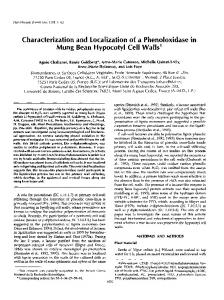
undertaken in which excess xanthophylls were added to solubilized thylakoids before their fractionation. No change in pigment composition of the protein complexes was observed after fractionation, suggesting that no nonspecific binding of the xanthophylls was caused by the solubilization conditions. DEAE Chromatography of the PSII Core The Suc-gradient fraction containing the PSII core was purified using DEAE chromatography as described previously (Giuffra et al., 1996). RESULTS Characterization of Pigment Content and Distribution prior to Photoinhibitory Treatment The pigment content of V. major var variegata Loud. was examined in control conditions from both isolated thylakoids and whole-leaf extracts (Table I). In control conditions the differences in carotenoid levels, relative to Chl a, from plants grown in the GC relative to those grown OD involved predominantly the fraction of VAZ (Table I). In both isolated thylakoids and leaf extracts from plants grown OD, the content of V and VAZ was approximately twice that of the plants grown in GCs. A comparison of the pigment composition of whole-leaf extracts versus isolated thylakoids demonstrates that the ratios of total carotenoid and of VAZ to Chl were not significantly changed following thylakoid isolation. Distribution of VAZ among Pigment Proteins In a first approach to examining the location of pigments within the protein complexes, thylakoid membranes were solubilized with dodecyl maltoside, and Suc-gradient fractionation was performed, followed by analysis of the pigment and protein content of the fractions (Fig. 1; Tables II and III). Five major fractions were obtained. Near the top of the gradient a distinct yellow band was evident (fraction 1). This fraction contained free pigments,
Figure 1. Fully denaturing Tris-sulfate SDS-PAGE of the fractions obtained from Suc-gradient ultracentrifugation of V. major thylakoids collected from dark-adapted (at least 12 h) leaves of GC plants. Fractions from stressed and recovered thylakoids, as well as the fractions obtained from plants grown OD, were very similar and are therefore not depicted. Whole thylakoids (T) were loaded onto the gel in addition to the four fractions (F2–F5) collected; fraction 2 contained the LHCII monomer (B), the minor Chl proteins CP29 (A), CP26 (comigrating with LHCII; B), and CP24 (C); fraction 3 contained the LHCII trimer (D); fraction 4 contained the PSII core with the D1/D2 heterodimer (E), CP47 (F), CP43 (G), and D1/D2 monomer (H); fraction 5 contained the PSI core (I) and LHCI (J). Fraction 1 contained the free pigments (Table III) and is not depicted here.
and V was the major component; only very low levels of Chl were present (Table II). Quantification of pigments in this fraction, relative to the fractions containing protein, showed that 80% to 88% of V in isolated thylakoids was bound to proteins (Table II). This fraction was not examined for proteins using SDS-PAGE; however, in a parallel experiment solubilized thylakoids from all treatments were applied to a nondenaturing deriphat gel and then to SDSPAGE gels in the second dimension. No proteins were visible in the free-pigment fraction in any of the treatments when the gels were stained with Coomassie Blue. The second fraction from the top (fraction 2) was heterogeneous, containing LHCII monomer and the minor Chl proteins (CP24, CP26, and CP29) of PSII; the fraction mi-
Table I. Pigment composition from both thylakoids and leaves of V. major grown either in GCs or OD under natural sunlight Leaves were collected following 12 h of darkness (control), at the end of the photoinhibitory treatment (stress), or following 2.5 h of recovery at low light (recovery). The leaf data are in parentheses, except b-carotene for which the leaf data are not available. See “Materials and Methods” for growth conditions and stress treatments. Pigment
V. major from GCs Control
Stress
V. major from OD Recovery
Control
Stress
Recovery
40.1 (42.0) 6.3 (5.9) 21.8 (21.7) 6.8 2.9 (3.5) 2.6 (2.9) 5.4 (5.3) 10.9 (11.8) 0.74 (0.70)
38.4 (35.2) 6.1 (5.6) 20.1 (19.6) 6.4 4.3 (4.6) 2.5 (3.2) 3.2 (3.8) 10.1 (11.7) 0.56 (0.60)
mol/100 mol Chl a
Chl b Neoxanthin Lutein b-Carotene V A Z VAZ (Z1A)/(VAZ)
39.8 (38.5) 5.5 (5.6) 18.0 (18.3) 6.2 4.5 (4.4) 0 (0) 0 (0) 4.5 (4.4) 0 (0)
39.1 (38.9) 5.0 (4.8) 21.0 (23.6) 5.8 1.8 (2.4) 1.2 (1.4) 4.2 (5.2) 7.2 (9.0) 0.75 (0.73)
37.1 (38.4) 5.3 (5.7) 19.4 (19.6) 5.8 2.2 (2.3) 1.3 (1.3) 3.5 (3.6) 7.0 (7.2) 0.69 (0.68)
39.1 (34.4) 5.9 (5.9) 18.8 (18.8) 6.4 9.1 (9.6) 0 (0) 0 (0) 9.1 (9.6) 0 (0)
730
Verhoeven et al.
Plant Physiol. Vol. 120, 1999
Table II. Pigment composition of the free-pigment fractions (fraction 1) after Suc-gradient ultracentrifugation of solubilized thylakoid membranes Pigment
V. major from GCs Control
Stress
V. major from OD Recovery
Control
% in free pigment band
Chl a Chl b Neoxanthin Lutein b-Carotene V A Z VAZ (Z1A)/(VAZ)
1.4 0.8 4.2 9.3 0 19.5 16.6 1.3 18.4 0.02
1.4 0.6 2.2 11.9 0 6.9 24.8 23.3 19.0 0.90
grating below it (fraction 3) contained pure LHCII trimer. The higher LHCII content in fraction 2 (derived from monomerization of trimeric LHCII) compared with findings from previous reports (Bassi and Dainese, 1992) was due to the relatively high detergent concentration used here to ensure fractionation of relatively pure PSI-LHCI and PSII core complexes. The composition of the xanthophyll cycle pigments in fractions 2 and 3 were strikingly different in fractions isolated from plants grown OD in the sun versus low-light GCs: the V content was 3 times higher in the LHCII trimer fraction (fraction 3) from plants grown OD
Stress
Recovery
% in free pigment band
1.1 0.4 2.0 8.7 0 8.7 20.8 16.3 14.6 0.81
0.7 0.3 4.4 5.2 4.7 12.4 0 0 12.4 0.00
1.3 0.4 2.5 8.6 4.0 8.0 18.3 16.3 14.4 0.82
1.4 0.5 2.9 9.2 6.5 13.0 18.7 13.4 14.5 0.59
(Table III). Although a 40% increase in V content was also observed in fraction 2 (minor CPs), this increase was possibly due entirely to the LHCII monomers present in this fraction (Fig. 1; Table III). The PSII core fraction (fraction 4), migrating below LHCII, was also relatively pure (the Chl a/b ratio was .20). The bottom fraction (fraction 5) contained pure PSI-LHCI with a Chl a/b ratio of about 9 and was the only gradient fraction that contained PSI-LHCI components. VAZ was present in all of the pigment-protein fractions, although in very different amounts. The major V-binding
Table III. Pigment composition of the fractions collected from Suc-gradient ultracentrifugation of solubilized thylakoids from V. major grown in GCs or OD under natural sunlight The pigments Chl b, neoxanthin, lutein, and b-carotene are means 6 SD of the control, stress (48 h of continuous light at 15°C for leaves from GC plants and 122 h of continuous light at 10°C for leaves from plants grown OD), and recovery (2.5 h in low light at 22°C) treatments. Pigment and Treatment
Minor CPs 1 LHCII Monomer (Fraction 2) GC
OD
LHCII Trimer (Fraction 3) GC
OD
PSII Core (Fraction 4)
PSI-LHCI (Fraction 5)
GC
GC
OD
OD
mol/100 mol Chl a
Chl b Neoxanthin Lutein b-Carotene V Control Stress Recovery A Control Stress Recovery Z Control Stress Recovery VAZ Control Stress Recovery (Z1A)/(VAZ) Control Stress Recovery
68.0 6 1.4 10.6 6 0.7 28.8 6 0.4 0
67.8 6 0.3 10.0 6 0.7 27.6 6 1.3 0.4 6 0.3
78.8 6 2.0 12.7 6 0.6 34.8 6 0.5 0
74.8 6 2.7 11.3 6 0.9 31.4 6 1.7 0
5.5 6 1.3 0.6 6 0.5 4.7 6 0.5 17.0 6 0.1
2.8 6 0.8 0.7 6 0.3 3.1 6 0.4 12.0 6 2.9
11.5 6 0.5 0 6.5 6 0.3 9.9 6 0.7
11.5 6 0.2 0.3 6 0.1 5.7 6 0.6 13.6 6 0.5
4.8 2.1 2.5
9.1 3.2 5.0
3.4 0.6 1.0
10.4 2.0 3.3
1.7 1.3 1.0
1.4 0.3 0.7
3.2 2.0 2.3
3.6 1.9 2.4
0.2 1.1 1.4
0 2.1 3.0
0 0.9 1.0
0 1.9 1.9
0 0.7 0.9
0 0.6 0.8
0 0.5 0.6
0 0.8 0.8
0.7 5.2 4.8
0 5.9 4.3
0 2.8 2.7
0 4.8 2.5
0 1.2 1.0
0 1.2 0.9
0 1.3 1.3
0 1.3 1.7
5.7 8.5 8.7
9.1 11.2 12.3
3.4 4.3 4.7
10.4 8.8 7.6
1.7 3.2 2.8
1.4 2.2 2.4
3.2 3.8 4.2
3.6 4.0 4.8
0 0.61 0.66
0 0.82 0.69
0 0.48 0.44
0 0.47 0.46
0.15 0.75 0.72
0 0.68 0.55
0 0.86 0.78
0 0.74 0.53
Zeaxanthin Localization and Dynamics during Photoinhibition
Table IV. PSII efficiency of V. major grown in GCs or OD under natural sunlight Measurements were taken after 12 h of darkness (control), at the end of the high-light treatment (stress), and during recovery in darkness at room temperature. PSII efficiency is either that of open PSII units in darkness (Fv /Fm; D) or that at the actual degree of closure in the light ([Fm9 2 F]/Fm9, where Fm9 is the maximal fluorescence measured in the light and F is the actual fluorescence measured in the light; L). The number of leaves sampled at each time is indicated in parentheses. Data are means 6 SD. V. major
GCs Control (5) Stress (7) 1.5 h of recovery (12) 2.5 h of recovery (19) OD Control (11) Stress (13) 1 h of recovery (14) 2.5 h of recovery (22)
Efficiency of PSII Units
0.72 6 0.03 (D) 0.04 6 0.04 (L) 0.33 6 0.12 (D) 0.37 6 0.12 (D) 0.77 6 0.02 (D) 0.15 6 0.08 (L) 0.38 6 0.08 (D) 0.47 6 0.13 (D)
complexes were PSI-LHCI, which bound 40% of Chl a, 12% to 15% of Chl b, and 24% to 26% of V in both sets of control plants, and PSII LHCs (minor CPs plus LHCII), which together bound 50% of Chl a, 86% of Chl b, and 49% of V in GC plants; the corresponding values in plants grown OD were 39%, 83%, and 59%. Effect of Photoinhibitory Treatment V. major plants were subjected to treatments of continuous light (48 or 122 h for plants grown in GCs and OD, respectively) and chilling temperatures (15°C and 10°C for plants grown in GCs and OD, respectively) to induce photoinhibition, after which plants were allowed to recover at room temperature in darkness for 2.5 h. Plants were monitored during the treatment to ensure that photoinhibitory conditions (i.e. persistent reductions in Fv/Fm) were achieved. The different treatment conditions reflect different requirements necessary to photoinhibit the plants, with the plants acclimated to the lower-light environment becoming photoinhibited much more rapidly than the plants grown in full sunlight. Fluorescence parameters ascertained at different times during the experimental treatment are depicted in Table IV. At the end of the stress treatment (after continuous high light and chilling temperatures) PSII efficiency at the actual degree of reaction center closure was quite low in both sets of plants. Increases in the efficiency of open PSII units (Fv/Fm) upon leaf darkening were very slow (Table IV), i.e. sustained decreases in PSII efficiency (photoinhibition) were present. The photoinhibitory (stress) treatment induced changes in pigment composition measured in extracts from both isolated thylakoids and whole leaves (Table I). At the end of the high-light treatment VAZ content (expressed per total Chl a) had increased in both sets of plants, with the increase being more pronounced in the plants grown in GCs (an increase of 53% versus 20%, calculated from the
731
thylakoid data). A comparison of the pigment content measured from isolated thylakoids versus leaf extracts in the stressed plants showed that the ratios of carotenoids to Chl a were fairly consistent in both sets of data. A possible exception is the VAZ content of the GC plants subjected to photoinhibitory treatment, in which an approximate 20% decrease in VAZ occurred following thylakoid isolation. The de-epoxidation state of VAZ at the end of the stress treatment was high in both sets of thylakoids. The reconversion of Z plus A to V was somewhat less in the thylakoids from GC plants versus the plants grown OD after 2.5 h in darkness (the change in [Z plus A]/[VAZ] upon recovery was 20.06 versus 20.18 in thylakoids from the GC versus OD, respectively), which correlated with a slower increase in Fv/Fm in the GC plants (Table IV). Thylakoids from leaves collected before and after the photoinhibitory treatment were analyzed for the presence of ELIPs. A possible role of ELIPs as xanthophyll-binding proteins, expressed when leaves were exposed to excessive irradiance, has been discussed (Adamska, 1997). Western blots, using double labeling with an antibody raised against fully denatured LHCII (which also weakly recognizes ELIPs) and the maize anti-ELIPs, indicated no induction of ELIPs following the photoinhibitory treatment. A faint band at approximately 17 kD (recognized by both antibodies) was apparent both before and after photoinhibition. In a control experiment with maize, cold stress induced the appearance of a 17-kD band reactive to polyclonal antibodies directed against an ELIPs epitope. However, because of the unusually high degree of species specificity of the ELIP antibody (especially the lack of cross-reactivity between monocot and dicot antibody to ELIP; B. Andersson, I. Adamska, and K. Kloppstech, unpublished observations and personal communication), it is likely that the maize antibody did not cross-react in V. major. Suc-Gradient Fractionation of Thylakoid Membranes upon Stress and Recovery During photoinhibitory stress the relative percentage of VAZ in the free-pigment fraction increased only slightly in thylakoids isolated from both sets of plants (Table II). However, the percentage of total Z plus A present in the free-pigment fraction (after stress) was higher than that of V under nonstressed conditions, indicating a relative enrichment of Z plus A in the free-pigment fraction following the stress treatment. Increases in bound VAZ (relative to Chl a) following high-light treatment were apparent in all of the fractions, except the LHCII trimer in high-lightacclimated samples in which some decrease in bound VAZ was observed (Table III). The de-epoxidation of bound VAZ was generally higher in samples from GC plants compared with plants grown OD, except in the PSII core fraction and the fraction bound to PSI-LHCI. Within a treatment, (Z plus A)/(VAZ) was generally similar in all fractions except PSI-LHCI, which had a lower conversion state (approximately 0.46 in PSI-LHCI compared with the 0.61–0.86 in the other fractions; Table III). The free-pigment fraction had the highest degree of de-epoxidation following
732
Verhoeven et al.
Plant Physiol. Vol. 120, 1999
Table V. Analysis of pigments in the fraction containing the PSII core (fraction 4) Comparison of the core fraction after Suc-gradient ultracentrifugation with values calculated based on previously published results for pure PSII core and core contaminated with 7% LHCII, in addition to pigment content after purification with DEAE-Fractogel chromatography. The data are means 6 SD of the three treatments. Calculations were based on data presented by Yamamoto and Bassi (1996), except the VAZ content per LCHII was based on data from Table III. Pigment
PSII Core Suc Gradient (Fraction 4)
Calculated Value if 100% PSII Core
Chl b Neoxanthin Lutein b-Carotene VAZ
2.7 6 0.8 0.8 6 0.3 3.1 6 0.4 12.1 6 2.9 2.0 6 0.5
0 0 2.5 8.9 0
Calculated Values if 7% Contamination with LHCII
Actual/Calculated
Repurified Core with DEAE
0.92 1.53 0.86 1.41 11.17
1.7 6 0.3 0 1.0 6 0.4 13.4 6 0.9 0.3 6 0.2
mol/100 mol Chl a
photoinhibitory stress (0.90 in the GC plants and 0.82 in the leaves of plants grown OD; Table II). The extent of reconversion of bound Z plus A to V following 2.5 h of darkness was consistently less in GC plants relative to plants grown OD, but the relative differences among proteins in the extent of reconversion were the same. The greatest extent of reconversion was in the free-pigment fraction (D[Z plus A]/[VAZ] of 20.09 and 20.23 in fractions from the GC and OD plants, respectively], with the LHCII trimer fraction exhibiting only slightly less reconversion (D[Z plus A]/[VAZ] of 20.08 and 20.21). The fractions containing the PSII core and the LHCII monomer/minor CPs showed approximately one-half the extent of reconversion (D[Z plus A]/[VAZ] of 20.05 and 20.03 in the core and minor CPs of the GC leaves and of 20.13 in both the core and minor CPs of the leaves of plants grown OD). The PSI-LHCI fraction showed the least reconversion in the plants grown OD (20.01), whereas in the GC plants the reconversion was similar to the core and minor CPs (20.04).
2.8 0.5 3.55 8.55 0.18
amounts of LHCII were present (data not shown). After the core was repurified with DEAE, there were decreases in all of the xanthophylls in addition to Chl b (Table V). The only small amount of VAZ still present (0.3 mol/100 mol Chl a) may indicate that VAZ was bound loosely to the core, because it was largely removed upon DEAE chromatography.
Analysis of the PSII Core-Containing Fraction Immunoblot analysis of the PSII core-containing fraction, isolated from the plants grown OD, indicated that LHCII was the principal contaminant and that minor CPs or PSILHCI were not present. To quantify the level of contamination high amounts of the core fraction (protein equivalent to 10 mg of Chl) in addition to known amounts of pure LHCII trimer were subjected to SDS-PAGE. Densitometric analysis of the Coomassie Blue-stained gels revealed the level of contamination at a maximum of 7% LHCII. Calculations of expected pigment content (moles per 100 moles of Chl a) assuming pure PSII core and 7% contamination with LHCII were compared with the actual data in Table V. Although LHCII contamination accounted for all of the Chl b, neoxanthin, and lutein present, there was a greater concentration of VAZ in the core than could be accounted for by LHCII contamination, suggesting that some VAZ may be bound to the PSII core. The PSII core fraction was subjected to further purification using DEAE-Fractogel (EM Science, Gibbstown, NJ) chromatography (Table V). Immunoblot analysis of the purified core verified that after purification only trace
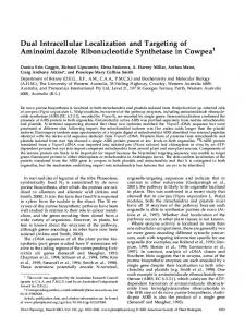
Figure 2. Fully denaturing Tris-Tricine SDS-PAGE of the bands (bands 1–8) collected following glycerol gradients of IEF fractions from separation of LHCII monomer and the minor Chl proteins of samples obtained from V. major plants grown OD. The three gels depict samples from the control, stress, and recovery sets of experiments, as indicated. Thylakoids were loaded as a standard (CT, ST, and RT). CP29 (A), CP26 (B), LHCII (C), and CP24 (D) are indicated.
Zeaxanthin Localization and Dynamics during Photoinhibition
733
Table VI. Average pigment composition of combined fractions (6SD) from flat-bed IEF of the Suc-gradient fraction containing LHCII and the minor Chl proteins from the plants grown OD The fractions combined are indicated below each treatment and correspond to lanes from the Tris-Tricine gels depicted in Figure 3. ND, The fraction is not depicted in Figure 3. LHCII Combined Fractions Pigment
Minor CP Combined Fractions
Control (C1, C3, C5)
Stress (S1, S4, ND)
Recovery (R1, R4, ND)
80.4 6 2.6 11.9 6 0.1 27.1 6 1.6 2.8 6 0.6 0 0 2.8 6 0.6 0
82.8 6 5.2 11.8 6 1.1 27.5 6 2.3 1.2 6 0.6 1.0 6 0.4 2.2 6 0.7 4.3 6 1.7 0.70 6 0.06
80.1 6 6.7 11.8 6 1.1 28.1 6 2.0 1.8 6 0.3 1.4 6 0.6 2.1 6 0.3 5.4 6 0.9 0.62 6 0.07
Control (C6, C7)
Stress (S6, S8, S9)
Recovery (R6, R8, R9)
54.5 6 4.5 9.24 6 1.2 22.2 6 4.1 8.3 6 2.1 3.5 6 1.4 8.3 6 2.8 20.1 6 5.9 0.54 6 0.07
53.4 6 4.0 9.29 6 1.8 20.9 6 3.1 8.2 6 1.1 3.1 6 0.4 5.9 6 0.8 17.2 6 0.9 0.48 6 0.04
mol/100 mol Chl a
Chl b Neoxanthin Lutein V A Z VAZ (Z1A)/(VAZ)
Flat-Bed IEF Fractionation of the LHCII Monomer and Minor CP-Containing Fraction from High-LightAcclimated Leaves The Suc-gradient fraction from the high-light OD samples containing LHCII monomer and the minor Chl proteins was subjected to flat-bed IEF. Fractions from IEF were applied to glycerol gradients (15%–40% glycerol) and ultracentrifuged, and two bands were collected in each case. Bands were analyzed by SDS-PAGE, and pigment analysis was performed (Fig. 2; Table VI). Although fully purified minor CPs were not obtained, the minor CPs were separated from LHCII, except for some contamination of the minor CPs by LHCII in the control sample (Fig. 2). The content of VAZ bound to LHCII following IEF (Table VI) was significantly lower than that bound to the LHCII trimer following Suc-gradient fractionation (Table II), which suggests that a portion of the VAZ bound to LHCII was stripped off during IEF (possibly due to the acidic pI of the LHCII bands that migrated between pH 3.5 and 4.5). The amount of VAZ bound to the LHCII fraction obtained after IEF was similar to that in the Suc-gradient fraction of the LHCII trimer from the GC plants (2.8–5.4 versus 3.4–4.7 mol VAZ/100 mol Chl a in the IEF fraction versus the LHCII trimer fraction of GC plants, respectively). The photoinhibitory treatment induced a considerable increase in VAZ bound to the minor CPs (11–20 mol VAZ/ 100 mol Chl a; Table VI), as well as an increase in the VAZ still bound to LHCII (2.8–4.3 mol VAZ/100 mol Chl a), in contrast to the apparent decrease in total (tightly plus loosely bound) VAZ associated with LHCII trimers from plants grown OD (Table II). At the end of the stress treatment the conversion state of the xanthophyll cycle was higher in the LHCII versus the minor CP fraction (Table VI), whereas the extent of reconversion of Z plus A to V upon 2.5-h recovery was similar in both fractions (20.08 versus 20.06 in LHCII and the minor CPs, respectively). When the VAZ pool that was apparently loosely bound to LHCII (present in the LHCII trimer fraction following Suc-gradient fractionation but not present in the LHCII monomer following IEF) was examined, the quantity of loosely bound VAZ had decreased after photoinhibitory
54.4 11.25 25.5 11.1 0 0 11.1 0
treatment (7.6–4.3 mol VAZ/100 mol Chl a), whereas there was a corresponding increase in the VAZ bound to the minor CPs (Tables II and VI). This may indicate a redistribution of loosely bound VAZ (particularly the Z plus A formed) from LHCII to the minor CPs during the photoinhibitory treatment. The presumably loosely bound VAZ had a higher degree of de-epoxidation relative to VAZ that was more tightly bound to LHCII (0.82 versus 0.70, respectively) and reconverted to a greater extent following recovery (20.45 versus 20.08, respectively), suggesting that this loosely bound VAZ was more accessible to the xanthophyll cycle enzymes than the more tightly bound VAZ. Nondenaturing Green Gel of the Minor CP-Containing Fraction IEF fractions containing the minor CPs were solubilized with 1.9% dodecyl maltoside and run on a nondenaturing
Figure 3. A, Densitometric analysis of Coomassie Blue-stained gels of bands from nondenaturing green gels of the minor CPs and LHCII monomer from samples obtained from plants grown OD (two bands were apparent in each lane). A, Relative percentages of CP29, LHCII, and CP26 in each band. B, The contents of the xanthophyll cycle pigments analyzed for each band.
734
Verhoeven et al.
Plant Physiol. Vol. 120, 1999
Table VII. Xanthophyll content of LHCII as calculated on the basis of 12 Chl (a1b) per polypeptide Values in parentheses refer to xanthophyll content following acid treatment during IEF. Values are from V. major grown in GCs versus OD. Pigment
Control GC
Stress OD
GC
Recovery OD
GC
OD
12 0.85 2.3 0.31 3.46
12 0.78 (0.78) 2.2 (1.9) 0.52 (0.36) 3.50 (3.04)
mol/mol polypeptide
Chl a1b Neoxanthin Lutein VAZ Total xanthophylls
12 0.85 2.3 0.23 3.38
12 0.78 (0.79) 2.2 (1.8) 0.71 (0.18) 3.64 (2.77)
green gel. Each of the fractions resulted in two green bands, which were excised, eluted from the gel, and analyzed. Although pure CPs were not obtained, SDS-PAGE analysis indicated that there was significant enrichment in CP26 or CP29 in two of the bands excised from both the stress samples and the recovery samples. Results of the densitometric analysis of the Coomassie Blue-stained gels, indicating the relative quantities of the proteins, in addition to the xanthophyll cycle pigment content for each fraction, is presented in Figure 3. A relative enrichment in V correlated with CP29, whereas enrichment with Z correlated with CP26. This suggests that the conversion state of the xanthophyll cycle was not uniform among the minor CPs and that the pigments bound to CP29 de-epoxidized to a lesser extent compared with either CP26 or the LHCIIs. DISCUSSION The results of this study demonstrate that at least 80% of the xanthophyll cycle pigments (VAZ) that were present in isolated thylakoids were bound to protein independently of the de-epoxidation state (Table II). These data are consistent with those of Thayer and Bjo¨rkman (1992) who found that 14% to 24% of the VAZ pool was in the pigment front following nondenaturing deriphat electrophoresis of solubilized thylakoids. The comparison of pigment content of extracts from both whole leaves and isolated thylakoids demonstrates a high degree of pigment conservation during the thylakoid isolation procedure (Table I). Of the VAZ found in the free-pigment fractions, the de-epoxidation state was higher in the samples collected during the stress treatment than for protein-bound VAZ, and upon 2.5 h of recovery this portion was reconverted to V to a greater extent than the bound VAZ. These data may indicate that loosely bound and/or free VAZ is the most accessible to the enzymes responsible for their interconversions. In addition, because the leaf VAZ content increased during stress treatment (Table I), there may be relatively more Z in this fraction because of the presence of newly synthesized pigment. An important, novel finding of the present study is that, unlike any other pigment component, the levels of VAZ associated with a given PSII protein fraction are variable and can be altered by environmental factors such as growth conditions (e.g. sun-exposed plants grown OD versus GC plants grown at low PPFD) and/or photoinhibitory treat-
12 0.85 2.3 0.28 3.43
12 0.78 (0.78) 2.2 (1.8) 0.60 (0.28) 3.58 (2.86)
ment. Our data suggest that an increasing demand for thermal energy dissipation results in increased levels of Z plus A bound to additional sites on, or associated with, PSII proteins. It is an attractive possibility that these additional sites may be additional, loose binding sites on given PSII proteins. But it cannot be excluded at this time that stressinduced additional proteins with binding sites for Z plus A may become associated with PSII proteins. Whereas such a possibility has been suggested for ELIPs (that can bind Chl and lutein; Adamska, 1997), no actual evidence has been obtained that ELIPs can in fact bind Z plus A. Moreover, the possibility that, at least in isolated extracts, ELIPs may associate with other proteins particularly easily should also be considered (V. Ebbert, B. Demmig-Adams, and W. Adams, unpublished observations). The distribution of the VAZ that was bound to protein was as follows. LHCII In leaves acclimated to different environmental conditions, the fraction containing LHCII exhibited an altered VAZ content. In Table VII the xanthophyll content per LHCII polypeptide was computed assuming 12 Chl a1b bound (Dainese and Bassi, 1991; Ku¨hlbrandt et al., 1994) and using the data from Table III and VI. Thus, VAZ per polypeptide was greater in plants grown OD versus V. major grown in GCs. The additional VAZ found in the plants grown OD was mostly loosely bound, because it was removed upon IEF. In addition, there were 2 luteins and 0.8 neoxanthin per LHCII polypeptide, both of which were not
Table VIII. Distribution of Chl and VAZ in the Chl-binding complexes Values are expressed as the percentages of total pigment found in each complex. Complex
V. major from OD Chl
V. major from GCs
VAZ
Chl
VAZ
1.2 10.2 43.6 13 32
17.6 27.3 22.6 7.5 25
%
Free pigment PSII minor CPs LHCII PSII core PSI-LHCI
0.6 12.1 37.3 16 34
12.4 11 48 4.6 24
Zeaxanthin Localization and Dynamics during Photoinhibition altered by growth conditions or stress treatment (Tables II, VI, and VII). These data therefore indicate approximately 3.5 carotenoids per LHCII monomer in the bands from Suc gradients (Table II) and close to 3 carotenoids per LHCII following IEF (Tables VI and VII), which is in agreement with the value of 3 xanthophylls per LHCII polypeptide consistently found for highly purified LHCII (Bassi et al., 1993; Ruban et al., 1994). We propose that LHCII possesses an additional xanthophyll-binding site (in addition to the three usually found) that binds additional VAZ in leaves acclimated to high light. Since loosely bound VAZ was found to be associated with the LHCII complex, it could be hypothesized that the VAZ found in the free-pigment fraction actually derives from LHCII, which would account for 0.2 mol VAZ per mol LHCII in the case of the sample from the plants grown OD. With this complement each LHCII would bind 3.7 xanthophyll molecules of which approximately 1 would bind to the new binding site postulated above. These data have important implications with regard to the distribution of xanthophyll cycle pigments among CPs. It was previously proposed that most of the VAZ was bound to the minor CPs, whereas a minor fraction was LHCII bound in maize and spinach (Bassi et al., 1993; Ruban et al., 1994). On the basis of Suc-gradient ultracentrifugation data (Table II), VAZ can be attributed to the different CPs (Table VIII), with the conclusion that a significant portion of VAZ is actually bound to the LHCII fraction, although in a low-affinity site. Moreover, upon acclimation to different environmental conditions, the distribution of V undergoes a dramatic change. In GC-grown plants, 28% of VAZ was bound to the minor CP fraction and 24% was bound to the LHCII fraction, whereas in thylakoids from plants grown OD only 11% of VAZ was bound to minor CPs and 48% was bound to LHCII. If it is assumed that the VAZ in the free-pigment band was stripped off of LHCII (see above), then LHCII would bind 60% of VAZ in the plants grown OD and 42% in V. major grown in GCs at low PPFD. Upon photoinhibitory treatment, the loosely bound fraction of VAZ decreased (0.53–0.30 VAZ per polypeptide), whereas the more tightly bound fraction increased slightly (0.2–0.3 VAZ per polypeptide). This may be indicative of a redistribution of xanthophyll cycle pigments under light stress, which differs from their localization in darkened leaves. It is possible that the V-binding site of LHCII has a low affinity for Z. The presence of such a V-binding but not Z-binding site is supported by the recent finding that the aba-3 mutant of Arabidopsis, lacking epoxy-xanthophylls, contained one less xanthophyll molecule per LHCII polypeptide (Connelly et al., 1997). It has previously been proposed that upon de-epoxidation Z and A are released from LHCII and become free in the membrane lipids (Krupa et al., 1987; Tardy and Havaux, 1997), where they may modify membrane fluidity (Gruszecky and Strzalka, 1991; Tardy and Havaux, 1997). Although our findings are consistent with some release of VAZ from LHCII, the amount of VAZ recovered in the free-pigment band does not change significantly following de-epoxidation (Table II). The constancy of free VAZ in spite of its apparent
735
release from LHCII suggests that newly formed Z becomes bound to one or more pigment-binding protein(s). Minor CPs are the best candidate, since (a) their VAZ content increased upon de-epoxidation and (b) their degree of deepoxidation was less than that of the free-pigment fraction, which is consistent with the hypothesis that they bind Z that has been de-epoxidized in the free-lipid phase. Consistent with this, high degrees of de-epoxidation have been found in chlorina mutants of barley (Dainese et al., 1992), which are enriched in xanthophylls found in the freepigment fraction (Bassi et al., 1985a).
Minor Chl Proteins The pigment-binding properties of the minor Chl proteins (CP29, CP26, and CP24) were relatively constant during purification, and total VAZ content of the minor CP fraction from darkened control leaves did not seem to differ in response to the acclimation to low- versus highgrowth PPFD. Assuming approximately equal proportions of the three minor CPs (CP29, CP26, and CP24), the pigment composition indicated by these data are in close agreement with data from highly purified proteins (Bassi et al., 1993; Ruban et al., 1994). Considering 6 Chl a per polypeptide, there were an average of 3.2 Chl b, 1.3 lutein, 0.5 neoxanthin, and between 0.7 and 1.2 VAZ per polypeptide. There were 2.5 carotenoids per polypeptide in the control leaves and a considerable increase in the VAZ bound to the minor CP fraction during stress, resulting in 3.2 carotenoids per polypeptide during stress. It is therefore possible that 3 carotenoid-binding sites are present in the minor CPs, one-third of which is only partially occupied in control leaves. Whereas the existence of a variably occupied site was also postulated for LHCII (in control GC plants versus those grown OD in full sun), in the case of minor CPs bound VAZ increased only during stress treatment rather than as an acclimation response, thus implying that the CP site has a higher affinity for Z than for V. Thus, affinities for Z versus V appear to be opposite for these newly postulated sites on the minor CPs versus LHCII. The present results confirm that the degree of deepoxidation is not the same among the different minor CPs. Although CP29 bound considerable amounts of V (Bassi et al., 1993; Giuffra et al., 1996), this pigment could be deepoxidized to a much lower extent compared with CP26 (Fig. 3; it was not possible to assess the de-epoxidation state of CP24). Low xanthophyll de-epoxidation in CP29 following high-light treatment was previously reported (Ruban et al., 1994; Croce et al., 1996a). These results are in contrast to those of Fa¨rber et al. (1997), who found that, when spinach was treated with 3 h of photoinhibition, there was no increased binding of VAZ to any of the pigment-protein complexes and no redistribution of pigments. This difference may be due to the very different experimental conditions used: a 3-h photoinhibitory treatment versus a 48- or 122-h treatment used in this study.
736
Verhoeven et al.
PSII Core Suc-gradient fractionation yielded PSII core fractions with a maximum of 7% contamination by LHCII. Assuming 56 Chls per PSII core, between 1 and 1.8 xanthophylls per PSII core were detected in samples from GC plants and 0.8 to 1.3 xanthophylls per PSII core were detected in samples from plants grown OD in full sunlight. The LHCII contamination accounted for less than 0.1 xanthophyll molecule per PSII core (Table V), suggesting that 1 to 2 VAZ molecules may be bound to the PSII core. This was not previously recognized in a study of maize leaves (Bassi et al., 1993) in which a multistep purification procedure was used, which would remove any loosely bound pigment. A loose binding site for VAZ in the PSII core is suggested by the results of ion-exchange chromatography; when the complex was bound to the column and the column was washed extensively, the xanthophyll content decreased to 0.17 per PSII core. The presence of VAZ in PSII core fractions has also been reported in barley (Lee and Thornber, 1995) and lettuce (Phillip and Young, 1995). There was some increase in VAZ present in the PSII core fraction following the stress treatment (Table III). Binding of Z to the PSII core upon photoinhibition was postulated previously (Jahns and Miehe, 1996; Fa¨rber et al., 1997). When expressed per polypeptide, the increase in apparent VAZ content was actually similar in magnitude for the PSII cores and minor CPs (an increase of 0.8 or 0.5 VAZ per PSII core complex in leaves from GCs or OD, respectively, versus an increase of 0.5 VAZ per minor CP in leaves from plants grown OD). However, it should be noted that the absolute content of VAZ detected in the PSII core was very small (Table II), thus precluding any firm conclusions. Again, the association of stress-induced proteins with the PSII core fraction, leading to the binding of additional VAZ, cannot be excluded.
PSI-LHCI We confirm the presence of VAZ in PSI-LHCI (Thayer and Bjo¨rkman, 1992; Lee and Thornber, 1995; Zhu et al., 1997). However, the VAZ bound to PSI-LHCI was deepoxidized to a lesser extent upon photoinhibitory treatment than that bound to the PSII complexes, and the extent of reconversion following 2.5 h recovery was also lower in PSI-LHCI. The presence of photoconvertible VAZ in PSILHCI raises the question of whether thermal energydissipation mechanisms similar to those observed in PSII take place in PSI. This seemed unlikely previously, since PSI was believed to be a deep trap, which would make energy dissipation in LHCI inefficient (in decreasing Chl a excited state concentrations at the level of the PSI reaction center). More recently, however, it was recognized that PSI, like PSII, is a shallow trap and that, therefore, PSI and its antenna are essentially equilibrated (Croce et al., 1996b), implying that thermal dissipation in the LHCI antenna could be an efficient regulatory mechanism for Chl a excitation in PSI. This could provide protection from the adverse effects of excess excitation on PSI as well (Terashima et al., 1994; Tjus et al., 1998).
Plant Physiol. Vol. 120, 1999 CONCLUSIONS
In this study we have shown that xanthophyll cycle pigments undergo dynamic changes not only in their epoxidation state but also in their association with CPs. Upon acclimation to growth OD in full sunlight, the V content of control thylakoids was greatly increased. This additional V was bound to the major LHCII fraction. Following photoinhibitory treatment, the VAZ content of the LHCII fraction decreased and the minor CP fraction of PSII (CP29, CP26, and CP24), and to a lesser extent the PSII core complex fraction, bound increased amounts of Z plus A. This is interpreted in terms of the presence of an additional, lowaffinity V-binding site in LHCII, in equilibrium with V free in the lipid phase, and of an additional high-affinity Z-binding site in the minor CPs. The higher degree of de-epoxidation in the free-pigment fraction suggests that the preferred substrate for V de-epoxidase is the pigment free in the lipid phase, which, upon conversion, becomes bound to minor CPs and PSII core CPs. ACKNOWLEDGMENT We gratefully acknowledge Paolo Pesaresi for his assistance in performing some of the experimental techniques. Received December 15, 1998; accepted March 22, 1999. LITERATURE CITED Adams WW III, Demmig-Adams B, Verhoeven AS, Barker DH (1995) ’Photoinhibition’ during winter stress: involvement of sustained xanthophyll cycle-dependent energy dissipation. Aust J Plant Physiol 22: 261–276 Adamska I (1997) ELIPs: light-induced stress proteins. Physiol Plant 100: 794–805 Bassi R, Dainese P (1992) A supramolecular light-harvesting complex from chloroplast photosystem-II membranes. Eur J Biochem 204: 317–326 Bassi R, Giacometti GM, Simpson D (1988) Changes in the composition of stroma lamellae following state I–state II transitions. Biochim Biophys Acta 935: 152–165 Bassi R, Hinz U, Barbato R (1985) The role of light harvesting complex and photosystem II in thylakoid stacking in the chlorina-f2 barley mutant. Carlsberg Res Commun 50: 347–367 Bassi R, Pineau B, Dainese P, Marquardt J (1993) Carotenoidbinding proteins of photosystem II. Eur J Biochem 212: 297–303 Connelly JP, Mu¨ller MG, Bassi R, Croce R, Holzwarth AR (1997) Femtosecond transient absorption study of carotenoid to chlorophyll energy transfer in the light-harvesting complex II of photosystem II. Biochemistry 36: 281–287 Croce R, Breton J, Bassi R (1996a) Conformational changes induced by phosphorylation in the CP29 subunit of photosystem II. Biochemistry 35: 11142–11148 Croce R, Zucchelli G, Garlaschi F, Bassi R, Jennings RC (1996b) Excited state equilibration in the photosystem I light harvesting complex: P700 is almost isoenergetic with its antenna. Biochemistry 35: 8572–8579 Dainese P, Bassi R (1991) Stoichiometry of the chloroplast photosystem II antenna system and aggregation state of the component Chl a/b proteins. J Biol Chem 266: 8136–8142 Dainese P, Hoyer-Hansen G, Bassi R (1990) The resolution of chlorophyll a/b binding proteins by a preparative method based on flat bed isoelectric focusing. Photochem Photobiol 51: 693–703 Dainese P, Marquardt J, Pineau B, Bassi R (1992) Identification of violaxanthin and zeaxanthin binding proteins in maize photosystem II. In N Murata ed, Developments in Photosynthesis
Zeaxanthin Localization and Dynamics during Photoinhibition Research. Kluwer Academic Publishers, Dordrecht, The Netherlands, pp 287–290 Demmig B, Winter K, Kru¨ger A, Czygan F-C (1988) Zeaxanthin and the heat dissipation of excess light energy in Nerium oleander exposed to a combination of high light and water stress. Plant Physiol 87: 17–24 Demmig-Adams B, Adams WW III (1996) Xanthophyll cycle and light stress in nature: uniform response to excess direct sunlight among higher plant species. Planta 198: 460–470 Demmig-Adams B, Adams WW III, Barker DH, Logan BA, Bowling DR, Verhoeven AS (1996) Using chlorophyll fluorescence to assess the fraction of absorbed light allocated to thermal dissipation of excess excitation. Physiol Plant 98: 254–264 Demmig-Adams B, Moeller DL, Logan BA, Adams WW III (1998) Positive correlation between levels of retained zeaxanthin and antheraxanthin and degree of photoinhibition in shade leaves of Schefflera arboricola. Planta 205: 367–374 Di Paolo ML, Dal Belin-Peruffo A, Bassi R (1990) Immunological studies on chlorophyll a/b proteins and their location in thylakoid membrane domains. Planta 181: 275–286 Eskling M, Arvidsson P-O, Åkerlund H-A (1997) The xanthophyll cycle, its regulation and components. Physiol Plant 100: 806–816 Fa¨rber A, Young AJ, Ruban AV, Horton P, Jahns P (1997) Dynamics of xanthophyll-cycle activity in different antenna subcomplexes in the photosynthetic membranes of higher plants. The relationship between zeaxanthin conversion and nonphotochemical fluorescence quenching. Plant Physiol 115: 1609–1618 Gilmore AM (1997) Mechanistic aspects of xanthophyll cycledependent photoprotection in higher plant chloroplasts and leaves. Physiol Plant 99: 197–209 Gilmore AM, Yamamoto HY (1991) Resolution of lutein and zeaxanthin using a nonendcapped, lightly carbon-loaded C-18 high-performance liquid chromatographic column. J Chromatogr 543: 137–145 Giuffra E, Cugini D, Croce R, Bassi R (1996) Reconstitution and pigment-binding properties of recombinant CP29. Eur J Biochem 238: 112–120 Goss R, Richter M, Wild A (1997) Pigment composition of PSII pigment protein complexes purified by anion exchange chromatography: identification of xanthophyll cycle pigment binding proteins. J Plant Physiol 151: 115–119 Gruszecki WI, Strzalka K (1991) Does the xanthophyll cycle take part in the regulation of fluidity of the thylakoid membrane? Biochim Biophys Acta 1060: 310–314 Jahns P, Miehe B (1996) Kinetic correlation of recovery from photoinhibition and zeaxanthin epoxidation. Planta 198: 202–210 Kilian R, Bassi R, Schaefer C (1997) Identification and characterization of photosystem II chlorophyll a/b binding proteins in Marcantia polimorpha L. Planta 204: 260–267 Knoetzel J, Simpson D (1991) Expression and organization of antenna proteins in the light- and temperature-sensitive barley mutant chlorina-104. Planta 185: 111–123 Krause GH (1994) Photoinhibition induced by low temperatures. In NR Baker, JR Bowyer, eds, Photoinhibition of Photosynthesis: Molecular Mechanisms to the Field. Bios Scientific Publishers, Oxford, UK, pp 331–348
737
Krupa Z, Huner NPA, Williams JP, Maissan E, James DR (1987) Development at cold-hardening temperatures. The structure and composition of purified rye light-harvesting complex II. Plant Physiol 84: 19–24 Ku¨hlbrandt W, Wang DN, Fujiyoshi Y (1994) Atomic model of plant light-harvesting complex by electron crystallography. Nature 367: 614–621 Lee AL-C, Thornber JP (1995) Analysis of the pigment stoichiometry of pigment-protein complexes from barley (Hordeum vulgare). Plant Physiol 107: 565–574 Nitsch JP, Nitsch C (1969) Haploid plants from pollen grains. Science 163: 85 Osmond CB (1994) What is photoinhibition? Some insights from comparisons of shade and sun plants. In NR Baker, JR Bowyer, eds, Photoinhibition of Photosynthesis: Molecular Mechanisms to the Field. Bios Scientific Publishers, Oxford, UK, pp 1–24 Phillip D, Young A (1995) Occurrence of the carotenoid lactucaxanthin in higher plant LHCII. Photosynth Res 43: 273–282 Ruban AV, Young AJ, Pascal AA, Horton P (1994) The effects of illumination on the xanthophyll composition of the photosystem II light-harvesting complexes of spinach thylakoid membranes. Plant Physiol 104: 227–234 Santini C, Tidu V, Tognon G, Ghiretti-Magaldi A, Bassi R (1994) Three dimensional structure of higher plants photosystem II reaction center: evidences for its dimeric organization in vivo. Eur J Biochem 221: 307–315 Scha¨gger H, von Jagow G (1987) Tricine sodium dodecyl sulfatepolyacrylamide gel electrophoresis for the separation of proteins in the range from 1 to 100 kDa. Anal Biochem 166: 368–379 Tardy F, Havaux M (1997) Thylakoid membrane fluidity and thermostability during the operation of the xanthophyll cycle in higher-plant chloroplasts. Biochim Biophys Acta 1330: 179–193 Terashima I, Funayama S, Sonoike K (1994) The site of photoinhibition in leaves of Cucumis sativus L. at low temperatures is photosystem I, not photosystem II. Planta 193: 300–306 Thayer SS, Bjo¨rkman O (1990) Leaf xanthophyll content and composition in sun and shade determined by HPLC. Photosynth Res 23: 331–343 Thayer SS, Bjo¨rkman O (1992) Carotenoid distribution and deepoxidation in thylakoid pigment-protein complexes from cotton leaves and bundle-sheath cells of maize. Photosynth Res 33: 213–225 Tjus SE, Møller BL, Scheller HV (1998) Photosystem I is an early target of photoinhibition in barley illuminated at chilling temperatures. Plant Physiol 116: 755–764 Verhoeven AS, Adams WW III, Demmig-Adams B (1996) Close relationship between the state of the xanthophyll cycle pigments and photosystem II efficiency during recovery from winter stress. Physiol Plant 96: 567–576 Zhu J, Gomez SM, Mawson BT, Jin X, Zeiger E (1997) The coleoptile chloroplast: distinct distribution of xanthophyll cycle pigments, and enrichment in photosystem II. Photosynth Res 51: 137–147