Zootaxa 1788: 1–20 (2008) www.mapress.com / zootaxa/
ISSN 1175-5326 (print edition)
Copyright © 2008 · Magnolia Press
ISSN 1175-5334 (online edition)
ZOOTAXA
Juvenile development of Callinectes ornatus Ordway, 1863 (Crustacea: Decapoda: Portunidae), from megalopae obtained in the neuston EDUARDO ANTONIO BOLLA JÚNIOR, MARIA LUCIA NEGREIROS-FRANSOZO1 & ADILSON FRANSOZO Nebecc (Crustacean Biology, Ecology and Culture Study Group), Departamento de Zoologia, Instituto de Biociências, C.P. 510, Universidade Estadual Paulista, 18618-000 Botucatu, SP, Brazil 1 Corresponding author. E-mail:
[email protected]
Abstract The juvenile development of Callinectes ornatus was studied from megalopae collected in the neuston off Ubatuba, São Paulo State, Brazil. The animals were raised in the laboratory under constant temperature (25 ± 1ºC), filtered sea water (35‰) from the sampling location, and the natural photoperiod. Eleven stages of the juvenile phase were obtained. The main features of the first juvenile stage diagnostic of the species are: the number of segments in the antenna; number of setae on the exopod, endopod, basal endite and coxal endite of the maxilla, on the exopod, endopod, basal endite, coxal endite and epipod of the 1st maxilliped, and on the exopod, endopod and epipod of the 2nd maxilliped. Sexual dimorphism becomes apparent from the fourth juvenile stage onwards. Key words: Portunidae, Callinectes, swimming crab, juvenile stages, morphological characters, sexual dimorphism
Introduction Descriptions of species of worldwide brachyurans are based on the morphological characters of adult specimens, because earlier stages of their life cycles are insufficiently known (Fransozo & Negreiros-Fransozo, 1987). Description of larval or even juvenile stages is commonly done by other specialists. Because of the difficulty of raising and maintaining young individuals in the laboratory (Fransozo, 1986/ 87), researchers have paid little attention to larval or juvenile development. However, studies of the early stages of crab life cycles are important to improve knowledge of their taxonomy and ecology (NegreirosFransozo et al., 2002), physiology (Anger, 2003) and phylogeny (Marques et al., 2003). Secondary sexual characters appear, through modification of the abdomen and pleopods, during the juvenile stages (Fransozo, 1986/87; Fransozo & Negreiros-Fransozo, 1987 and Barutot et al., 2001). Portunids are an important fishery resource, and their commercial exploitation is relevant in the economies of several countries in Europe, Asia and the Americas. People consume these crabs either wild-caught or produced in captivity, such as soft-shell crabs, just after the molt, or ecdysis. In Brazil, only a few wild-caught crabs are consumed (Fernandes et al., 2006). The family Portunidae Rafinesque, 1815 is represented on the Brazilian coast by 20 native species (Melo, 1996) and an exotic one, Charybdis hellerii (A. Milne-Edwards, 1867) (Negreiros-Fransozo, 1996) in the genera Arenaeus Dana, 1851; Callinectes Stimpson, 1860; Coenophtalmus Milne-Edwards, 1879; Cronius Stimpson, 1860; Laleonectes Manning & Chace, 1990; Ovalipes Rathbun, 1898; Portunus Weber, 1795; Scylla De Haan, 1833; and Charybdis De Haan, 1833. All of the six local species of genus Callinectes occur off the coast of the state of São Paulo (Melo, 1996). Accepted by P. Castro: 7 May 2008; published: 9 Jun. 2008
1
Of the portunids found on the Brazilian coast, only Callinectes sapidus Rathbun, 1896 has had its juvenile development until the 11th stage studied by Barutot et al. (2001). These authors observed that sexual dimorphism is apparent from the fourth juvenile stage on. Juveniles of portunids can be easily distinguished from species belonging to other families, as early as the first stage (Barutot et al. 2001). However, because of the lack of research on juvenile development of portunid species on the Brazilian coast, it is difficult to define their diagnostic characters at this life phase. Only with detailed studies from laboratory rearing can comparisons be made and distinctive characters established for the local species. This study aimed to describe and present morphological details of the first stage of juvenile development of C. ornatus, from megalopae obtained in the neuston and maintained under laboratory conditions. Morphological details of the most significant changes that occur in subsequent stages are described, particularly concerning the pleopods and abdomen changes.
Methods The geographical distribution of C. ornatus includes much of the Western Atlantic: North Carolina to Florida, Gulf of Mexico, the Caribbean, Colombia, Venezuela, Guyana and Brazil (Amapá to Rio Grande do Sul). This species is found on sand and mud bottoms, from the intertidal zone to depths of 75 meters, and also in less saline waters (Melo, 1996). The juvenile development of C. ornatus was studied from materials obtained through collections of megalopae from the neuston in the region of Ubatuba, São Paulo State (Brazil), for subsequent rearing in the laboratory. The megalopae were collected with a neuston net (500 µm mesh) at night, with at least 12 trawls, of 10 minutes each, during November/2005 through January/2006. The megalopae were sorted and selected for rearing. Portunid megalopae were selected with reference to morphological characteristics reported in the literature, including the presence of a long acute rostrum, a paddle-shaped 5th pereopod, and sternal spines on the 7th thoracic somite (Kurata, 1975). Only after they reached advanced juvenile stages, they could be separated in different species, such as C. ornatus, C. danae and Portunus spinimanus. After the megalopae were collected and selected, they were individually packed in acrylic containers (30 ml capacity) with screw caps, containing pre-filtered sea water (35‰) from the collection locality, and transported to the crustacean culture laboratory. In general, the culture techniques were similar to those of Barutot et al. (2001), Guimarães & NegreirosFransozo (2005) and Negreiros-Fransozo et al. (2007). Megalopae and young crabs were raised separately in acrylic containers of 30 ml capacity containing filtered and aerated sea water. The containers were inspected daily, and debris, probable exuviae and dead individuals were removed. The water was partly renewed for two consecutive days, and completely on the third day with pre-filtered and aerated sea water. After inspection, individuals were fed with newly hatched nauplii of Artemia sp., offered ad libitum. For advanced juveniles (approximately from the 5th juvenile stage), commercial food for ornamental fish was offered as a supplement. Dead individuals and exuviae were fixed in 80% ethanol and glycerin, in a proportion of 2:1. After approximately 10 molts, the juvenile crabs showed external morphology similar to adults, and also the typical color pattern of the species. The diagnostic characters used to separate genera in the family Portunidae (subfamily Portuninae) and to separate species of Callinectes can be used to distinguish crabs in these advanced stages (Melo, 1996). Thus, the identity of the megalopae was established by identifying the reared crabs. Exuviae of initial stages of specimens that reached advanced stages (approximately 10) were dissected, and the characteristics noted were used to identify other individuals that died before they reached these advanced stages.
2 · Zootaxa 1788 © 2008 Magnolia Press
BOLLA JR ET AL.
Specimens of C. ornatus were dissected, drawn (using a camera lucida), measured and described from fixed exuviae and dead individuals, with the use of a stereoscopic microscope (SV6) and micrometer rule. The stages were designated as “juveniles” because they did not show fully mature gonads. According to Mantelatto and Fransozo (1996), sexual maturity is reached in C. ornatus at larger sizes than those obtained in the laboratory. The terminology for descriptions of setae types is based on Pohle and Telford (1981) and Clark et al. (1998). The setae were observed with the use of a microscope fitted with Nomarski differential interference contrast optics (magnification = 1000).
Results Analysis of growth and duration of stages obtained for the juvenile phase (Tables 1 and 2) In the laboratory, we obtained up to 11 stages of the juvenile phase. The duration and survival of the stages are shown in Table 1. Only the age of the last stage of each individual was disregarded, because these individuals did not die from natural causes. Only four individuals reached the tenth-first stage, and most deaths occurred in the sixth and seventh stages. Table 2 presents the measurements for carapace width and length of all stages obtained. TABLE 1. Callinectes ornatus Ordway, 1863. Duration and survival of juvenile stages, from megalopae collected in the neuston, salinity 35‰. A, average duration (in days); mD and MD, minimum and maximum duration (in days), respectively; N, number of live individuals; †, number of dead individuals; %S, survival percentage. Initial N = 64. Stage
JI
JII
JIII
JIV
JV
JVI
JVII
JVIII
JIX
JX
JXI
A
05
06
08
09
11
14
19
21
28
39
-
mD
02
03
04
05
06
06
12
08
22
32
-
MD
08
09
14
16
17
29
41
35
41
49
-
N
64
64
64
64
63
59
41
19
14
09
04
†
00
00
00
01
04
18
22
05
05
05
04
%S
100.00
100.00
100.00
98.44
92.19
64.06
29.69
21.88
14.06
6.25
0.00
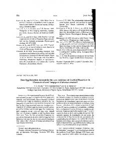
Morphology of the first juvenile stage (Figs. 1 through 5) The main setae and structures found on different parts of the body of the first juvenile stage are illustrated in Figure 1. The general form of the first juvenile stage is already similar to that of an adult swimming crab (Fig. 2 a): the carapace is wider than long, dorso-ventrally flattened and slightly convex; the rostrum has very small subterminal projections and a slightly median notch (Fig. 2 b); it bears 8 lateral teeth (finely serrate) (Fig. 2 c), as well as the pair of lateral spines common in portunids of the genus Callinectes. The chelipeds (Fig. 5) are symmetrical, with a few inner marginal spines on the merus and carpus, and small protuberances on the merus, carpus and propodus, in addition to sparse simple and plumose setae. The second, third and fourth pereopods (Fig. 3 c, d, e) are similar to each other and have sparse simple and plumose setae. The last pair of pereopods (Fig. 3 f) has the definitive portunids character, a flat paddle-shaped
JUVENILE STAGES OF C. ORNATUS (PORTUNIDAE)
Zootaxa 1788 © 2008 Magnolia Press ·
3
dactyl with many marginal plumose setae. The eyes are pedunculate and well developed. The sternum (Fig. 2 d) has sparse simple setae over its surface. On the 7th thoracic sternite, near the lateral projection, there is a protuberance like a concentric semicircle. The 5th thoracic sternite has an abdominal locking mechanism from the first juvenile stage. TABLE 2. Callinectes ornatus Ordway, 1863. Measurements (in millimeters) of carapace width (CW) and length (CL), of first to tenth-first juvenile stages. A, average; s, standard deviation; CW/CL, width and length ratio. Stage JI JII JIII JIV JV JVI JVII JVIII JIX JX JXI
A
Maximum
Minimum
s
CW/CL
CW
2.42
2.63
2.05
0.13
1.28
CL
1.90
2.07
1.53
0.11
CW
3.50
4.11
2.84
0.23
CL
2.41
2.76
2.05
0.15
CW
4.72
5.35
3.98
0.36
CL
3.10
3.45
2.62
0.22
CW
6.07
7.36
5.15
0.52
CL
3.84
4.58
3.29
0.29
CW
7.65
9.56
6.12
0.73
CL
4.76
5.78
3.90
0.40
CW
9.38
10.98
7.06
0.89
CL
5.77
6.74
4.32
0.47
CW
11.18
12.92
8.37
1.00
CL
6.89
7.79
5.21
0.53
CW
13.45
15.49
11.57
1.03
CL
8.13
9.11
7.05
0.57
CW
16.22
18.66
13.28
1.97
CL
9.84
11.26
8.27
0.95
CW
19.69
22.29
15.96
2.18
CL
11.96
13.17
9.88
1.04
CW
22.96
25.85
20.96
2.27
CL
13.63
15.25
12.87
1.12
1.45 1.52 1.58 1.61 1.63 1.62 1.66 1.65 1.65 1.68
The abdomen (Fig. 2 e) has six somites, all wider than long; each of the first four somites has horizontal grooves. The telson is smooth. The ventral face of the abdomen has four pairs of rudimentary pleopods from the 2nd to 5th somite; only the 4th and 5th somite pairs have an endopod (Fig. 7, JI). The antennule (Fig. 4 b) has a well-developed basal segment provided with various plumose and simple setae, in addition to numerous small protuberances; a bi-segmented peduncle with sparse simple setae; and a bi-segmented endopod (ventral flagellum) with several simple setae on the two segments; the exopod (dorsal flagellum) consists of seven segments with 0, 10, 9, 8, 5, 0 and 0 aesthetascs on the proximal to distal segments respectively. The exopod bears 0, 0, 1, 2, 0, 4 and 3 simple setae on the proximal to distal segments. Antenna (Fig. 4 a)—tri-segmented antennal peduncle provided with sparse plumose setae on the two proximal segments, besides simple setae and marginal and terminal small protuberances on the first and second segments, respectively; the third segment has only simple setae. The antennal flagellum has 10 (11) segments with sparse simple setae.
4 · Zootaxa 1788 © 2008 Magnolia Press
BOLLA JR ET AL.
FIGURE 1. Scheme of main ornamentation found in different parts of the bodies of juvenile stages of Callinectes ornatus Ordway, 1863; a, simple setae; b, plumose setae; c, serrate setae; d, denticulate harpoon-shaped setae; e, aesthetascs; f, brush-shaped setae; g, denticulate cuspidate setae; h, spine; i, protuberance.
Mandible (Fig. 4 d)—with a strongly chitinized cutting blade; palp tri-segmented, with 13 plumose setae and 2 serrate setae on the distal segment, and 1 simple seta on the middle segment. Maxillule (Fig. 4 f)—coxal endite with 3 plumose setae, 12 simple setae of various sizes and 5 serrate setae; basal endite provided with 5 small simple setae on the proximal margin, 11 simple setae and 9 serrate setae on the distal margin, besides 2 distal plumose setae; endopod bi-segmented, with 3 plumose setae on the proximal segment and 5 (4) simple setae on the distal segment. The protopod also has 3 simple setae on its margin. JUVENILE STAGES OF C. ORNATUS (PORTUNIDAE)
Zootaxa 1788 © 2008 Magnolia Press ·
5
FIGURE 2. Callinectes ornatus Ordway, 1863. Juvenile I: a, dorsal view; b, rostrum detail; c, lateral spines detail; d, sternum, dorsal view; e, abdomen, dorsal view. Scales: a = 0.5 mm; b, c, d, e = 0.2 mm.
Maxilla (Fig. 4 e)—coxal endite bilobed, with 6 (7) plumose setae on the proximal segment, and 4 simple setae and 3 plumose setae on the distal segment; the basal endite, also bilobed, bears 7 (8) plumose setae and 1 simple seta on the proximal segment, 12 (13) plumose setae and 1 simple seta on the distal segment; exopod (scaphognathite) provided with 88 to 90 marginal plumose setae and approximately 57 surface setae; endopod with 4 (3) simple setae on its distal margin.
6 · Zootaxa 1788 © 2008 Magnolia Press
BOLLA JR ET AL.
FIGURE 3. Callinectes ornatus Ordway, 1863. Juvenile I: pereopods; a, cheliped dorsal view; b, cheliped, lateral view; c, second pereopod (P2); d, third pereopod (P3); e, fourth pereopod (P4); f, fifth pereopod (P5). Scale: a, b, c, d, e, f = 0.2 mm.
First Maxilliped (Fig. 5 a)—coxal endite with approximately 22 plumose setae and 4 serrate setae; basal endite with approximately 28 plumose setae, 2 serrate and 37 simple; endopod unsegmented, with 10 (9) plumose setae and 23 to 25 simple; exopod bi-segmented, with 14 plumose setae and 4 simple setae on the proxJUVENILE STAGES OF C. ORNATUS (PORTUNIDAE)
Zootaxa 1788 © 2008 Magnolia Press ·
7
imal segment, 9 plumose setae and 2 simple setae on the distal segment; epipod well developed, with approximately 49 simple setae.
FIGURE 4. Callinectes ornatus Ordway, 1863. Juvenile I: a, antenna; b, antennule; c, mandible palp detail; d, mandible; e, maxilla; f, maxillule. Scales: a, d, e = 0.1 mm; b, c, f = 0.05 mm.
Second Maxilliped (Fig. 5 b)—endopod four-segmented, with 8, 2, 0 and 0 plumose setae on the proximal to distal segments, 1 and 10 serrate setae on the penultimate and terminal segments respectively, besides several simple setae on all segments; exopod bi-segmented, with several rows of simple setae, 6 plumose setae
8 · Zootaxa 1788 © 2008 Magnolia Press
BOLLA JR ET AL.
and 9 serrate setae on the proximal segment, and 2 simple setae and 8 plumose setae on the distal segment; rudimentary epipod with approximately 4 simple and 2 plumose setae.
FIGURE 5. Callinectes ornatus Ordway, 1863. Juvenile I: a, first maxilliped; b, second maxilliped; c, third maxilliped. Scale: a, b, c = 0.1 mm.
Third Maxilliped (Fig. 5 c)—endopod five-segmented, with 12, 8, 4, 5 and 4 serrate setae on the proximal to distal segments, besides several simple setae on all segments; exopod bi-segmented, with approximately 21 plumose setae and 24 simple setae on the proximal segment, and 10 plumose setae and 1 simple seta on the JUVENILE STAGES OF C. ORNATUS (PORTUNIDAE)
Zootaxa 1788 © 2008 Magnolia Press ·
9
distal segment; protopod with 16 plumose setae and many simple setae; epipod with 6 plumose setae, besides numerous simple setae on the proximal and 22 simple setae on the distal portion.
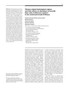
Morphology of second to tenth-first juvenile stages (Table 3; Figs. 6 through 10)] The proportions of the carapace growth (Table 2) is maintained with the development of the individual in the following stages, which reaches similar proportions as found in the adults from the first to the tenth-first stage. The abdomen increases in size during growth, but until the tenth-first stage does not show any change in its form that might allow identification of the sex. In adult males, the abdomen is shaped as an inverted "T" and in adult females is semicircular. From the second juvenile stage, the third and fourth abdominal somites are fused (Fig. 6 d). The abdominal locking mechanism, present from the first juvenile stage, becomes more evident in the fourth juvenile stage, being possible its observation even without specimens dissection (Fig. 6 e). Regarding secondary sexual characters, the rudimentary pleopods existing in the first juvenile stage, disappear in the second stage. In the fourth stage (Fig. 7, JIV) in the male, two pleopods appear on the first and second abdominal segments, where the second pair of pleopods is now slightly biramous. In the female, four pairs of biramous pleopods appear on the second to fifth abdominal segments. In the fifth juvenile stage (Fig. 7, JV), the female’s pleopods increase in size, and simple setae appear on the pleopods of the second and third abdominal segments; whereas in males the pleopods only increase in size. In the sixth stage (Fig. 7, JVI) of the females, all the pleopods increase in size and acquire simple setae; male’s pleopods only increase in size. In the seventh juvenile stage, a brush-shaped setae appear on the pleopods of the second and third abdominal segments of females (Fig. 9, JVII); in males, the second pleopod becomes totally uniramous and both pleopods pairs begin to take on the characteristic adult shape (Fig. 8, JVII). From the eighth stage (Figs. 8 and 9, JVIII), the changes in pleopods are limited to a gradual increase in size, increase in number of setae and the inherent morphological changes of each sex. In the ninth stage (Fig. 8 and 11, JIX), the tip of the pleopod of second abdominal segment of the male, assumes its piston-like shape. In the tenth juvenile stage (Figs. 8 and 10, JX) the general morphology of pleopods is already very similar to that found in adults. The growth rate of female pleopods between juvenile stages is approximately 36% at each molt. In females, a slight sign of the gonopore begins to appear from the fourth juvenile stage (Fig. 6 e). In males, it was not possible to verify in which stage this structure arises, because the exuviae were accidentally broken during dissection and the gonopores could not be observed. In other structures, the major changes that occur are: •The endopod of the first maxilliped, from the second stage (Fig. 6 b), shows a bend in the apical region. In the following stages, it begins to acquire a foliaceous aspect, particularly in the marginal region. •The first segment of the antennal peduncle (Fig. 6 c) bears a projection in the apical region, forming a structure probably reminiscent of the antennal scale. •The endopod of the maxillule is modified in relation to the previous stages, as shown in Figure 6 a. The other appendices did not undergo significant morphological changes, but there was an increase in the number of setae per segment of each appendix. From the fourth juvenile stage, denticulate cuspidate setae appear on the epipod of the third maxilliped. From the fifth stage, these setae are found on the epipod of the first, second and third maxillipeds, and harpoon-shaped setae with denticulate ends on the epipod of the second maxilliped. The main morphological characters that allow the identification of the first 11 stages of juvenile development of C. ornatus are presented in Table 3.
10 · Zootaxa 1788 © 2008 Magnolia Press
BOLLA JR ET AL.
FIGURE 6. Callinectes ornatus Ordway, 1863. Main morphological changes in the second to tenth juvenile stages; a, juvenile X: maxillule endopod detail; b, juvenile II: first maxilliped endopod detail; c, juvenile X: first segment, dorsal view, detail of antennal peduncle; d, juvenile II: abdomen, dorsal view; e, juvenile IV: trace of female gonopore (arrow). Scales: a, b, c, d = 0.2 mm; e = 0.4 mm.
JUVENILE STAGES OF C. ORNATUS (PORTUNIDAE)
Zootaxa 1788 © 2008 Magnolia Press ·
11
FIGURE 7. Callinectes ornatus Ordway, 1863. Pleopods of 1st to 5th abdominal somites (Pl1 to Pl5) of male and female: juvenile I (JI); juvenile IV (JIV); juvenile V (JV); juvenile VI (JVI). Scales: JI = 0.1 mm; JIV, JV, JVI = 0.2 mm.
12 · Zootaxa 1788 © 2008 Magnolia Press
BOLLA JR ET AL.
FIGURE 8. Callinectes ornatus Ordway, 1863. Male pleopods (Pl1 and Pl2): juvenile VII (JVII); juvenile VIII (JVIII); juvenile IX (JIX); juvenile X (JX); a, region detail (arrow). Scales: JVII, JVIII, JIX = 0.2 mm; a = 0.1 mm; JX = 0.5 mm.
JUVENILE STAGES OF C. ORNATUS (PORTUNIDAE)
Zootaxa 1788 © 2008 Magnolia Press ·
13
FIGURE 9. Callinectes ornatus Ordway, 1863. Female pleopod (Pl2 to Pl5): juvenile VII (JVII); juvenile VIII (JVIII). Scale: JVII, JVIII = 0.2 mm.
14 · Zootaxa 1788 © 2008 Magnolia Press
BOLLA JR ET AL.
FIGURE 10. Callinectes ornatus Ordway, 1863. Female pleopods (Pl2 to Pl5): juvenile IX (JIX); juvenile X (JX). Scales: JIX = 0.2 mm; JX = 0.4 mm.
JUVENILE STAGES OF C. ORNATUS (PORTUNIDAE)
Zootaxa 1788 © 2008 Magnolia Press ·
15
TABLE 3. Callinectes ornatus Ordway, 1863. Main morphological characters that allow the identification of the first 11 juvenile stages. Length measurements were taken in millimeters and represent average values; numbers in parentheses represent alternative values, of lower occurrence. JI
JII
JIII
JIV
JV
JVI
JVII
JVIII
JIX
JX
JXI
Segments on antennule 2 endopod
3
3
3
4(3)
4
4
4
5
5
5
Segments on antennule 7 exopod
8
8 (9)
10
11
12
13
14
15
16
18
Segments on antennal flagellum
11
12
12 (13) 14
16
18
23 (26) 27 (30) 30 (33) 33
34
Antenna length
1.69
1.71
1.72
2.26
2.82
3.41
4.14
5.42
6.26
7.04
8.33
Setae on maxillule protopod base
3
4 (3)
4(3)
5(4)
5
6(5)
6
8
9
11
12
Marginal setae on maxilla exopod
±89
±109
±126
±155
±179
±212
±241
±298
±317
±348
±411
Maxilla exopod length 0.67
0.86
1.01
1.29
1.51
1.84
2.31
2.69
3.16
3.64
4.78
Apical setae on 1º maxilliped
11
14
17
±23
±32
±32
±35
±45
±49
±53
±68
Length of basal segment exopod of 1st maxilliped
0.58
0.73
0.89
1.13
1.38
1.61
1.99
2.42
2.72
3.13
3.89
Length of basal segment exopod of 2nd maxilliped
0.64
0.79
0.96
1.20
1.46
1.71
2.12
2.51
2.90
3.28
4.07
Length of basal segment exopod of 3rd maxilliped
0.60
0.75
0.92
1.17
1.44
1.64
2.01
2.43
2.73
3.12
3.91
Length of 2nd pereopod merus
0.93
1.25
1.58
2.07
2.53
3.14
3.84
4.50
5.32
6.36
7.84
± = approximated number
Discussion Establishment of the secondary sexual characters in brachyurans occurs during the early juvenile stages, but the number of molts required before gender can be established can vary among crab species. There are very few published reports on the morphology of juvenile development. The eubrachyuran families, whose representatives in the Brazilian fauna have been studied in this respect, are listed in Table 4. The growth of Callinectes sapidus was discussed by Barutot et al. (2001). The variation in the ratio between the width and length of the carapace, in this species, is large (1.23) during the first to tenth-first stage, compared with C. ornatus (0.4). In C. ornatus, this ratio does not increase significantly, demonstrating that this species shows, from the juvenile stage, a proportion (1.28 in JI to 1.68 in JXI) similar to that seen in the adult (1.75; according to Williams, 1984). Barutot et al. (2001) described the rostrum of C. sapidus as smooth until the 5th juvenile stage, with the spines characteristic of the adult appearing only from the 6th stage onwards; and suggested that this character can be used to distinguish the first juvenile stage of species of this family, because no other species in this family has had its juvenile development studied yet. In C. ornatus, however, the rostrum already has very
16 · Zootaxa 1788 © 2008 Magnolia Press
BOLLA JR ET AL.
small subterminal projections and a slightly median notch, from the first stage of this phase, which refutes the above hypothesis; we therefore suggest that this feature can be used only to differentiate species in the genus Callinectes, but even this need to be confirmed through study of other species in this genus. TABLE 4. List of Brachyuran species of the Brazilian fauna for which the juvenile development is known. Family
Species
Last juvenile Stage which occurs the stage obtained sexual differentiation
Author(s)
Xanthidae
Eriphia gonagra
10th
4th
Fransozo and NegreirosFransozo (1987)
Eurypanopeus abbreviatus
7th
4th
Fransozo and NegreirosFransozo (1987)
Menippe nodifrons
8th
4th
Fransozo et al. (1988)
Eurytium limosum
10th
4th
Guimarães and NegreirosFransozo (2005)
Panopeus austrobesus (described 15th as Panopeus herbstii)
4th
Hebling et al. (1982)
Hexapanopeus heblingi
14th
6th
Rodrigues (1997)
th
th
Panopeus rugosus
16
6
Rodrigues (1997)
Panopeus occidentalis
16th
6th
Rodrigues (1997)
Panopeus bermudensis
5th
?
Martin et al. (1984)
Hexapanopeus caribbaeus Grapsidae
11
Vieira (2000)
th
4
Fransozo (1986/87)
Sesarma rectum
th
15
12
Armases rubripes
10th
5th
Negreiros-Fransozo et al., in prep
Percnon gibbesi
1st
?
Paula and Hartnoll (1989)
1
st
?
Pachygrapsus transversus
8
th
Goniopsis cruentata
Metasesarma rubripes
Varunidae
th
th
Diaz and Ewald (1968)
3
Flores et al. (1998)
10th
5th
Negreiros-Fransozo et al., in prep
Chasmagnathus granulata
8th
3rd
Rieger and Nakagawa (1995)
Cyrtograpsus angulatus
13th
4th
Rieger and Beltrão (2000)
rd
3
Rieger (1986)
9th
2nd
Flores et al. (2002)
Callinectes sapidus
11th
4th
Barutot et al. (2001)
Arenaeus cribrarius
3
rd
?
Stuck and Truesdale (1988)
3
rd
?
Dineen et al. (2001)
5th
Negreiros-Fransozo et al., submitted
Hepatidae
Hepatus pudibundus
8
Majidae
Pyromaia tuberculata
Portunidae
Charybdis hellerii Ocypodidae Uca maracoani
th
10th
rd
? = was not provided by authors
The antennal flagellum consists of 8 segments in C. sapidus, whereas in C. ornatus it consists of 10 (11) segments. This characteristic is important for the identification of juvenile C. ornatus, because the flagellum can be easily observed and there is no need to dissect specimens.
JUVENILE STAGES OF C. ORNATUS (PORTUNIDAE)
Zootaxa 1788 © 2008 Magnolia Press ·
17
The presence of rudimentary pleopods since the first juvenile stage in C. ornatus could also serve to differentiate between C. ornatus and C. sapidus, the latter showing them only from the 4th stage of this phase, although it was probably missed by Barutot et al. (2001). In relation to pleopods, there is also another differential feature: all pleopods of males, in C. sapidus, are biramous at least until the 7th stage, whereas in C. ornatus the pleopods are uniramous throughout almost development, with exception for the pleopods of the second somite that becomes fully uniramous from the seventh juvenile stage on. Regarding to the sternum (7th thoracic sternite), the presence of protuberances in concentric semicircle shape at the first juvenile stage could be suggested as remaining from the sternal spine present in the megalopa stage, which is characteristic of Portunidae larvae in such phase (Kurata, 1975). These spines disappear in the subsequent stages, but it can be seen as protuberances at the first juvenile stage. Nevertheless, further studies should be accomplished in order to prove these protuberances origin. The abdominal locking mechanism verified from the first juvenile stage on for C. ornatus in the sternite 5 is in accordance with Guinot & Bouchard (1998), who mentioned that it is a most common holding system typical for Eubrachyura. Table 5 lists the comparative features between C. ornatus and C. sapidus. The main features that can be used to differentiate these species in the first juvenile stage are the following: number of segments on the antenna; number of marginal setae on the maxilla exopod, endopod, basal endite and coxal endite; number of setae on the 1st maxilliped exopod, endopod, basal endite, coxal endite and epipod; number of setae on the 2nd maxilliped exopod, endopod and epipod. TABLE 5. Diagnostic characters that allow differentiation and identification of the first juvenile stages of Callinectes ornatus Ordway, 1863 and Callinectes sapidus Rathbun, 1896. JUVENILE I
Callinectes ornatus
Callinectes sapidus
References
Present Study
Barutot et al. (2001)
Segments on antennule endopod
2
2
Segments on antennule exopod
7
7
Segments on antenna
3 + 10(11)
3+8
Segments on mandible palp
3
3
Segments on maxillule endopod
2
2
*
Setae on maxillule endopod (E), basal endite (BE), 3, 4 (E); ±29 (BE); coxal endite (CE) and protopod (P) ±20 (CE); 3 (P)
4, 2 (E); 23-25 (BE); 15-17 (CE); 2 (P)
Marginal setae on maxilla exopod (Ex), endopod (E), basal endite (BE) and coxal endite (CE)
88-90 (Ex); 3-4 (E); 21-22 (BE); 13-15 (CE)
60-70 (Ex); 0 (E); 20-23 (BE); 10-13 (CE)
Segments on 1st maxilliped exopod
2
2
Segments on 1 maxilliped endopod
1
1
Setae on 1st maxilliped exopod (Ex), endopod (E), basal endite (BE), coxal endite (CE) and epipod (Ep)
±29 (Ex); 32-35 (E); ±67 (BE); 26-28 (CE); ±49(Ep)
16-19 (Ex); 17 (E); 38-44 (BE); 17-21 (CE); 39(Ep)
Segments on 2nd maxilliped exopod
2
2
Setae on 2nd maxilliped exopod (Ex), endopod (E) and epipod (Ep)
±52 (Ex); 27-30, 6-8, 14-16, 14 (E); 6 (Ep)
25-27 (Ex); 12, 2, 3, 12-14 (E)
Segments on 3rd maxilliped exopod
2
2
±56 (Ex); 15-17 (P); >40 (Ep)
18-20 (Ex); 14-17 (P); 22-24 (Ep)
st
rd
Setae on 3 maxilliped exopod (Ex), protopod (P) and epipod (Ep)
*
*
*
* = more important features for species comparison; ± = approximated number; > = more than.
18 · Zootaxa 1788 © 2008 Magnolia Press
BOLLA JR ET AL.
Because of the lack of studies on juvenile development, it is still difficult to define diagnostic characters of this phase of life for species of the family Portunidae. However, these features are of great importance to identify specimens for future culture in captivity, and also to provide bases for ecological investigation.
Acknowledgments The authors are grateful to FAPESP for financial support (#2006/04262-6 to the first author as a student fellowship, and # 2004/15194-6 to the second author for laboratory infrastructure); to members of Nebecc for their assistance during sampling, and to Dr. Janet W. Reid for her help with the English revision. All sampling was performed according to state and federal laws concerning wild animals.
References Anger, K. (2003) Salinity as a key parameter in the larval biology of decapod crustaceans. Invertebrate Reproduction and Development, 43(1), 29–45. Barutot, R.A., Vieira, R.R.R. & Rieger, P.J. (2001) Desenvolvimento juvenil de Callinectes sapidus Rathbun, 1896 (Crustacea: Decapoda: Portunidae), em laboratório, a partir de megalopas coletadas no plâncton. Comunicações do Museu de Ciências e Tecnologia da PUCRS, 14(1), 23–42. Clark, P.F., Calazans, D.D. & Pohle, G.W. (1998) Accuracy and standardization of brachyuran larval descriptions. Invertebrate Reproduction and Development, 33, 127–144. Diaz, H. & Ewald, J.J. (1968) A comparison of the larval development of Metasesarma rubripes (Rathbun) and Sesarma ricordi H. Milne Edwards (Brachyura, Grapsidae) reared under laboratory conditions. Crustaceana Suppl., 11, 225– 248. Dineen, J.F., Clark, P.F., Hines, A.H., Reed, S.A. & Walton, H.P. (2001) Life history, larval description, and natural history of Charybdis hellerii (Decapoda, Brachyura, Portunidae), an invasive crab in the Western Atlantic. Journal of Crustacean Biology, 21(3), 774–805. Fernandes, J.M., Rosa, D.M., Araujo, C.C.V., Ripoli, L.V. & Santos, H.S. (2006) Biologia e distribuição temporal de Callinectes ornatus Ordway, 1863 (Crustacea, Portunidae) em uma praia arenosa da Ilha do Frade, Vitória-ES. Boletim do Museu de Biologia Mello Leitão, 20, 59–71. Flores, A.A.V., Negreiros-Fransozo, M.L. & Fransozo, A. (1998) The megalopa and juvenile development of Pachygrapsus transversus (Gibbes, 1850) (Decapoda, Brachyura), compared with other grapsid crabs. Crustaceana, 71(2), 197–222. Flores, A.A.V., Marques, F.P.L. & Negreiros-Fransozo, M.L. (2002) Postlarval stages and growth patterns of the spider crab Pyromaia tuberculata (Brachyura, Majidae) from laboratory-reared material. Journal of Crustacean Biology, 22(2), 314–327. Fransozo, A. (1986/87) Desenvolvimento dos estágios juvenis de Sesarma (Holometopus) rectum Randall, 1840 (Decapoda, Grapsidae) obtidos em laboratório. Naturalia, 12, 77–87. Fransozo, A. & Negreiros-Fransozo, M.L. (1987) Morfologia dos primeiros estágios juvenis de Eriphia gonagra (Fabricius, 1781) e Eurypanopeus abbreviatus (Stimpson, 1860) (Crustacea, Decapoda, Xanthidae), obtidos em laboratório. Papéis Avulsos de Zoologia, 36(22), 257–277. Fransozo, A., Negreiros-Fransozo, M.L. & Hiyodo, C.M. (1988) Développement juvénile de Menippe nodifrons Stimpson, 1859 (Crustacea, Decapoda, Xanthidae) au laboratoire. Revue d'Hydrobiologie Tropical, 21(4), 297–308. Guimarães, F.J. & Negreiros-Fransozo, M.L. (2005) Juvenile development and growth patterns in the mud crab Eurytium limosum (Say, 1818) (Decapoda, Brachyura, Xanthidae) under laboratory conditions. Journal of Natural History, 39(23), 2145–2161. Guinot, D. & Bouchard, J.-M. 1998. Evolution of the abdominal holding systems of brachyuran crabs (Crustacea, Decapoda, Brachyura). Zoosystema, 20(4), 613–694. Hebling, N.J., Fransozo, A. & Negreiros-Fransozo, M.L. (1982) Desenvolvimento dos primeiros estágios juvenis de Panopeus herbstii H. Milne-Edwards. 1834 (Crustacea, Decapoda, Xanthidae), criadas em laboratório. Naturalia, 7, 177–188. Kurata, H. (1975) Larvae of Decapoda Brachyura of Arasaki, Sagami Bay-V. The Swimming Crabs of Subfamily Portuninae. Bulletin of Nansei Regional Fisheries Research Laboratory, 8, 39–65. Mantelatto, F.L.M. & Fransozo, A. (1996) Size at sexual Maturity in Callinectes ornatus (Brachyura, Portunidae) from
JUVENILE STAGES OF C. ORNATUS (PORTUNIDAE)
Zootaxa 1788 © 2008 Magnolia Press ·
19
the Ubatuba region (SP), Brazil. Nauplius, 4, 29–38. Marques, F.P.L., Pohle, G.W. & Vrbova, L. (2003) On the larval stages of Macrocoeloma diplacanthum (Decapoda: Brachyura: Majidae), and a review of Mithracinae phylogenetic aspects. Journal of Crustacean Biology, 23(1), 187– 200. Martin, J.W., Felder, D.L. & Truesdale, F.M. (1984) A comparative study of morphology and ontogeny in juvenile stages of four western Atlantic xanthoid crabs (Crustacea: Decapoda: Brachyura). Philosophical Transactions of the Royal Society of London, 303, 537–604. Melo, G.A.S. (1996) Manual de Identificação dos Brachyura (Caranguejos e Siris) do Litoral Brasileiro. Plêiade/ FAPESP, São Paulo, 603pp. Negreiros-Fransozo, M.L. (1996) The zoea I of Charybdis helleri (A. Milne-Edwards, 1867) (Decapoda, Portunidae) obtained in laboratory. Nauplius, 4, 165–168. Negreiros-Fransozo, M.L., Fransozo, A., Gonzalez-Gordillo, J.I. & Bertini, G. (2002) First appraisal on releasing and reinvasion of decapod larvae in a subtropical estuary from Brazil. Acta Limnologica Brasiliensia, 14(3), 87–94. Negreiros-Fransozo, M.L., Wenner, E.L., Knott, D.M. & Fransozo, A. (2007) The megalopa and early juvenile stages of Calappa tortugae Rathbun, 1933 (Crustacea, Brachyura) reared in the laboratory from South Carolina neuston samples. Proceedings of the Biological Society of Washington, 120(4), 469-485. Paula, J. & Hartnoll, R.G. (1989) The larval and post-larval development of Percnon gibbesi (Crustacea, Brachyura, Grapsidae) and the identity of the larval genus Pluteocaris. Journal of Zoology, 218, 17–37. Pohle, G.W. & Telford, M. (1981) Morphology and classification of Decapod Crustacean larval setae: A scanning electron microscope study of Dissodactylus crinitichelis Moreira, 1901 (Brachyura: Pinnotheridae). Bulletin of Marine Science, 31, 736–752. Rieger, P.J. (1986) Desenvolvimento larval e juvenil de Hepatus pudibundus (Herbst, 1785) (Decapoda, Calappidae), em laboratório. Master Science Dissertation, Instituto de Biociências - Universidade Estadual Paulista, Rio Claro, 76pp. Rieger, P.J. & Nakagawa, C. (1995) Desenvolvimento juvenil de Chasmagnathus granulata Dana, 1851 (Crustacea, Decapoda, Grapsidae) em laboratório. Nauplius, 3, 59–74. Rieger, P.J. & Beltrão, R. (2000) Desenvolvimento juvenil de Cyrtograpsus angulatus Dana (Crustacea, Decapoda, Grapsidae), em laboratório. Revista Brasileira de Zoologia, 17(2), 405–420. Rodrigues, M.D. (1997) Desenvolvimento larval e juvenil de Panopeus rugosus A. Milne Edwards, 1889 e de Hexapanopeus heblingi sp. n. e juvenil de Panopeus occidentalis de Saussure, 1857 (Crustacea, Decapoda, Xanthidae) em laboratório. Doctoral thesis, Biological Sciences - Universidade Federal do Paraná, Curitiba, 153pp. Stuck, K.C. & Truesdale, F.M. (1988) Larval development of the speckled swimming crab, Arenaeus cribrarius (Decapoda: Brachyura: Portunidae) reared in the laboratory. Bulletin of Marine Science, 42(1), 101–132. Taissoun, E.N. (1973) Biogeografia y ecologia de los cangrejos de la familia “Portunidae” (Crustáceos Decápodos Brachyura) en la costa Atlántica de América. Boletin del Centro de Investigaciones Biológicas de la Universidad de Zulia, 7, 7–23. Vieira, R.R.R. (2000) Desenvolvimento larval e juvenil de Hexapanopeus caribbaeus (Stimpson, 1871) (Crustacea, Decapoda, Xanthidae), em laboratório. Master of Science Dissertation, Instituto de Biociências - Universidade Estadual Paulista, Botucatu, 103pp. Williams, A.B. (1984) Shrimps, lobsters, and crabs of the Atlantic Coast of the Eastern United States, Maine to Florida. Smithsonian Institution, Washington, xviii +550pp.
20 · Zootaxa 1788 © 2008 Magnolia Press
BOLLA JR ET AL.